Animals
Experiments were performed on pregnant Sprague-Dawley rats and their 8-16 week old male and female offspring.
CPS and CAS models
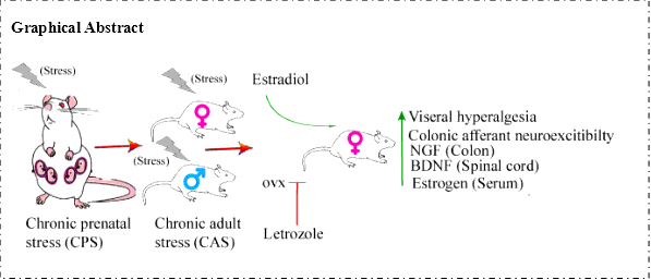
Pregnant dams were subjected to a CPS protocol that consisted of a random sequence of twice-daily applications of one of three stress sessions, one hour water avoidance stress, 45 min cold restraint stress or 20 min forced swim stress starting on 6th day and continuing until delivery (21st day). Male and female offspring from the stressed dams were designated CPS rats. Control dams received sham stress and their offspring were designated control rats. As adults (8-16 wks), control and prenatally stressed offspring were challenged by the same CAS protocol for nine days. Ovariectomy (OVX) or sham surgery was performed on female prenatal stress offspring in the 56th day. Daily Letrozole treatment was initiated on the 49th day, 2 weeks prior to initiation of adult stress. Treatment was continued through the stress protocol. Detail schematic diagram is present in Fig. 1A.
Rattreatment
Before the treatment of OVX or Letrozole, Vaginal smear test was used to identify the estrus cycle phases of female rats. The OVX or sham surgery was performed on female prenatal stress offspring in the 56th day. The aromatase inhibitor Letrozole (4,4’-(1H-1,2,4-triazol-l-yl-methylene)-bis-benzonitrile; 1.0 mg/kg, oral administration; Novartis) was used in experiment group; and Vehicle (hydroxypropyl cellulose 0.3% in water) was used in contral group once daily for 14 days. Direct transcutaneous intrathecal injections (i.t.) of estrogen and letrozole were performed respectively as described by Mestre et al. [21]
In vivo single fiber recording of L6-S2 DRG rootlets
Multiunit afferent discharges were recorded from the distal ends of L6-S2 dorsal rootlets decentralized close to their entry into the spinal cord. A bundle of multiunit fibers was distinguished into 2-6 single units off-line using wave mark template matching in Spike 2 software that differentiates spikes by shape and amplitude. Colonic afferent fibers were identified by their response to graded colorectal distention (CRD). Isoflurane, 2.5%, followed by 50 mg/kg, i.p. sodium pentobarbital induced general anesthesia that was maintained by infusing a mixture of pentobarbital sodium + pancuronium bromide + saline by intravenous infusion through the tail vein. Adequacy of anesthesia was confirmed by the absence of corneal and pupillary reflexes and stability of end-tidal CO2 level. A tracheotomy tube connected to a ventilator system provided a mixture of room air and oxygen. Expired CO2 was monitored and maintained at 3.5%. Body temperature was monitored and maintained at 37 °C by a servo-controlled heating blanket. A laminectomy from T12 to S2 exposed the spinal cord. The head was stabilized in a stereotaxic frame. The dura was gently opened and a warm mineral oil pool, contained by skin flaps, covered the exposed spinal cord and roots as described previously [22].
Invitropatch clamp recordings in colonic DRG neurons
Retrograde fluorescence label injections
Labeling of colon projecting DRG neurons was performed as previously described [20]. Under general 2% isoflurane anesthesia, the lipid soluble fluorescence dye,1,1’-dioleyl-3,3,3’,3’-tetramethylindocarbocyanine methane-sulfonate (9-DiI, Invitrogen, Carlsbad, CA) (50 mg/mL) was injected into muscularis externae on the exposed distal colon in 8 to 10 sites (2 µL each site). To prevent leakage, the needle was kept in place for 1 min following each injection.
Dissociation and culture of DRG neurons
Rats were deeply anesthetized with isoflurane followed by decapitation. Lumbosacral (L6–S2) DRGs were collected in ice-cold and oxygenated dissecting solution, containing (in mM): 130 NaCl, 5 KCl, 2 KH2PO4, 1.5 CaCl2, 6 MgSO4, 10 glucose, and 10 HEPES, pH 7.2 (305 mOsm) [23]. After removal of the connective tissue, the ganglia were transferred to a 5 ml dissecting solution containing collagenase D (1.8 mg/mL; Roche) and trypsin (1.0 mg/mL; Sigma, St Louis, MO), and incubated for 1.5 hours at 34.5°C. DRGs were then taken from the enzyme solution, washed, and put in 0.5 to 2 mL of the dissecting solution containing DNase (0.5 mg/mL; Sigma). Cells were subsequently dissociated by gentle trituration for 10–15 times with fire-polished glass pipettes and placed on acid-cleaned glass coverslips. The dissociated DRG neurons were kept in 1 mL DMEM (with 10% FBS) in an incubator (95% O2/5% CO2) at 37°C overnight.
Whole-cell patch clamp recordings from dissociated DRG neurons
Before each experiment, the glass coverslip with DRG neurons was transferred to recording chamber perfused (1.5 mL/min) with external solution containing (10 mM): 130 NaCl, 5 KCl, 2 KH2PO4, 2.5 CaCl2, 1 MgCl2, 10 HEPES, and 10 glucose, pH adjusted to 7.4 with NaOH (300 mOsm) at room temperature. Recording pipettes, pulled from borosilicate glass tubing, with resistance of 1–5 MΩ, were filled with solution containing (in mM): 100 KMeSO3, 40 KCl, and 10 HEPES, pH 7.25 adjusted with KOH (290 mOsm). DiI labeled neurons were identified under fluorescent microscope. Whole-cell currents and voltage were recorded from DiI-labeled neurons using Dagan 3911 patch clamp amplifier. Data were acquired and analyzed by pCLAMP 9.2 (Molecular Devices, Sunnyvale, CA). The currents were filtered at 2–5 kHz and sampled at 50 or 100s per point. While still under voltage clamp, the Clampex Membrane Test program (Molecular Devices) was used to determine membrane capacitance, Cm and membrane resistance, Rm, during a 10 ms, 5 mV depolarizing pulse form a holding potential of -60 mV. The configuration was then switched to current clamp (0 pA) for determining other electrophysiological properties. After stabilizing for 2–3 min, resting membrane potential was measured. The minimum acceptable resting membrane potential was -40 mV. Spontaneous activity (SA) was then recorded over two 30 second periods separated by 60 s without recording as described by Bedi and Chen [24].
Transient A-type K+ current (IA) recording method in Patch studies
To record voltage-gated K+ current (Kv), Na+ in control external solution was replaced with equimolar choline and the Ca2+ concentration was reduced to 0.03 mM to suppress Ca2+ currents and to prevent Ca2+ channels becoming Na+ conducting. The reduced external Ca2+ would also be expected to suppress Ca2+-activated K+ current. The current traces of Kv in DRG neurons were measured at different holding potentials. The membrane potential was held at -100 mV and voltage steps were from -40 to +30 mV to record the total Kv. The membrane potential was held at -50 mV to record the sustained Kv. The IA currents were calculated by subtracting the sustained current from the total current. The current density (in pA/pF) was calculated by dividing the current amplitude by cell membrane capacitance.
Real time RT-PCR
Total RNA was extracted using the RNeasy Mini Kit (QIAGEN, Valencia, CA). One microgram of total RNA was reverse-transcribed using SuperScriptTM III First-Strand Synthesis System. PCR was performed on a StepOnePlus thermal cycler with 18S as the normalizer using Applied Biosystems primer/probe set Rn02531967_s1 directed against the translated exon IX. Fold-change relative to control was calculated using the ΔΔCt method (Applied Biosystems).
Western Blot
Samples were lysed in RIPA buffer containing protease inhibitor cocktail and phenylmethanesulfonyl fluoride. Lysates were incubated for 30 min on ice and then centrifuged at 10 000×g for 10 min at 4 °C. The protein concentration in the supernatant was determined using BCA kits with bovine serum albumin as a standard. Equal amounts of protein (30 μg per lane) were separated with 10% SDS-PAGE and then transferred to nitrocellullose membranes (Bio-Rad, USA). The membrane was blocked in Li-Cor blocking buffer for 1 h at room temperature and then incubated with primary antibodies. BDNF antibody (Santa Cruz Biotechnologies, Santa Cruz, CA) was used at 1:200 dilution; Nerve growth factor (NGF) antibody (Abcam, MA) was used at 1:1000 dilution; β-actin antibody (Sigma Aldrich, St Louis) was used at 1:5000 dilution. Secondary antibodies used were donkey anti-rabbit alexa fluor 680 (Invitrogen) and goat anti-mouse IRDye 800 (Rockland). Images were acquired and band intensities measured using the Li-Cor Odyssey system (Li-Cor, Lincoln, Nebraska).
Immunofluorescence
Frozen sections of colon mounted on glass slides from control, CAS, CPS and CPS + CAS female rats were rehydrated in phosphate buffered saline at room temperature. All slides were treated for antigen retrieval and blocked with 10% normal goat serum (diluting in 0.3% phosphate buffered saline-Triton) for 1 h. Primary antibody NGF in antibody diluent (Renoir Red, Biocare Medical, Concord, CA) were incubated at 4oC overnight. The slides were exposed to fluorescent dye conjugated secondary antibody for 2 h at room temperature. Slides were counterstained with DAPI and coverslipped. Images were taken in fluorescence mode on an Olympus laser scanning confocal microscope and the average signal intensity was calculated by the bundled software.
Serum Estradiol and norepinephrine Levels
Serum estradiol, adrenocorticotropic hormone (ACTH), and norepinephrine levels were measured using specific ELISA kits for each analyte (CSB-E05110r, CSB-E06875r, CSB-E07022, Cusabio Bioteck CO.LTD, USA) according to the manufacturer’s instructions.
Data analyses
Single fiber responses (impulses/second) to CRD were calculated by subtracting SA from the mean of 30 seconds of the maximal activity during distension. Fibers were considered responsive when CRD increased their activity 30% greater than the baseline value. Mechanosensitive single units were classified into high-threshold (>20 mmHg) and low-threshold (≤20 mmHg) on the basis of their response threshold and profile during CRDs. Single fiber activity data were analyzed using ANOVA with repeated measures; CRD intensity was the repeated factor and experimental group as the between group factor. If significant main effects were present, the individual means were compared using the Fisher post-hoc test.