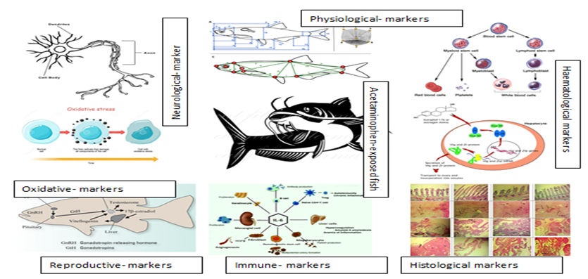
4.1 Effect of Acetaminophen on Physiological Condition Indices and Oxidative stress biomarkers
Exposure to acetaminophen at environmentally relevant concentrations had distinct effects in female post-juvenile Clarias gariepinus. Morphometric analyses demonstrated that the HIS and K of the acetaminophen-exposed fish did show any significant change and this was similar to the report of Stancova et al. (2014) who observed no changes in growth parameters of Tench (Tinca tinca) exposed to a combination of ibuprofen, diclofenac, and carbamazepine for 35 days. Acetaminophen significantly reduced gonadosomatic index of exposed female C. gariepinus and agrees with the report of Mills et al. 2011 who showed a significant decrease in the gonadosomatic index of marine fish Cunner (Tautogolabrus adspersus) exposed to an anticancer drug, Anastrozole.
The biotransformation pathway of APAP has showed that the toxicity is as a result of the formation of the very active metabolite N-acetyl-p-benzo-quinone Imine (NAPQ1) by cytochrome P450s (Macherey, 2015; Ramos-Tavar and Muriel, 2019) when consumed at a non-pharmacologic doses. At the high doses, the pathways for the transformation of APAA (sulfation and glucuronidation) becomes saturated and a portion of APAP is excreted in the parent form leading to excess NAPQ1 which further reacts with other protein groups to form protein adducts (McGill et al., 2013) leading to oxidative stress and mitochondrial dysfunction resulting in the production of free radicals.
The increased SOD enzyme activities must have been triggered by the presence of acetaminophen which produced reactive oxygen species (ROS) (Halliwell and Gutteridge, 1999). As the levels of ROS increases, the biological system creates a first-line defence mechanism by altering the behaviours of SOD (Roberts and Oris, 2004; Bagnyukova et al., 2006). Increased SOD activity is an indication of increased antioxidant status that attempts to neutralize the ROS effect (Kurutas, 2016). The elevated CAT activity in our study might be as a result of the functioning of the defence mechanism, which counteracts the oxidative stress induced by acetaminophen metabolism in exposed catfish (Li et al., 2010; Shukla et al., 2017). Acetaminophens have been reported to disturb the redox status of the organism (Trachootham et al. 2008) and CAT level is an excellent marker of protein oxidation and lipid peroxidation (Bohn, 2019).
Several authors hitherto detailed the significance of GST, GPx, and GSH in averting cellular destruction (Livingstone, 2001; Guiloski et al., 2015). The depletions in GPx activities observed in acetaminophen-exposed fish may have been a sign of the induced distress in the fish and agrees with the findings of Guiloski et al. (2015) who reported a decline in GPx activities in gonads of H. malabaricus exposed to dexamethasone.
GST is a phase II detoxifying enzyme that catalyzes the conjugation of the reduced form of glutathione to xenobiotics for detoxification (Stancova et al., 2017). GST is known to be the first line of defence preventing oxidative stress damage, decomposition of superoxide radicals and hydrogen peroxide before interacting to form reactive hydroxyl, which has many unfavourable biological consequences when present in high concentrations (Kaur et al., 2017). The increase in GST enzyme activity in exposed catfish may have represented the occurrence of a defensive mechanism to prevent the impact of acetaminophen, as indicated by Sayeed et al. (2003).
The increase in TAC activity in exposed fish could have been triggered by the oxidative stress in the exposed fish, which may be due to an increase in antioxidant activity employing a cellular defensive mechanism to achieve homeostasis and, in some way, adaptation in the management of antioxidant stress (Kohen et al., 2000, Castillo et al., 2006). A similar report of an increase in TAC activity is comparable to the report of Mantle et al. (2000) who observed increase in TAC activity in human administered with perioperative anaesthetics dopamine, propofol, dobutamine and noradrenaline.
4.2 Immunological Biomarkers Alteration
C-reactive protein (CRP) is an acute inflammatory protein that can increase its activity at infection or inflammation sites (Sproston and Ashworth, 2018). While it is mainly generated by hepatocytes in the liver, it can also be produced by smooth muscle cells, macrophages, and endothelial cells. CRP is involved in inflammatory processes and host responses to infection, such as the complement pathway, apoptosis, phagocytosis, and the development of cytokines (IL), specifically interleukin-6 and tumor necrosis factor-α (Sproston and Ashworth, 2018; Pathak and Agrawal 2019).
It has been established in previous paragraph that in the metabolism of APAP, abundant NAPQI use up the glutathione (GSH) of the cell and bond the proteins covalently, leading to mitochondrial dysfunction, necrosis of the hepatocyte (Fig. 7) and eventually the death of cells (Hinson et al., 2010; McGill et al., 2012). When parenchymal hepatocytes are impaired, innate immunity plays a role in the development and enhancement of liver injury triggered by APAP. The induction is marked by the release of pro-inflammatory cytokines and chemokines by activated Kupffer cells (resident hepatic macrophages), which leads to signal propagation marked by the facilitation of neutrophil adhesion and transmigration into the hepatic vasculature, which aggravates liver injury (Liu et al., 2004; Liao et al., 2016).
Increased C-reactive protein (CRP) in all acetaminophen-exposed fish may have resulted from the entrance of pro-inflammatory cytokines into the circulatory system of the exposed fish due to the presence of acetaminophen (Gani et al. 2009). Our study revealed that acetaminophen induces the expression of CRP in exposed fish, which may be an indication that the synthesis of the CRP is part of the host defence reaction to the toxic effect of Acetaminophen. In the recent past, the innate immune system of fish has drawn attention and it's considered to be crucial in primary defence and adaptive immunity in fish (Whyte, 2007). Acetaminophen exposure caused elevated hepatocyte injuries as seen in the histopathological analysis of the liver and the increased level of CRP shows the protective function (Sproston and Ashworth 2018). Similarly, Ghosh et al. (1993) and Kodama et al. (2004) documented a rise in CRP activity in fish exposed to Carbaryl and formalin.
Interleukin-6 (IL-6) is one of the most pleiotropic cytokines due to its function in both innate and adaptive immune responses and other physiological processes (Varela et al. 2012). Our study showed increased IL-6 activity in the acetaminophen-exposed fish. This increase may be an indication that IL-6 returned the exposed fishes to a homeostatic condition and it has been stated that IL-6 runs to regulate the magnitude of tissue inflammatory responses (Gabay, 2006, Hong et al., 2013). Interleukin (IL)-6 is developed at the site of inflammation and plays a significant role in the acute phase response as defined by a variety of diagnostic and biological characteristics, such as the production of acute-phase proteins (Gabay, 2006). IL-6 is an inflammatory cytokine that is freed from monocytes, lymphocytes at the sites of tissue injury and may have been freed from the severe liver, gill injuries observed in this study (Gani et al., 2009). Several authors (Kopf et al. 1994, Xing et al. 1994, Romani et al., 1996, Ruzek et al., 1997, Gabay, 2006) have noted that in chronic disorder, usually evidenced by immune stressors such as chronic intracellular infection and tumours, IL-6 not only induce acute phase reactions but also perform an important role in producing cellular immune responses to affected cells and mucosal humoral responses to re-infection.
4.4 Neurotransmitter Biomarker
The inhibition of AChE in all Acetaminophen-exposed fish suggests an adverse consequence in cholinergic neurotransmission, and probably in nervous and neuromuscular function (Ribeiro et al., 2017). The normalization of the enzyme activity in the exposed fish compared to the control can be due to the compensatory response of the fish cell to the toxic effects induced by acetaminophen exposures. This study has shown the susceptibility of AChE activities in plasma of acetaminophen-exposed C. gariepinus (Mdegela et al., 2010). These results indicate that C. gariepinus is a model organism for pollutants monitoring and evaluation (Mdegela et al., 2006) and the activity of AChE in C. gariepinus is a prospective diagnostic tool for environmental pollution assessment of anticholinesterases.
4.5 Haematological Indices
There were increased responses in most haematological parameters of exposed fish except MCV and MCH after the 28 days exposure period. Acetaminophen-exposed fish showed an increase in WBC counts which indicate immune and protective response (Saravanan et al., 2011) to the presence of acetaminophen. The increased WBC counts show that C. gariepinus develop a protective mechanism to control the toxic stress caused by acetaminophen exposure (Mohammod Mostakim et al., 2015). Similarly, ibuprofen and clofibric acid increased WBCs in exposed C. carpio and Cirrhinus mrigala (Saravanan et al., 2011, 2012). Again, the observed increased WBC may reveal physiological and immunological (inflammatory) challenges in response to the toxic impact of the acetaminophen (Alimba et al., 2019). The observed reduction in RBC counts at 25.50µg/L might be inhibition of RBC production by acetaminophen. The reduction in RBC counts in fish may show the anaemic state of the fish under stress conditions (Sigbojorn and Tatjana, 2015). Acetaminophen exposure resulted in increased HGB, HCT, Lymphocyte, platelets counts and this may be ascribed to the stimulation of the hematopoietic system of the fish, caused by the exposure.
4.6 Reproductive Markers
Vitellogenin (VTG) is a known biomarker for the confirmation of endocrine activity in fish and it is used in numerous OECD test guidelines (TG) to detect the activities of chemicals on hormonal pathways (Baumann et al. 2019). Acetaminophen-induced VTG in female fish with increasing concentration. Induction of VTG has been reported in fish following exposure to pharmaceuticals, heavy metals and pesticides (Versonnen et al. 2003, Hook et al. 2014). The increased VTG activities in all exposed catfish correspond to the fact that Estradiol induces high levels of VTG (Versonnen et al. 2003). This study agrees with the report of Hong et al. (2007) who stated that NSAID diclofenac increased VTG expression levels in male Japanese medaka following 96 hours of exposure to concentrations as low as 1µg/L. This study thus suggests that analgesic drugs (acetaminophen) are capable of endocrine disruption via estrogenic effects. Hepatotoxic chemicals, on the other hand, have been shown to dramatically disrupt VTG synthesis in trout liver cells in an in vitro research (Miller et al., 1999, Ayobaha et al., 2020). Baumann et al., 2020 reported similar significant increase (p<0.05) in vtg in female zebrafish exposed to acetaminophen. Furthermore, Wheeler and Coady, 2016 reported that if liver damage worsens, the functional capacity to process hormones may be diminished, potentially leading to higher circulating estrogen and vitellogenin levels. Again VTG production can be influenced not just by endocrine-related pathways, but also by non-endocrine-mediated mechanisms. Because VTG is formed in hepatocytes, hepatotoxicity, or toxicant-induced deterioration of liver structure and function, could affect VTG as a biomarker (Baumann et al., 2020).
Endocrine-disrupting compounds (EDCs) disrupt hormonal pathways in some organisms, negatively influencing reproductive performance (Martin and Voulvoulis, 2009). There was an anti-androgenic effect observed in all acetaminophen-exposed fish as a result of the increased aromatase activity which facilitated the transformation of Testosterone to Estradiol and therefore leads to a reduction of Testosterone (Fenske and Segner 2004, Baumann, 2012). There was a concentration-dependent increase in 17β-estradiol with the corresponding reduction of T. These alterations in Estradiol concentration and aromatase activity are significant as they can lead to the alterations in reproduction (Flippin et al. 2007; Han et al. 2010). Similarly, the report of Guiloski et al. (2015) showed a reduction in testosterone levels when male fish Hoplias malabaricus were exposed to anti-inflammatory drugs diclofenac and dexamethasone. Guiloski et al. (2017) revealed that the anti-androgenic impact of acetaminophen in male fish Rhamdia quelen, as the reduction in testosterone levels and a corresponding increase in the volume of 17β-estradiol were found in fish exposed to higher concentrations of acetaminophen.
4.7 Histopathology Biomarkers
Histological changes are associated with complex biochemical and physiological responses to any stressor (Lushchak et al. 2018). Tissue histology is seen as a sign of exposure to contaminants and is an essential apparatus for assessing pollution levels, especially for short-term exposure and prolonged effects (Mostakim et al. 2015). Changes in fish tissue histology have been commonly used, both in controlled experiments and field studies to assess fish health (Mela et al. 2007, Erhunmwunse et al. 2014, Alimba et al. 2015, Alimba et al. 2019).
“Injury to gill tissue can impede the gas exchange capacity of gill and cause respiratory disturbances, ion modulation and osmoregulation dysfunction, and inefficiency in the removal of waste nitrogen metabolites in exposed fish” (Nero et al. 2006; Cengiz and Unlu, 2006; Velmurugan et al. 2007; Banaee, 2013). The gill morphology showed there were disruptions in the lamellar epithelium, gill hyperplasia, cell proliferation, and lamellar fusion in the gills of acetaminophen-exposed fish. Besides, secondary lamellae often had vascular lesions, such as aneurysms, caused by cell disruption and haemorrhage. These findings indicate a reduced respiratory potential and the development of osmotic imbalances due to acetaminophen-induced stress. Lamellar fusion occurred as a result of the excessive proliferation of filament epithelial cells and is an innate defensive mechanism to shield the lamellar epithelium from direct interaction with acetaminophen (Flores-Lopes and Thomaz 2011; Hadi and Alwan 2012). “The fusion and hyperplasia of gill lamellae can be caused by the effects of acetaminophen altering glycoprotein in the cell-covered mucus, influencing the negative charge of the epithelium and favouring adhesion to adjacent lamellae” (Hadi and Alwan 2012; Malarvizhi et al., 2012). The presence of lamellar telangiectasia in the gills of exposed fishes must have resulted from the break-up of elongated bodies and capillaries under the effect of acetaminophen which leads to the accumulation of erythrocytes in the distal section of the secondary lamellae (Randi et al. 1996; Hadi and Alwan 2012). “Changes such as epithelial lifting, hyperplasia and hypertrophy of the epithelial cells, besides partial fusion of some secondary lamellae, are examples of defence mechanisms, since; in general, these result in the increase of the distance between the external milieu and the blood” (Hadi and Alwan 2012). Hoeger et al. (2005) reported severe histopathological changes in the gill of diclofenac treated fish Salmo trutta.
The liver is primarily involved in metabolism, detoxification, storage, and removal of xenobiotics and their metabolites (Mela et al. 2007, Alimba et al. 2019). Liver morphology of acetaminophen-exposed fish showed several alterations which were concentration-dependent. Degenerated vacuolar, Kuffer cells activation, hepatocellular necrosis, single-cell necrosis, coagulation necrosis, focal necrosis, necrosis with inflammatory cells, spotty necrosis, necrosis characterized by hyperplasia with loss of cellular details and necrosis of hepatocytes characterized the liver. Matos et al. 2007; Sepici-Dinçel et al. 2009; Banaee, 2013 “reported similar histopathological alterations in the liver tissue of O. niloticus and C. carpio exposed to sub-lethal concentrations of carbaryl and cyfluthrin, respectively". Acetaminophen cause liver injury in animals in large concentrations leading to increased liver enzymes (Kumar et al. 2004, Guiloski et al. 2017).
Gonad histological analysis proved useful in understanding and evaluating the impacts of possible endocrine-disrupting compounds on aquatic organisms (Leino et al. 2005). Acetaminophen-exposed female fish showed several altered ovarian structure with patches of degenerative follicles, follicular atresia, necrosis and degenerative nucleus. Atresia follicles were lower in lower concentrations of acetaminophen exposure. Increased follicle atresia in the ovaries of estrogen-exposed fish have been reported by some authors (Länge et al., 2001; Metcalfe et al., 2001; Zillioux et al., 2001; Kidd et al., 2007; Kaptaner and Ünal, 2011). Acetaminophen caused degenerative nucleus and follicle, necrosis atresia, eccentric nucleus, ovarian cell hyperplasia and destructive ovarian wall. Similarly some classes of phthalates have been shown to cause atresia of follicle, degenerative follicle in both human and fish (Hannon and Flaws 2015).
4.8 Multivariate Statistical Analysis
Cazenave et al., 2009 highlighted the significant use of arrays of biomarkers when assessing the biological effects in polluted environments, since one marker may not show the true status of exposed organisms. Beliaeff and Burgeot (2002) have stressed the need for researchers to carefully select suitable fusion of markers that can yield data about universal detrimental environmental effects. The statistical analyses of our results distinctly confirm that the use of a single biomarker analysis was not enough to detect alteration induced by acetaminophen in the in exposed fish. Nevertheless, the assessment of multiple-biomarkers in physiological, biochemical, oxidative, reproductive, immunological and neurological activities in the C. gariepinus validate to visibly differentiate between the control groups and the acetaminophen-exposed groups. Multivariate statistical analysis (Figure 9) indicated that fish in the control groups exhibited a distinctly response from the acetaminophen-exposed fish and that over 95% of the biomarkers significantly contribute to discriminate between the acetaminophen-exposed fish and the control group. Figure 9 denotes that acetaminophen-exposed fish may have undergone several stresses, possibly due to exposure to different acetaminophen concentrations.