The Ag/TA coatings were deposited in ambient aqueous conditions similar to other metal-phenolic coatings.11 Generally, the substrate was first immersed in 500 µL of ultra-pure water followed by the addition of 5 µL of 40 mg mL-1 phenolic solution (TA, or other phenolics) and subsequent mixing. Next, 5 µL to 60 µL of 10 mg mL-1 AgNO3 solution was added followed by vigorous mixing and incubation. The coatings were then rinsed with ultrapure water, dried by air or nitrogen, and then used for subsequent studies. After the addition of AgNO3, complexation rapidly occurred as seen by a near immediate (<10 s) size increase in dynamic light scattering results from ~2 nm (roughly the radius of gyration of TA) to ~100 nm (a similar size to Fe/TA complexes seen in solution) (Figure S1).14 Moreover, the silver peak in UV-Vis spectroscopy disappeared after complexation and the TA phenolic peak shifted to slightly lower wavelengths and no new peaks appeared, suggesting that films made out of these complexes would be transparent (Figure 1A). X-ray Photoelectron Spectroscopy (XPS) demonstrated shifts in the C1s spectra of TA to lower binding energies after interacting with Ag (from 533.1 eV for TA to 531.8 eV for Ag/TA complexes) and noticeable shifts to higher binding energies in the Ag3d spectra for the two silver peaks (from 374.1 eV for AgNO3 to 374.5 eV for Ag/TA complexes) (Figures 1B and S2).18 Fourier-transform infrared (FTIR) spectroscopy demonstrated a shift in the C–O peaks of TA (1011, 1070, and 1300 cm-1) to higher wavenumbers (1023, 1189, and 1303 cm-1, respectively), which is a signature of chelation (Figure 1C).19,20 Similarly, Raman spectroscopy demonstrated increased peak intensity of TA after interacting with Ag, which is common for metal chelation with phenolic compounds (e.g. catechol-metal binding peak at 649 cm-1) (Figure 1D).21,22
Importantly, the coatings made from Ag/TA had negligible absorbance in the visible light spectrum (i.e., were colorless as seen by eye) even after 2 h incubation, with a broad peak in the UV spectrum (Figures S3 and S4). Note that the pH was not raised after mixing to maintain the transparency of the coatings as both NaOH and buffers are known to induce the formation of Ag nanoparticles, which have a strong unappealing color in solution and on substrates (Figure S4). Atomic force microscopy (AFM) images of scratched coatings showed thicknesses of roughly 6–9 nm with root-mean squared roughness of ~1.6 nm after 10 seconds of deposition, with only a slight increase in thickness and roughness even after 20 min incubation (~10 nm and ~2.7 nm RMS roughness), which are both comparable to metal-phenolic coatings made from other metals (Figures 1E and S5).11,12 The thin nature of Ag/TA coatings suggests that coated textiles will remain breathable. The Ag/TA coatings exhibited over an order of magnitude more mass than a monolayer coating of TA as measured by quartz crystal microgravimetry (QCM) (Figure 2). Increasing concentrations of Ag (higher ratio of Ag to TA) led to coatings with more adsorbed mass up to a saturation point after which the mass deposited was fairly stable, likely due to the increased amounts of Ag in the resultant coatings until the free binding sites of TA were fully saturated (Figure S6).
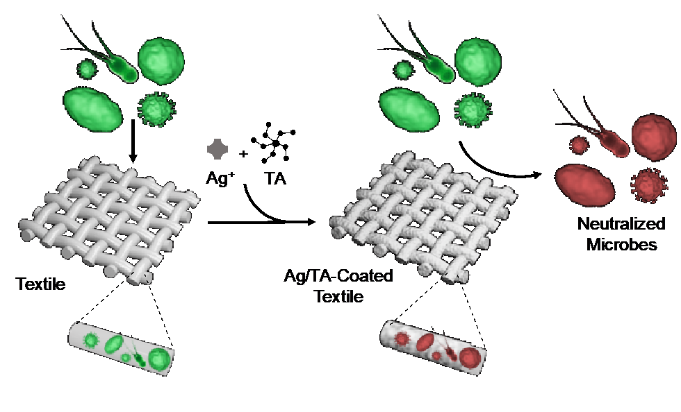
We recently demonstrated that metal-phenolic coatings can interact with lipids, proteins, and polysaccharides,11,16,23,24 suggesting they will be able to interact with the envelopes, capsids, cell membranes and cell walls of viruses, Gram negative and positive bacteria, and fungi. Therefore, the performance of the Ag/TA coatings to capture and neutralize lipid-enveloped viruses was first compared against coatings with different metals using phi6, a safe-to-study bacteriophage often used as a model for pathogens such as Zika and SARS-CoV-2.25,26 The Ag/TA coatings could adsorb a similar amount of virus to TA monolayers and Cu/TA coatings, and roughly 50% more virus mass than Fe/TA coatings (Figure 2A). In terms of virus neutralization, Ag/TA outperformed coatings made from TA alone and outperformed metal-phenolic coatings made from other metals (Cu, Zn, Al, Fe) and TA with a 3-log to 4-log reduction at lower virus concentrations (104 plaque forming units (PFU) mL-1) and 2-log to 3-log reduction at higher virus concentrations (106 PFU mL-1) (Figure 2B). Note that Phi6 was only detectable after incubation with Ag/TA-coated materials at concentrations above 104 PFU mL-1 as the detection limit of the assay was ~10 PFU mL-1. After removing bound virus with surfactant from the textiles incubated at high virus concentrations, their viability was checked and again Ag/TA had at least 2-log fewer viable virus than all of the other samples (Figure S7). Importantly, the antiviral performance of the Ag/TA coatings was maintained for at least 5 vigorous soap and water washing steps, suggesting that such coatings could be used for continued antiviral protection in everyday settings (Figure S8). Finally, other ligands, such as persimmon tannin, could also be used to form Ag/phenolic coatings with comparable antiviral performance to Ag/TA, demonstrating the versatility of this approach (Figure S9).
Although viruses are topically relevant due to the ongoing COVID-19 pandemic, bacteria and fungi pose constant threats to health and quality of life,27 and therefore the Ag/TA coatings were tested against these microbes using different textiles (i.e., cotton, silk, and polyester). The Ag/TA coatings showed significant inhibition on all textiles to E. coli, S. aureus, and S. cerevisiae, while generally the uncoated textiles showed negligible inhibition to these three microbes (Figure 3). Textiles soaked in AgNO3 showed partial inhibition, while uncoated silk also showed some partial inhibition in certain scenarios, as previously documented.28 Moreover, after incubation with E. coli, the Ag/TA-coated textile and uncoated textile were left to sit in LB growth media overnight, and the OD600 of the uncoated textile (high turbidity) was at least 2-log higher than that of the Ag/TA-coated textile (transparent), at OD600 values of 26.6 vs 0.1 for paper, 2.21 vs 0.01 for silk, and 27.3 vs 0.27 for polyester, respectively (Figure S10). Notably, textiles coated with Ag/TA via spraying (rather than immersion), also showed inhibition zones, though they were slightly smaller likely due to the lesser amount of silver available in small volumes used for spraying when compared to the far excess volumes used in immersion (Figure 3D).
Microbes can impact human health, but also play a big role in biological process such as body odor.29,30 Therefore, we further tested whether fabrics sprayed with Ag/TA coatings could reduce odor caused by normal daily life. By coating only one armpit of shirts (polyester and cotton), internal controls between coated (left) and uncoated (right) fabric could be maintained on a day-to-day basis by comparing smell on a 0 to 10 scale at the end of the day (after 10–12 h of wear). Roughly 350 µL of each precursor was sprayed sequentially onto the armpit (roughly 7 x 7 cm) of the shirts and left to dry, afterwards the shirt was worn and machine washed normally. Notably, the Ag/TA coated armpits were nearly odorless across 10 wear/wash cycles, and showed significantly less odor than the uncoated armpits in all scenarios (Figures 4 and S11). Specifically, the uncoated armpit averaged a pungent smell of 5.3 over 3 trials of 11 wears and 10 washes each, while the Ag/TA-coated armpit has a nearly imperceptible average smell of 1.2 over these same wears. The ability of the coatings to withstand machine washing with commercial detergent was expected as phenolic molecules in food and drink can cause stains (e.g., chocolate, wine, and coffee stains) that are notoriously difficult to remove.31 This ability to neutralize odor-causers, in combination to the colorless nature of the coatings, suggests that Ag/TA will be able to find use in various niches of everyday life.