Human patients and samples collection
This study was approved by the Institutional Review Board (IRB)/Independent Ethics Committee of Hospital de la Princesa, according to the Declaration of Helsinki Principles. All methods were performed in accordance with the relevant guidelines and regulations. All participants provided written informed consent. Thereafter, 20 control individuals and 30 patients with untreated plaque psoriasis were enrolled. Patients were eligible for the study if they were adult candidates to systemic therapy. The following washout periods were established: 14 days for topical corticosteroids, 28 days for systemic treatment including corticosteroids, methotrexate, cyclosporine, acitretin or phototherapy and 3 months for biologic agents. From each psoriasis patient, two non-sun-exposed cutaneous biopsies (10 mm) were taken, one from lesional psoriatic skin and another from apparently healthy skin (non-lesional skin). At the same time, 20 ml of peripheral venous blood were extracted. Normal leftover skin samples and peripheral venous blood samples were obtained from 10 surgical patients. Each biopsy was cut in half; one piece was snap frozen for RNA isolation, and the other one included in OCT and stored at -80ºC until processing for immunofluorescence stainings 17.
Quantitative RT-PCR
GADD45a, GADD45b, UCHL1 and IFN-γ mRNA expression levels were determined by quantitative reverse transcription polymerase chain reaction (RT-PCR). Total RNA was isolated from skin samples, peripheral blood CD4+ T cells and moDCs using the TRIzol reagent (Invitrogen) following the manufacturer's instructions. One microgram of RNA was reverse-transcribed to cDNA and amplified with the specific primers pairs using GoTaq qPCR Master Mix (Promega, WI USA). Real‐time (RT)–PCR was performed in a CFX384 Real‐time System (Bio‐Rad) using SYBR Green PCR Master Mix (Applied Biosystems). The data were analysed using StepOne Plus Software (Applied Biosystems®, Carlsbad, CA). GADD45a, GADD45b, UCHL1 and IFN-γ mRNA levels were normalized to GAPDH levels and expressed as relative levels.
Immunofluorescence staining
Skin OCT sections of 5 μm were fixed (formaldehyde 4%), permeabilized (Triton X-100 0,2%) and blocked with 100 µg/ml human γ-globulin (Sigma-Aldrich, St Louis MO, USA) and a 1:100 dilution of donkey serum (Sigma-Aldrich) in phosphate buffer solution (PBS). Skin sections were then incubated over-night with 5µg/ml goat anti-human GADD45a and mouse anti-human CD45 antibodies (Abcam), followed by donkey anti-goat (DAG) Alexa Fluor 488 and DAM Alexa Fluor 555. Finally, cell nuclei were counterstained with DAPI. Negative controls were performed with omission of the primary antibody. Sections were examined with a Leica DMR immunofluorescence microscopy under the same acquisition conditions. Images were analysed using the ImageJ sowftware (http://imagej.softonic.com) GAD45a levels were analysed on regions of interest (ROIs) drawn for CD3+ cells.
Peripheral blood CD4+ T cells and monocyte derived DCs (moDCs) isolation and culture
Peripheral blood mononuclear cells (PBMCs) were obtained by density gradient and CD4+ T cells were isolated by negative selection using magnetic microbeads (Miltenyi Biotec Bergisch Gladbach, Germany). Where indicated, CD4+ T cells were incubated for 24 h in the presence of IL-12 (10ng/ml) plus IL-18 (10ng/ml). For moDCs, PBMCs were allowed to adhere for 30 min at 37ºC, and plastic adhered cells were cultured for 5 days in complete RPMI medium supplemented with 500 U/ml GM-CSF (Peprotech) and 10 ng/ml IL-4 (R&D systems). On day 6, 10ng/ml LPS were added and after 24 h cells were harvested for analysis.
Expression of p38 (pTryr180/pThr183) in CD4+ T cells analysis by flow cytometry
Isolated CD4+ T cells were incubated with anti-CD3/CD28 (10 µg/ml and 5 µg/ml respectively) during 30 min at 4ºC and then with anti-mouse Fc for additional 30 min at 4º and then immediately incubated at 37ºC. After 15min, cells were fixed, permeabilized and stained with mouse anti-human p38 (Becton-Dickinson®) following manufacturer’s instructions and analyzed in a FACScanto flow cytometer (BD Bioscience).
Methylation datasets
Datasets from DNA methylation arrays were obtained from the repository Gene Expression Omnibus (https://www.ncbi.nlm.nih.gov/geo/). We searched for available methylation datasets performed in psoriasis patients. Three datasets of psoriasis skin samples fulfilled these criteria: GSE6331518, GSE7389419 and GSE11579720. Skin punch biopsies of 4 mm diameter were collected. Methylation was analysed with an Illumina Infinium Human Methylation 450k BeadChip array following manufacturing protocol.
Although the three selected studies fulfil the inclusion criteria, and were performed with the same sampling and technology, we would like to point out the heterogeneity regarding the objectives of the three studies, and their differences: (1) GSE63315 dataset 1, data comes from 12 pre-UV irradiation moderate-to-severe psoriasis patients and 12 healthy controls; (2) GSE115797 dataset 3 contained lesional (L) and non-lesional (NL) samples from 24 moderate-to-severe plaque psoriasis patients; (3) GSE73894 dataset 2 combines both types of data including 114 samples of lesional skin, 41 of non-lesional skin and 62 of healthy subjects 18-22.
Statistical analyses
Data were analysed with GraphPad Prism (GraphPad Software, San Diego, CA, USA). The Kruskal-Wallis and Mann-Whitney U-tests were used, as appropriate. Where indicated, Wilcoxon signed rank test was used to analyze paired data. The Spearman test was used for correlation analysis. Significance was set at *p < 0.05, **p < 0.01, ***p < 0.001.
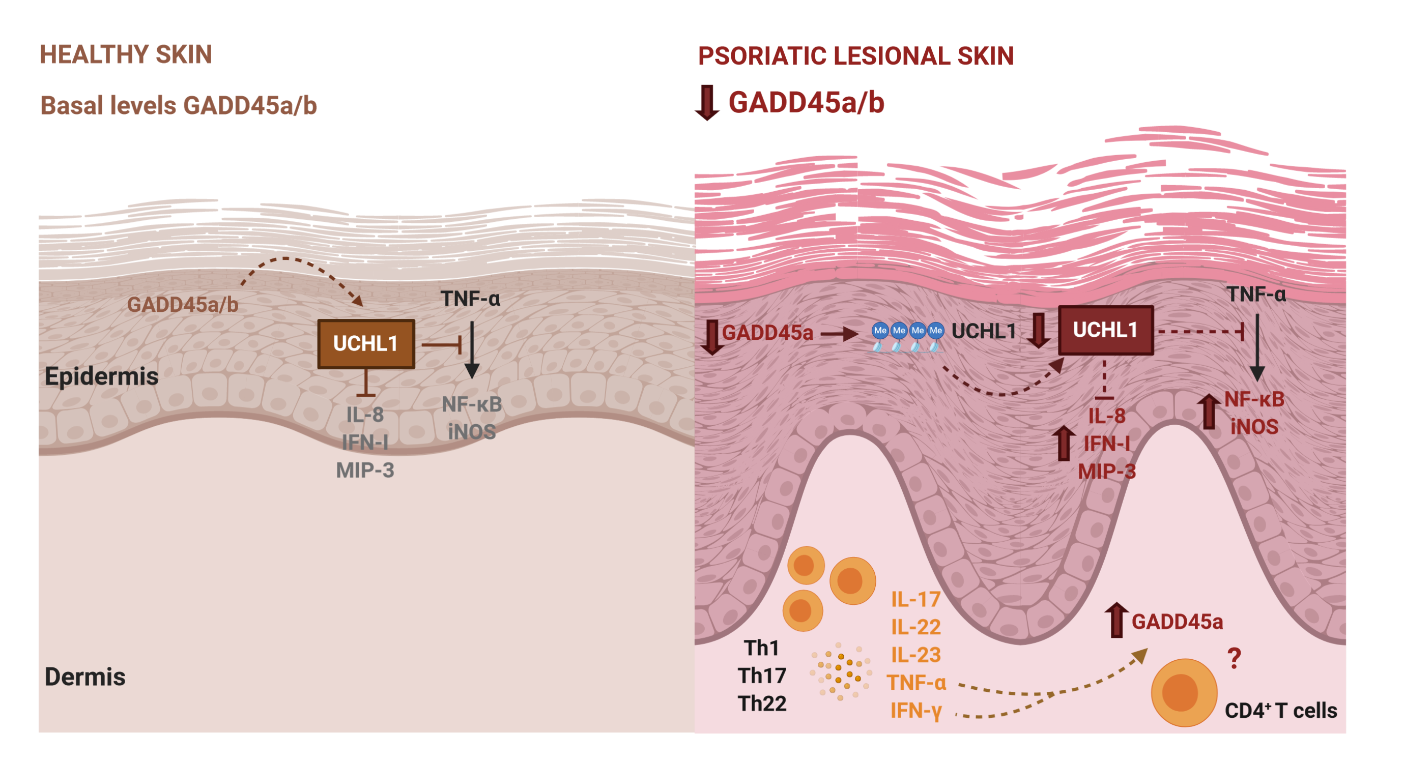
Differentially methylated CpG sites between psoriasis and controls were detected by GEO2R analysis tool (https://www.ncbi.nlm.nih.gov/geo/geo2r/) a web-based program that employs the Bioconductor packages GEOQuery 23 and limma21 in R, with the Benjamini-Hochberg false-discovery rate (FDR). Log2Fold-change of methylation was calculated in psoriasis lesional skin referred to control (psoriasis non-lesional skin or healthy controls’ skin). Thus, positive values mean that UCHL1 is hypermethylated in psoriasis skin with respect to controls and negative values, hypomethylated. Although we have analysed epigenetic differences in all the methylation sites included in this array (485,000), we have focused on those sites located on CpG islands of UCHL1 promoter that present a FDR adjusted p-value lower than 0.05. We have selected this region since CpG island hypermethylation is commonly associated with gene repression.