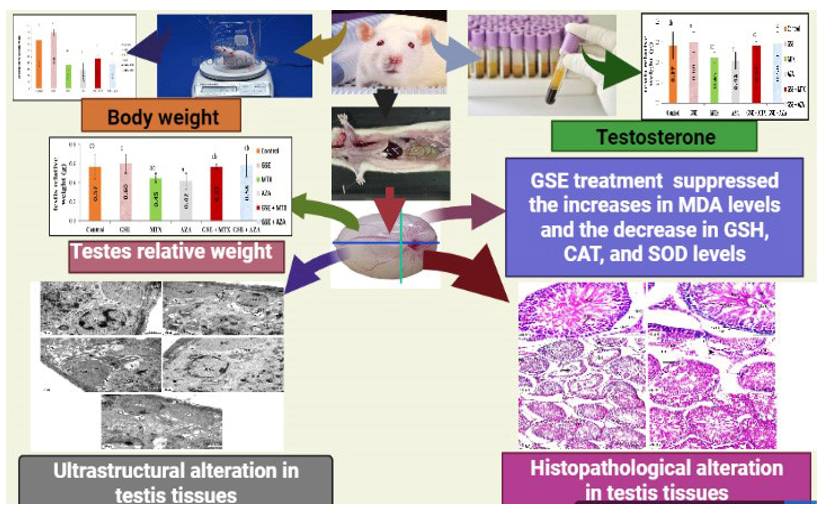
3.4 Testis antioxidant parameters and oxidative stress
Lipid peroxidation (LP), articulated as MDA concentration was measured as a biomarker of testis OS state. It is demonstrated a significant increase in MTX and AZA treated groups when compared to control and GSE treated ones. GSE treatment agent produced a potential improvement of the LP level for both MTX and AZA treated groups. Concerning the enzymatic and non-enzymatic antioxidant defense system, CAT, SOD, and GSH all revealed a noticeable reduction in MTX and AZA groups (P< 0.05). On the other hand, GSE improved the activities of these antioxidant enzymes and increased GSH levels in the treated groups (Table 1).
Table 1
The potential preventive effect of GSE against MTX and AZA induced changes in MDA level, CAT, SOD activity and GSH concentration in testis tissue of all experimental groups.
|
Parameters
|
MDA
(nmol/g tissue)
|
CAT
(K × 102)
|
(GSH)
(nmol/g tissue)
|
(SOD)
(U/g tissue)
|
|
Control
|
22.861 ± 1.9d
|
107.60 ± 2.8c
|
20.84 ± 0.93cd
|
121.97 ± 2.2c
|
|
GSE
|
20.83 ± 1.05d
|
112.49 ± 5c
|
22.14 ± 0.82d
|
132.34 ± 1.9c
|
|
MTX
|
78.33 ± 2.4a
|
51.93 ± 2a
|
9.29 ± 1.3a
|
59.87 ± 3.1a
|
|
AZA
|
86.55 ± 1.7a
|
41.40 ± 3.7a
|
6.97 ± 1.2a
|
53.52 ± 1.3a
|
|
GSE+MTX
|
40.95 ± 2.1c
|
77.75 ± 1.8b
|
16.78 ± 0.34cb
|
94.68 ± 1.3b
|
|
GSE + AZA
|
56.4 ±5b
|
71.44 ± 3b
|
14.95 ± 0.61b
|
84.03 ± 2.5b
|
| Each value represents the Mean ± standard error (SE) |
| Values with different superscript letters are considered significantly different (P< 0.05) |
| Values with letter (a) is considered significantly different (P< 0.05). |
| b,c,d not significantly different from control at p< 0.05. |
3.5 Histopathological changes
Examination of testicular tissue of both control and GSE-treated animals revealed normal seminiferous tubules with active spermatogenesis. Spermatogonia and triangular Sertoli cell resting upon the basement membrane. Leydig cell and cluster of spermatozoa were seen in lumen. Primary spermatocytes were recognized by their large nuclei containing coarse clumps of chromatin, and spermatids appeared with rounded nuclei in control (Fig. 4a and b) and GSE (Fig. 4c and d).
Animals treated with MTX showed severe degenerated and variable-sized seminiferous tubules, where many tubules appeared with a marked decrease in the spermatogenic cells and few or no sperms. Detachment of spermatogenic cells from the basal lamina and vacuolated cytoplasm was observed. There was sloughing of spermatogenic cells into the lumen of seminiferous tubules (Fig. 4e).
Animals treated with AZA revealed degenerated seminiferous tubules with ruptured basement membrane were observed. Notice the detachment of spermatogenic cells from the basal lamina and vacuolated cytoplasm. The reduction of spermatogenic cells and pyknosis of some nuclei were also seen (Fig. 4f).
Examination of testes of animals treated with GSE plus MTX revealed approximate recovery of seminiferous tubules amelioration of spermatogenic cells, and spermatozoa except few vacuoles were observed. Normal Leydig cells were seen in the interstitial tissue (Fig. 4g). Examination of testes of animals treated with GSE and AZA showed improvement of spermatogenesis, nearly normal structure in most seminiferous tubules. Compact spermatogenic layers with few degenerative germ cells and normal Leydig cells were seen (Fig. 4h).
3.6 Ultrastructural changes
The electron microscopic examination of the control testis revealed the spermatogonia rested on the basement membrane, myoid cell, the Sertoli cell had a triangular nucleus, primary spermatocyte with mitochondria, and large spherical nucleus (Fig. 5a). There was a Rounded Spermatid with spherical nuclei, acrosomal cap, and peripherally located mitochondria were observed (Fig. 5b). In addition to lumen contains normal spermatozoan at the midpiece and tail region (Fig. 5c). Normal interstitial tissue containing Leydig cell with a large nucleus, thin rim of chromatin, prominent nucleolus, lipid droplet, and capillaries (Fig. 5d).
The ultrastructure observation of testes of animals administered with MTX revealed many vacuoles and vacuolated mitochondria in spermatogonia, Sertoli cell, and primary spermatocyte. Presence of some lysosome in the Sertoli cells and spermatogonia (Fig. 6a). Distorted spermatid with marked cytoplasmic vacuolation and rarified cytoplasm surrounded by a large lytiareashe presence of some lysosome and some degenerated mitochondria was observed (Fig. 6b). A marked decrease in the number of sperms in the lumen of the seminiferous tubule (Fig. 6c). Abnormal interstitial tissue with degenerated Leydig cell with an irregular nucleus having a thick rim of heterochromatin, few lipids drop and dilated smooth endoplasmic reticulum were also seen (Fig. 6d).
The ultrastructure observation of testes of animals administered with AZA showed many vacuoles in both Sertoli cells and spermatogonia cells and an overall decrease in cytoplasmic ground substance (Fig. 7a). Thick ruptured irregular basement membrane, vacuolated primary spermatocyte, and severe degenerated Sertoli cells with many vacuoles and some lysosomes were detected (Fig. 7b). There was distorted and vacuolated early spermatid. Notice the dissolution of some cells (Fig. 7c). Degenerated vacuolated spermatid and dissolution of some cells were observed (Fig. 7d). In addition to degenerated Leydig cell. Notice the irregular nuclear envelope with dark clumps of heterochromatin adjacent to the nuclear membrane and notice presence of few lysosomes (Fig. 7e).
The ultrastructure observation of testes of animals administered with GSE and MTX showed amelioration in the structure of spermatogonium, Sertoli cell and primary spermatocyte except few degenerated mitochondrial (Fig. 8a). Normal spermatid appeared with a rounded nucleus and peripherally located mitochondria except a few cytoplasmic vacuolations and a few lysosomes (Fig. 8b). Lumen with marked recovery in transverse sections of normal sperms at mid piece and tail region (Fig. 8c). Leydig cells with a large nucleus, smooth endoplasmic reticulum, lipid droplets, and blood capillaries were seen (Fig. 8d).
The ultrastructure observation of testes of animals administered with GSE and AZA revealed a slightly normal basement membrane, improvement in the structure of cells except vacuolation in Sertoli cell, spermatogonia, primary spermatocyte, and spermatid which with a rounded nucleus and normal acrosomal cap (Fig. 9a). In addition, around spermatid appeared with an acrosomal cap, peripherally located mitochondria, and few cytoplasmic vacuolations (Fig. 9b). Round spermatid and a moderate number of cross-sections at the midpiece and tail region of sperms were seen in the lumen (Fig. 9c). Interstitial tissue and Leydig cell with a normal nucleus and a moderate number of lipid droplets were observed (Fig. 9d).