Dry eye is a multifactorial disease with complicated pathophysiological process, various symptoms and causes. Dry eye is a disease of ocular surface, which including the cornea, conjunctiva, eyelids, tear film, lacrimal glands, and the meibomian glands[22]. The disorder of the function or injury of different parts will cause the change of ocular surface state. Disrupted tear film homeostasis is the key link of the onset of dry eye[5]. Trouble is, there are also many underlying causes result in disrupted tear film homeostasis[23, 24]. The clinical symptoms of dry eye are various, from redness, painful, gritty eyes to visual impairment[25]. The difference between patients is huge. Therefore, dry eye is lack of a unified standard for diagnosis and classification which was a stumbling block for the diagnosis. Above all, it is urgent to identify effective biomarkers for diagnosis.
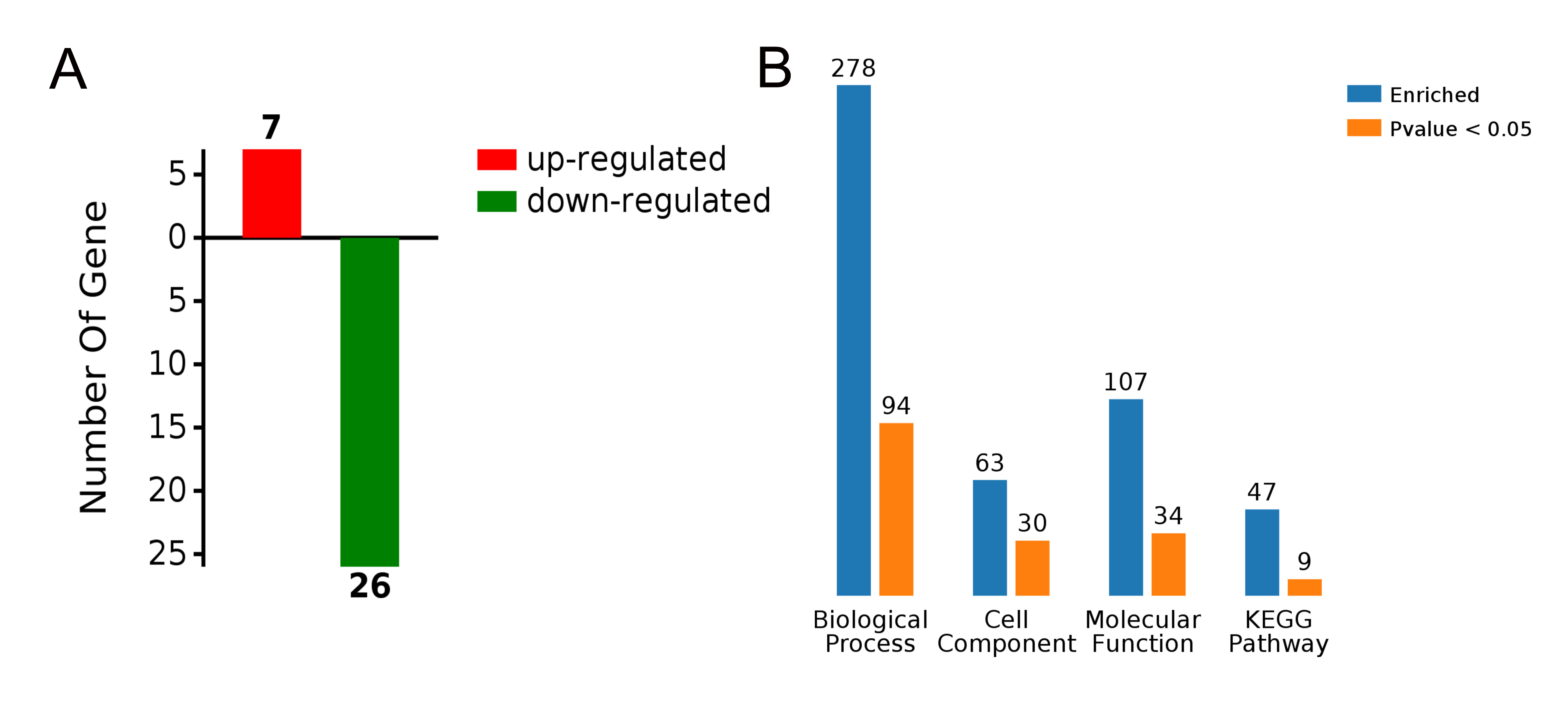
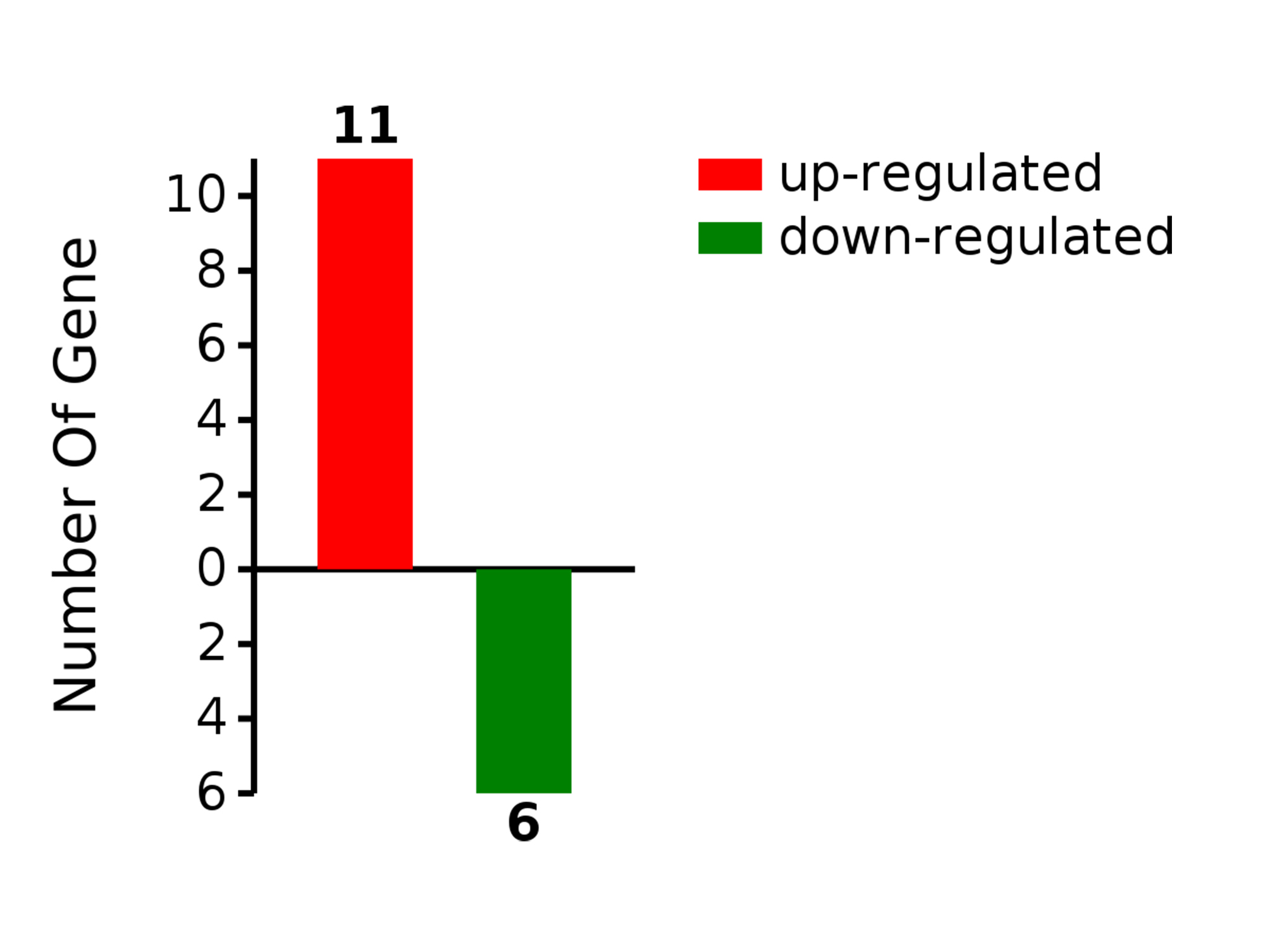
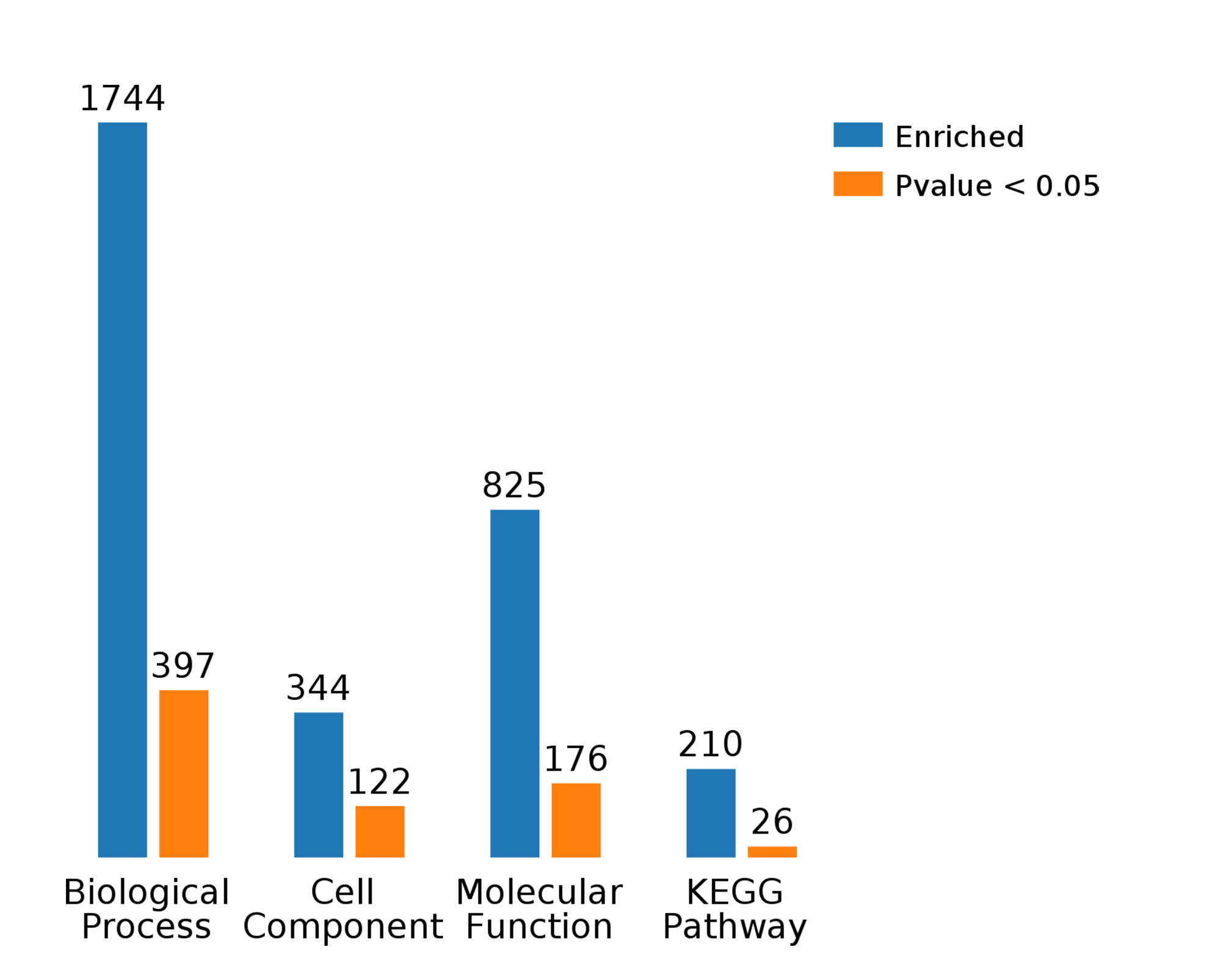
Proteomic analysis can provide theoretical basis of pathogenesis of disease. In the study, 33 DEPs have been identified in tears. 7 of DEPs in tears are up-regulated proteins and the rest are down-regulated proteins. Similarly, 17 differentially expressed proteins have been identified in serum. 11 of DEPs in serum are up-regulated proteins and the rest are down-regulated proteins. KEGG enrichment analysis in tears has discovered Glucagon signaling pathway and neurotrophin signaling pathway may play an important role in the pathogenesis of dry eye. Gene ontology (GO) enrichment analysis in serum has discovered insulin-like growth factor binding and growth factor binding in molecular function probably make effort in pathogenesis of dry eye. KEGG analysis in serum has discovered salivary secretion may be the key pathway in pathogenesis of dry eye.
Glucagon is a hormone that secreted by pancreatic cells. Its function is increasing blood glucose, by promoting glycogenolysis and gluconeogenesis. Glucagon regulates insulin and sustains glucose homeostasis in human body[26]. The glucagon binds to a G Protein Coupled Receptor (GPCR) in the cell membrane. Then, adenylate cyclase is activated to increase cAMP in cells which activates the protein kinase A to phosphorylate and activates enzymes in the target cell[27]. Protein G7PCH4 and Q2PG17 were involved in the pathway. First of all, high glucose is toxic to epithelial cells of meibomian gland[24]. The damage of meibomian gland is one of the important causes of dry eye[28]. Secondly, glucagon induced phosphorylation of insulin receptors. Insulin receptors were found in lacrimal and ocular surface tissue[29]. At the same time, animal studies have shown that insulin loss causes lacrimal gland shrinkage, corneal innervation, and decreased tear volume[30, 31]. In human, insulin resistance was observed with oral estrogen and progesterone replacement therapy in menopause women[32]. Glucagon signaling pathway play an important role in dry eye.
Neurotrophins is a family of proteins that regulates the differentiation and survival of nerve cells. It is activated by engaging special receptors, such as Trk tyrosine kinase receptors and p75 neurotrophin receptor[33]. The neurotrophin signaling pathway is regulated by connecting multiple intracellular signaling cascades[34]. Protein G7PT55 and Q2PG17 were involved in the pathway. Neurotrophin (e.g. NGF, GDNF) is one of the chemical mediators responsible for the inflammatory environment of the ocular surface[35]. Increasing sustained neural activity by reducing the stimulation threshold of sensory at nerve endings. At the same time, these neurotrophins are key reverse messengers that induce changes in gene expression[36]. Inflammatory environment of the ocular surface plays an important role in the pathogenesis and long-term course of DED and is a major driver of injury, and regeneration of peripheral sensory nerves[37].
Growth factor is a protein that stimulates cell growth, proliferation, healing, and differentiation. It's a signal molecule between cells. It works by binding to receptors[38]. Protein G7P1T1 and G7PUN9 were involved in both insulin-like growth factor binding and growth factor binding molecule function. Among them, insulin-like growth factor (IGF) has been studied in the pathogenesis of dry eye. The IGF-binding protein also known as a carrier protein for IGF-1. It mediates growth hormone (GH). The receptors of GH and IGF-1 and associated signaling pathways have been found in lacrimal and ocular surface tissues[39]. There is evidence that these hormones can affect tissue development and wound healing[40].
GH binds to a predimerized GH receptor on the cell membrane to activate the intracellular Janus-kinase 2 (JAK2) pathway, which can further activate Signal Transducers and Activators of Transcription (STATs)[29]. It regulates the transcription of GH, including IGF-1. GH induces cells to secrete IGF-1, which can enhance and supplement the effect of GH. IGF-1 signaling is initiated by binding to membrane bound insulin-like growth factor receptor (IGF-1R). IGF-1R phosphorylates and activates insulin receptor substrate (IRS)-1 which actives the cascade of phosphoinositide 3-kinase (PI3K)/Akt pathway. It is an important regulator of cell cycle progression and cell survival[41].
Studies have shown that GH/IGF-1 axis regulates the growth and function of the meibomian gland[42]. IGF-1 may be an autoimmune target in Sjogren's syndrome[18]. Sex hormones play an important role in regulating GH and IGF-1. There are a lot of kinds of growth factors. Markedly, nerve growth factor and its receptor levels are upregulated after ocular injury[43]. Epidermal growth factor and transforming growth factor were found elevated in the protein level of hydro-deficient dry eye.
Salivary secretion is a nerve-mediated reflex. It is stimulated by neurotransmitters released from autonomic nerve endings. Protein G8F302 was involved in the pathway. Sympathetic fibers are found surrounding the arteries and arterioles in the lacrimal stroma[44]. The sympathetic nerve relaxes the blood vessels in lacrimal gland with the purpose of changing the blood flow and increasing the secretion at the same time[45]. Sympathetic neurotransmitters can also directly induce the secretion, including proteins, electrolytes and water. Glycosylation of mucin was observed in dry eye disease[46]. Salivary acidification of MUC16 detected in patients with dry eyes is correlated with severity of the disease[47].
In this study, we constructed a dry-eye animal model of deovariated cynomolgus monkeys and performed proteomic analysis in the level of tears and serum. Protein G7PCH4, Q2PG17 and G7PT55 in tears may be the key protein in pathogenesis of dry eyes. Protein G7P1T1, G7PUN9 and G8F302 in serum may play an important role in pathogenesis of dry eyes. Similar pathways and molecular function have been found in human being. It may provide suspicious biomarkers for the diagnosis and reveal the potential pathogenesis of dry eyes in menopausal women. Notably, the differences between cynomolgus monkeys and menopausal women should be taken into consideration. Our study provides potential biomarkers in protein level and useful dry-eye animal models for the further study.