Characterization of Major Components of PM2.5 Particulates
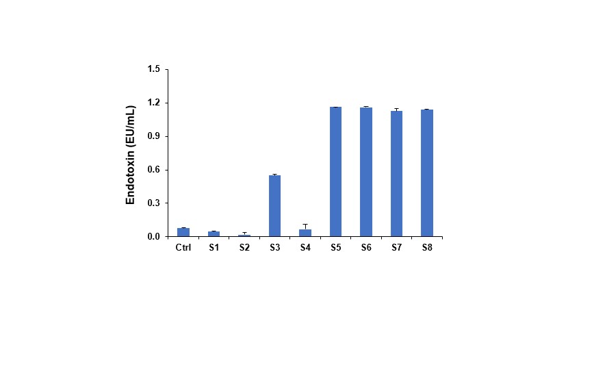
Eight PM2.5 samples were collected on filters (labeled as S1 to S8, PM2.5) on different days in November 2017 in Zhengzhou, Henan, China. The control (Ctrl) is the blank filters without PM2.5 samples, (Table 1). The collected filters were cut in small pieces and gently rinsed in DI H2O for three times to wash off the PM2.5 particles on the filters. The metal and metalloid components of each sample were determined by inductively coupled plasma mass spectrometer (ICP-MS). Total 14 metal and metalloid elements (Table 2) were analyzed, where aluminum (Al), barium (Ba), zinc (Zn), iron (Fe) and sulfur (S) were the most abundant elements (Table 2). We carried out physicochemical characterization of the particle suspensions. We assessed the primary size, hydrodynamic size and zeta potential of these PM2.5 particles in DI water as well as in tissue culture medium (RPMI 1640) prior to biological experimentation (Table 3). The results showed heterogeneous sizes of these PM2.5 samples in DI water and RPMI 1640 ranging from 20.8 nm to 66.5 μm (Table 3). All the particles showed negative zeta-potentials in DI water and the media tested ranging from -5.8 mV to -45.7 mV (Table 3). The endotoxin content of all the particles was lower than 1.2 EU/mL, as determined by the Limulus Amebocyte Lysate (LAL) assay (Figure S1).
The Ability of PM2.5 Particulates to Induce NLRP3 Inflammasome Activation and IL-1β Production in THP-1 cells
Because our ultimate goal is to assess the profibrogenic effects of PM2.5 particles in the lung, we used a well-established in vitro platform to gain an understanding of the cooperation between macrophages and cellular elements of the epithelial–mesenchymal trophic (EMT) as the basis for pulmonary fibrosis.[26-28] The synergy between macrophage released IL-1β and TGF-β1 production by the EMT unit plays a major role in collagen production and deposition in the lung.[29, 30] In order to model the events leading to IL-1β production, we used a human monomyelocytic leukemia cell line, THP-1, which differentiates into a myeloid phenotype in response to a phorbol 12-myristate 13-acetate (PMA) treatment.[31] THP-1 cells have been used successfully to look at the effect of nanomaterials, such as CNTs, graphene, GO, rare earth oxides and nanowires, on the induction of IL-1β release by phagocytic cells.[16, 17, 19-23, 26-28, 31-38] This pro-inflammatory response is dependent on lysosomal damage and the assembly of the NLRP3 inflammasome, which is responsible for pro-IL-1β cleavage and IL-1β release.[12, 17, 23] While exposure of THP-1 cells to PM2.5 particles did not induce significant loss of cell viability in cells (Figure 2A and B) as determined by conducting MTS (metabolic activity) and ATP (cell energy) assay; but did trigger robust IL-1β release into the THP-1 culture medium (Figure 2C). The overall abundance of IL-1β production was S6 > S7 > S5 > S8 > S3 > S2 > S1 > S4 (Figure 2C). In addition, the exposure to PM2.5 particles also showed the significant increase of TNF-α level in THP-1 cells at similar trend (Figure 3). This confirmed the ability of these PM2.5 particles to induce pro-inflammatory effects in vitro.
In order to confirm the involvement of the NLRP3 inflammasome, we used NLRP3-deficient (NLRP3–/–) as well as ASC-deficient (ASC–/–) THP-1 cells to demonstrate that gene knockout leads to abrogation of the IL-1β response to PM2.5 particles (Figure 2D and 2E). The initiation of this response by PM2.5 particles is dependent on lysosomal damage, which leads to cathepsin B release and provides a robust signal for assembly of the NLRP3 subunits in phagocytic cells. The role of cathepsin B was confirmed by using a cathepsin B inhibitor, CA-074 methyl ester, to show interference in IL-1β production (Figure 2F).
The Ability of PM2.5 particles to Induce TGF-β1 Production in BEAS-2B Cells
The epithelial cell is an important component of the trophic cell unit undergoing epithelial–mesenchymal transition in the lung during development of fibrosis.[29, 30] The production of TGF-β1 by the epithelial cells contributes in synergistic fashion to the establishment of a matrix synthesis phenotype and collagen synthesis by the participating mesenchymal cellular elements and other pro-fibrogenic cytokines such as PDGF-AA.[29, 30] We chose BEAS-2B cells, a human lung epithelial cell line, to determine whether the PM2.5 particles impact the synthesis of this growth factor, as determined by conducting an ELISA of the supernatant. The results showed that similar to THP-1 cells, none of the PM2.5 samples were associated with cytotoxicity in BEAS-2B cells (Figure 4A). Moreover, all these PM2.5 samples induced TGF-β1 production irrespective of the particle types (Figure 4B). Taken together with the IL-1β data, these results show that PM2.5 particles could induce pro-inflammatory and profibrogenic cytokine production in vitro.
Exposure to Mouse Lung to Determine the Fibrogenic Potential of PM2.5 Particulates
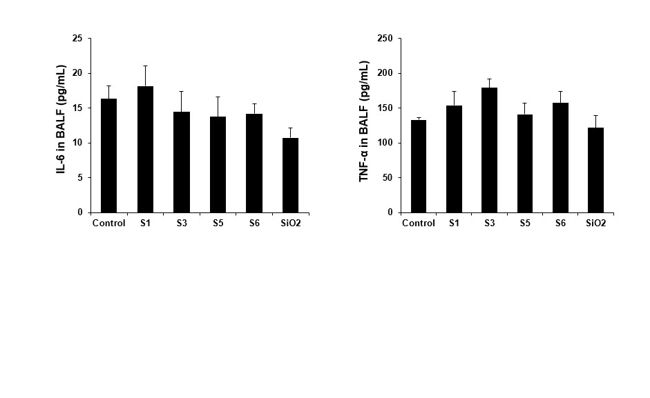
We have previously demonstrated for silica and carbonaceous nanomaterials that the in vitro profibrogenic responses in THP-1 and BEAS-2B cells largely reflect their ability to generate similar biomarker responses in the bronchoalveolar lavage fluid (BALF) of mice receiving oropharyngeal aspiration.[22, 26, 32] The C57BL/6 mice were used for exposure to S1, S3, S5 and S6 PM2.5 particles, which correspondence to low, medium and high response level of pro-inflammatory and profibrogenic effects in vitro. The mice were sacrificed at 21 days after the oropharyngeal inspiration of the PM2.5 particles. Aspiration of Min-U-Sil (quartz or crystalline SiO2) was used as positive control. Examination of the BALF fluid showed that all the particles could induce an increase in eosinophil cell counts at the order of S1 and S3 <S5 and S6, with an association with allergic response. The eosinophil cell counts for S5 and S6 were significantly higher than S1 and S3 (Figure 4A and B). Although this effect was accompanied by a nonresponsive effect in macrophage cell count (Figure 4C), neutrophil cell count (Figure 4D), LIX levels for all particles tested (Figure 4E) as well as IL-6 and TNF-α (Figure S2), there was a significant increase of TGF-β1 for all PM2.5 particles tested (Figure 4F) with an order of S1 <S3 <S5 <S6. This particle dependent profibrogenic effect was consistent with the IL-1β production data in vitro (Figure 2A). The trend toward more inflammation by S6 was confirmed by haematoxylin and eosin (H&E) staining, which showed visual evidence of more inflammatory infiltrates around small- and medium-sized airways in the lung (Figure 5). Again, the effect of the S6 was most pronounced (Figure 5). SiO2 induced significant increases in neutrophil counts, LIX and TGF-β1 levels in the BALF along with inflammatory changes in the lung (Figure 4 and 5).
Moreover, the S6 but not S1, S3 or S5 induced apparent increase in collagen deposition around airways and alveolar spaces of mice, as determined by the Masson’s trichrome staining (Figure 6A). In contrast, no trichrome staining was seen in the lungs of animals receiving S1, S3 or S5. This was confirmed by the Sircol collagen assay, which showed that the exposure to S6 significantly increased the collagen production in the mouse lung (Figure 6B). The exposure to S1, S3 or S5 could also induce an increase in collagen production, but was not statistically significant if compared to the none-treated control; while SiO2 (positive control) could induce a significant effect both in the Masson’s trichrome staining and Sircol collagen assay (Figure 6A and B). The data above demonstrate that PM2.5 particles can cause pro-inflammatory and profibrogenic effects in vitro and in vivo. In addition, the ranking of in vitro IL-1β productions relatively correlate with the in vivo TGF-β1 production and collagen deposition.