In December 2019, Wuhan, the capital of Hubei Province in China, became the center of the outbreak of pneumonia with an unknown etiology. By January 7, 2020, Сhinese scientists identified the causative infectious agent as a novel strain of coronavirus and named it SARS-CoV-2. SARS-CoV-2 causes COVID-19, a very complex disease with a wide spectrum of symptoms. Now it is known for sure that at first SARS-CoV-2 invades the epithelium of the upper and lower airways, but in the case of severe course, it reaches the gas exchange units of the lungs and infects alveolar type II cells29.
Clinical observations quickly established that the main cause of the severe course of COVID-19 is not the lung damage itself, but hyperactive immune response leading to cytokine storm, systemic inflammation, and downstream acute respiratory distress syndrome (ARDS)30. It was shown that the virus has complex interaction with the innate and adaptive immunity and can directly infect the immune cells. Viral particles and SARS-CoV-2 RNA were found in monocytes, CD4 + T cells, CD8 + T-cells, and B-cells31, 32, 33.
Many reports show that besides the lungs and the immune system, an important target of SARS-CoV-2 is the vascular endothelium34. After the beginning of the disease outbreak, quickly accumulated autopsy data revealed thromboembolic complications and microthrombosis in COVID-19 patients35, 36. Recently, Monteil et al. reported successful SARS-CoV-2 inoculation in blood vessel organoids37. Furthermore, some researchers directly observed the presence of viral particles in endothelial cells of patients38, 39. In sum, these data show that the vasculature involvement may be the key link in the pathogenesis of COVID-19 because endothelium is a master regulator of hemodynamics, hemostasis, and immune cells homing.
Another tissue classically involved in the coronavirus infection is the nervous system. Clinical observations of COVID-19 patients have shown neurological complications such as headache, loss of smell, encephalopathy, cognitive impairment, and ischemic stroke40. Most likely, these symptoms are caused by the systemic inflammatory syndrome or direct SARS-CoV-2 replication in the nervous system. Using human brain organoids, mice overexpressing human ACE2, and brain autopsy, Song et al. have shown neuroinvasive activity and direct infection of neurons by SARS-CoV-2 through the ACE2-dependent pathway41.
Unfortunately, the list of potential targets for COVID-19 is growing. There are reports of the involvement of other organs with the high expression of ACE2, such as the intestines, kidneys, and heart42. As ACE2 is highly expressed in tissues of the male reproductive system, there are concerns that SARS-CoV-2 can cause infertility in men43, 44. These concerns are partially confirmed by cases of orchitis following the outbreak of SARS-CoV in 200245 and sex hormones imbalance in male patients with COVID-1946.
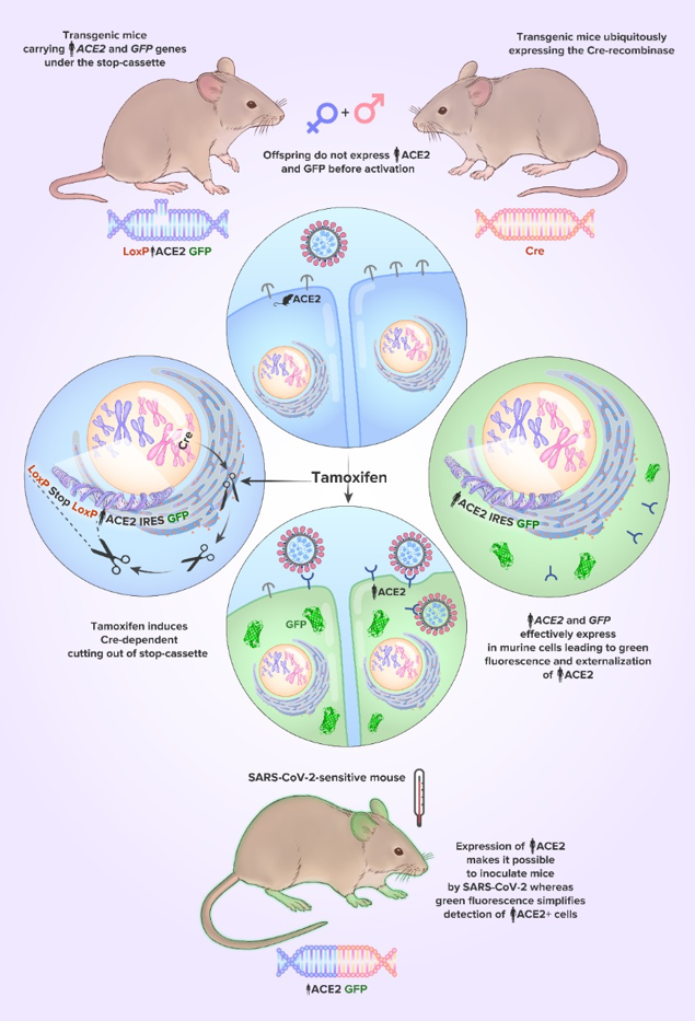
Thus, COVID-19 has very complex and multifaceted pathogenesis. The revelation of its pathways is an essential way in the search for effective approaches to therapy. Herein, we describe novel transgenic mice with the Cre-dependent co-expression of hACE2 and GFP, designed to study the role of different tissues in SARS-CoV-2 infection. For instance, specific activation of hACE2 in endothelial, immune, or alveolar cells, can help to reveal the primary causative tissue, which involvement leads to the acute respiratory distress syndrome in COVID-19. Another option, for example, is to study the fertility of SARS-CoV-2-infected male mice with testicular-specific activation of hACE2. In this way the fertility will depend only on testicular but not on systemic and behavioral changes caused by COVID-19.
In our study, we examined F1 generation, obtained after crossing the hACE2(LoxP-Stop) F0 mouse with the Ubi-Cre mouse. The human ubiquitin C (UBC) promoter that drives Cre expression in this strain provides a relatively consistent expression level across different cell types. One of the offspring carried both hACE2GFP and Cre alleles. In this mouse, the expression of hACE2 and GFP was then successfully triggered by tamoxifen administration at least in blood mononuclears and skin.
After activation, we detected the STOP-cassette excision, hACE2 mRNA presence, and hACE2 protein assembling. Westernblot analysis found hACE2 with the molecular weight about 120 kDa, indicating proper protein glycosylation. Using FACS, we confirmed that hACE2 is located on the cell surface, which means that transgenic protein successfully underwent the surface translocation and can interact with SARS-CoV-2. Interestingly, our dosage scheme of tamoxifen administration led to relatively modest activation of the transgene, but it can be increased in further experiments if required47. Thus, tamoxifen-dependent activation is an additional option to adjust the transgene expression and, therefore, the SARS-CoV-2 sensitivity.
We think that use of Ubi-Cre/ERT2 strain is an important point in our research. We chose this approach because tamoxifen-dependent activation is a two-step defense against SARS-CoV-2 infection, simplifying work with animals. Before activation, the mice are epidemiologically safe for laboratory staff, and workers are safe for the mice. Thus, it minimizes all the risks related to undesired infection of the hACE2-expressing mice by SARS-CoV-2 before placing them in specific conditions of a virological laboratory.
Developing our strategy for the generation of mice with Cre-dependent activation of hACE2, we were guided by several points. Firstly, we decided to co-express hACE2 with GFP because we think it simplifies visualization procedures. GFP fluorescence confirms the transgene activation allowing to visualize hACE2-expressing tissues without the use of complicated and expensive immunostaining protocols. In our study we demonstrated that activation of hACE2/GFP expression can be confirmed just by the fluorescence in the transgenic mouse skin. Noteworthy, some authors notice that floxed transgenes can be expressed at a small level even before Cre-dependent activation48, so co-expression with GFP also allows immediately detecting this leak.