Clinical samples
Our sample cohort contained a total of 60 pairs of matched normal and GC tissues, which were obtained from Affiliated Dongguan People's Hospital, Southern Medical University (Dongguan People's Hospital). All the tissue samples after liquid nitrogen refrigeration were saved in the-80°C cryogenic refrigerator. All subjects have written informed consent. This study was approved by the Ethics Committee of Dongguan People's Hospital, Southern Medical University (KYKT-2020-026) and that this was conducted in accordance with the Declaration of Helsinki.
Quantitative qRT-PCR (qRT-PCR) assay
Cellular RNAs were extracted using Trizol Reagent (Invitrogen, CA, USA) according to the manufacturer’s instructions. PrimeScript RT Reagent Kit (TaKaRa Biotechnology, Dalian, China) was used for total RNA reverse transcription. The expression of NORAD and KMT2D was detected by a LightCycler 480 Instrument (Roche, Basel, Switzerland), using SYBR Green PCR Kit (TaKaRa Biotechnology) and GAPDH was used as the internal control. Relative expression of miR-204-5p was measured using TaqMan MicroRNA Assays (Applied Biosystems) and U6 was treated as an internal control. All the primer sequences were listed in Table 1.
Table 1
The sequences of the primers.
|
Gene name
|
Primer sequences
|
|
NORAD forward primer
|
5`-CTGGATTGAAGGCAGAGAAGGAAGG-3`
|
|
NORAD reverse primer
|
5`-TTGTCCACCACATACACAGCACTG-3`
|
|
GAPDH forward primer
|
5`-GTCAACGGATTTGGTCTGTATT-3`
|
|
GAPDH reverse primer
|
5`-AGTCTTCTGGGTGGCAGTGAT-3`
|
|
miR-204-5p forward primer
|
5`-ACTATGGCTTCCCTTTGTCATCC-3`
|
|
miR-204-5p reverse primer
|
5`-AGTGCAGGGTCCGAGGTATT-3`
|
|
U6 forward primer
|
5`-CTCGCTTCGGCAGCACA-3`
|
|
U6 reverse primer
|
5`-AACGCTTCACGAATTTGCGT-3`
|
|
KMT2D forward primer
|
5`-TGACAAGTGTGAATCCCGTGAAG-3`
|
|
KMT2D reverse primer
|
5`-CATTTCATCCGTTGTTACGAAG-3`
|
Cell culture
Cell lines including MGC-803, SGC-7901, BGC-823, MKN-45, AGS and GES-1 were purchased from Chinese Academy of Sciences (Shanghai, China). MGC-803, BGC-823 and MKN-45 cells were cultured with RPMI 1640. SGC-7901, AGS and GES-1 cells were cultured with DMEM supplemented with 10% fetal bovine serum (FBS; Gibco, Rockville, MD, USA), 100 U/ml penicillin, and 100 mg/ml streptomycin (Invitrogen,) at 37 °C in 5% CO2.
Cell transfection
Short hairpin RNA (shRNA) and a NC shRNA specific for NORAD were obtained from Sangon Biotech (Guangzhou, China). They infected GC cells with a mixture of the lentiviruses (multiplicity of infection [MOI], 100) and 5 mg/mL polybrene. miR-204-5p angomir/NC and miR-204-5p antangomir/NC were synthesized by GenePharma Co., Ltd. (Shanghai, China). They were transfected into GC cells using Lipofectamine 3000 (Invitrogen, Carlsbad, CA, USA) according to the manufacturer's instructions.
Cell Counting Kit (CCK-8) assay
Cells were placed in 96-well plates at cell density of 2×10³ cells/well. At the appointed time points (24, 48, 72 h), 10 μL of Cell Counting Kit-8 solution (CCK-8; Dojindo, Kumamoto, Japan) was added in each well. Absorbance at 450 nm was taken for each sample using a micro-plate reader (Bio-Rad, Hercules, CA, USA).
Cell cycle assay
Cells were fixed using 70% cold anhydrous ethanol. Then, cells were treated with PI (KeyGen Biotech, Nanjing, China) with RNase A. Cell cycle distribution was analyzed using a flow cytometer (BD Biosciences, San Diego, CA, USA).
In vivo tumor formation assay
Lentiviral-mediated stably NORAD-knockdown cells and negative control cells were subcutaneously injected into the flanks of BALB/c nude mice (SPF grade, 3-4 weeks old, male). The tumor volume was measured every 4 days. The volume of xenografted tumor was monitored every 3 days using the following formula: Volume = (length × width × width)/2. The animal study was conducted with the approval of the Institutional Animal Care and Use Committee of the Affiliated Dongguan People's Hospital, Southern Medical University (Dongguan People's Hospital) (KYKT-2020-026).
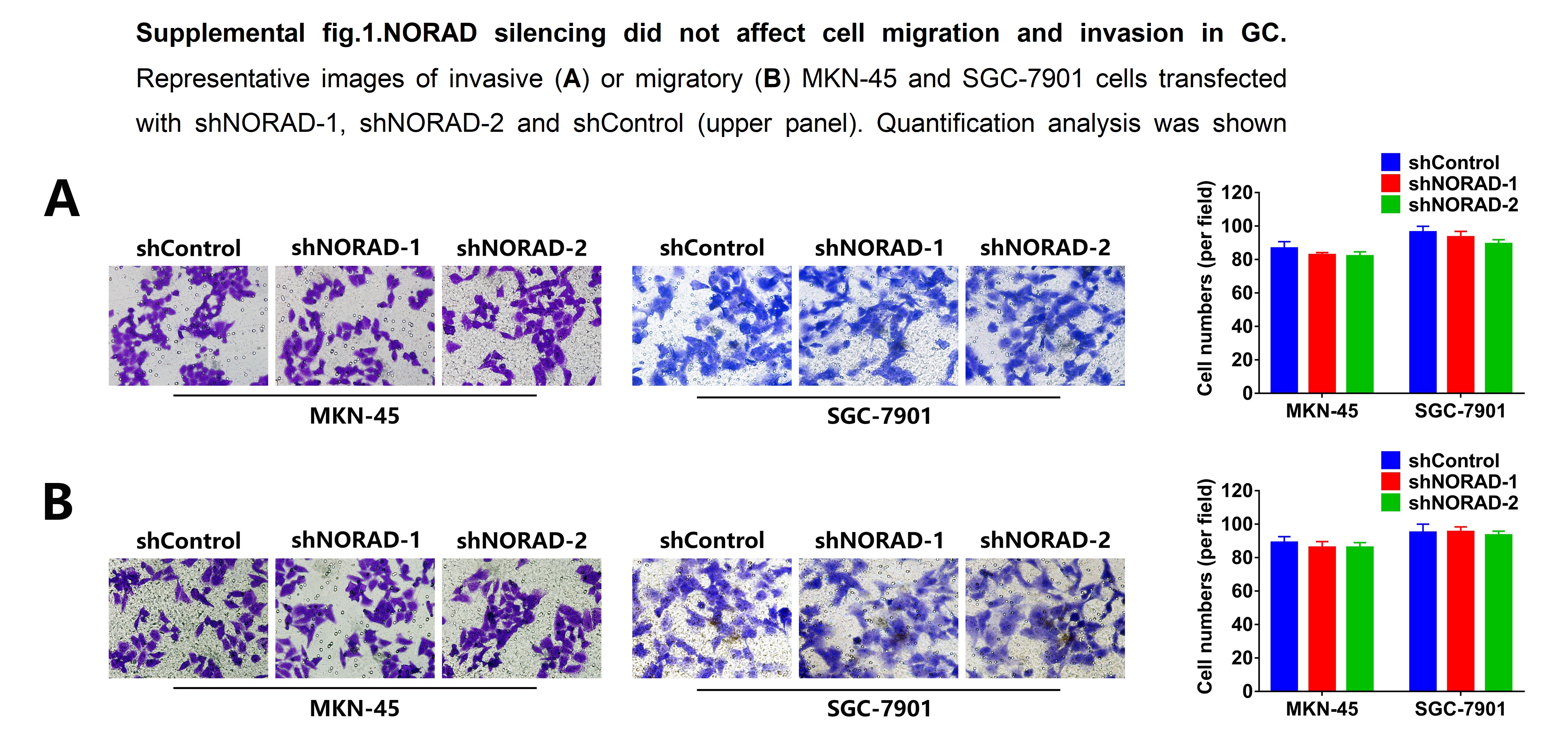
Transwell assay
Cells were added to the top chamber without or with Matrigel coating while lower chamber was added with culture medium containing 10% FBS only. Following 24 h cell culture, cells migrating or invading the lower chamber were fixed using 4% paraformaldehyde, stained with 0.1% crystal violet solution and subsequently visualized using inverted light microscope.
Nucleocytoplasmic fractionation assay
The cytoplasmic and nuclear components were separates using PARIS Kit (Life Technologies, MA, USA). The nuclear and cytoplasmic fractions was detected by qRT-PCR assay to determine NORAD distribution using GAPDH and U6 as the cytoplasmic reference and nuclear reference, respectively.
Bioinformatics analysis and dual-luciferase reporter gene assay
The binding sites of miR-204-5p with NORAD-3`UTR and KMT2D-3`UTR were obtained from Starbase. Putative wild-type (WT) and mutant (MUT) miR-204-5p -binding sites in the 3ʹ-UTR of NORAD or KMT2D mRNA, termed NORAD-WT or NORAD-MUT and KMT2D-WT or KMT2D-MUT, were sub-cloned into pGL3 Basic vector (Promega, Madison, WI, USA). miR-204-5p angomir or angomir NC were co-transfected with NORAD-WT or NORAD-MUT and KMT2D-WT or KMT2D-MUT reporter constructs using Lipofectamine™3000 agent. Luciferase activity was determined using the Multimode Detector reporter assay system (Beckman Coulter, WI, USA).
RNA immunoprecipitation (RIP) assay
RIP was performed using a Magna RIP RNA-Binding Protein Immunoprecipitation kit (Millipore, Billerica, MA, USA) according to the manufacturer’s instructions. An anti-Argonaute-2 (Ago-2) or anti-IgG antibodies was also used. Enrichment of NORAD and miR-204-5p was determined by qRT-PCR assay.
Western blot
Cells lysates extracted using radioimmunoprecipitation assay (RIPA) (Beyotime, Shanghai, China) were fractionated by electrophoresis and transferred on a polyvinylidene fluoride (PVDF) membrane (Millipore). The membrane was blocked in 5% nonfat skim milk and incubated with primary antibodies and secondary antibodies. Bands on the membrane were visualized by electrochemiluminescence (ECL; Pierce, Rockford, IL, USA). The primary antibodies used were as follows: anti-PTEN (1:1000), anti-PTEN (1:1000), anti-PI3K (1:1000), anti-AKT (1:1000), anti-phosphorylated-AKT (pAKT; 1:1000), (Cell Signaling Technology, Danvers, CO, USA).
Statistical analysis
All statistical analysis were performed using SPSS 21.0 (SPSS, Chicago, IL, USA) or GraphPad Prism (GraphPad Prism, Inc., La Jolla, CA, USA). Each experiment was performed in three independent replicates and the data are presented as the mean ± standard deviation (SD). Student’s t-test or one-way ANOVA was used to compare the means of two or three groups. P<0.05 was considered statistically significant.