Culture and identification of UC-MSCs
As shown in Fig. 1A, UC-MSCs were identified as a population of fibroblast-like cells. And their surface positively expressed CD73, CD90, CD29, CD106 and CD44, and negatively expressed CD34, CD45 and HLA-DR (Fig. 1B). After specific incubation for 14 days or 21 days, respectively, the cells could differentiate into adipocytes or osteocytes by staining with Oil red O or crystal violet solution (Fig. 1C).
The Biological Features And Viability Of Hs Pretreated Uc-mscs
As shown in Fig. 2B and 2C, pretreating HS 1 h and subsequently culturing in common condition for 24 h did not affect the surface characterizations and the potentials of multi-differentiation of UC-MSCs. CCK-8 assay was conducted to determine the proliferative ability of UC-MSCs from these two groups according to the OD value. The results of the cell growth curve showed that there was no significant difference of the cell proliferation rate between UC-MSCs culturing in common condition and HS-pretreated UC-MSCs (Fig. 2D). And the results from flow cytometry analysis revealed that the apoptotic rate of HS pretreated UC-MSCs did not statistical difference from that in the control group (Fig. 2E).
Administration Of Hs-pretreated Uc-mscs Attenuates Lps-induced Ali
We assessed the severity of lung injury in each group to result whether HS-pretreated UC-MSCs can improve pulmonary performance after LPS administration. A mouse model of ALI was established following to intratracheally injected with LPS. After 24 h, the lung injury was pathologically confirmed which characterized by lung edema and widespread septal thickening as well as filled with neutrophil/mononuclear infiltration in alveolar spaces, while the changes were significantly improved after administration of UC-MSCs, and treatment with HS-pretreated UC-MSCs showed the further recovery of histopathological characterizations (Fig. 3A). The lung injury score which was evaluated from the staining of lung sections with H&E showed the consistent results (Fig. 3B). Meanwhile, the results found that LPS induced an increased lung wet/dry weight ratio of mice, while UC-MSCs could significantly improve the pulmonary edema (Fig. 3C). Interestingly, the better therapeutic effect was observed in the HS-pretreated UC-MSCs, compared with that in the UC-MSCs group (Fig. 3C). Finally, the changes in BALF are important indicator for assessing lung function. The results from the cell smear showed that HS pretreatment could further improve UC-MSCs-caused the decreased BALF inflammatory cell count (Fig. 3D). In addition, a TUNEL assay was also performed to evaluate endothelial apoptosis from each group. As shown in Fig. 3E, the apoptotic index of the lung endothelium obviously increased after exposed to LPS, whereas the increased index was remarkably decreased when treated with UC-MSCs. Moreover, in UC-MSCs with HS pretreatment, the apoptotic value of the lung endothelium was significantly lower than that in the UC-MSCs group. These results exhibited that HS pretreatment play a critical role in strengthening lung-protective roles of UC-MSCs.
HS-pretreated UC-MSCs modulate the functions of alveolar macrophages in vivo and in vitro
Growing evidence demonstrated that the polarization of alveolar macrophages is a critical factor to affect the pathological process of ALI[28]. Thus, we next collected BALF and detected the change of alveolar macrophage phenotype from each group. As shown in Fig. 4A, the results of flow cytometry analysis showed that UC-MSCs reversed the increased expression of a pro-inflammatory marker, TNF-α, in alveolar macrophages (F4/80+) stimulating by LPS, and meanwhile, promoted the anti-inflammatory marker, CD206, expression on these cells. HS pretreatment could further strengthen the immunoregulative effect of UC-MSCs to induce M2 macrophage transformation compared to UC-MSCs cultured in common condition (Fig. 4A). Furthermore, we detected the concentrations of several cytokines to evaluate the secreted function of alveolar macrophages indirectly, and found that the roles of HS-pretreated UC-MSCs in reducing the increased pro-inflammatory cytokines, including TNF-α, IFN-γ, IL-1β, IL-6 and IL-18, in BALF by LPS stimulation was significant better than that in the UC-MSC group (Fig. 4B).
Moreover, we used the LPS-stimulated THP1 cells as an in vitro model. The level of M1 marker was significantly increased after treated with LPS compared with that in the control group, while co-cultured with non-pretreated or HS-pretreated UC-MSCs significantly reversed this phenomenon (Fig. 4C). Furthermore, we also detected the level of M2 marker in each group. After stimulation, the expression of CD206 was significantly increased in macrophages when co-cultured with non-pretreated UC-MSCs compared to that in the control group, and this change was further obviously upregulated on macrophages after co-cultured with HS-pretreated UC-MSCs (Fig. 4C). To evaluate the secretive function of macrophages from each group, we also measured the concentrations of pro-inflammatory cytokines in co-cultured supernatants using ELISA. As shown in Fig. 4D, LPS promoted macrophages to secrete various cytokines, including TNF-α, IFN-γ, IL-1β, IL-6 and IL-18, while UC-MSCs could reversed these changes of macrophages. As expect, HS pretreatment could further strengthen these immunoregulative roles of UC-MSCs. These results suggested that HS pretreatment act as an approach to enhance effects of UC-MSCs on modulating polarization and secretive functions of alveolar macrophages.
HS-pretreated UC-MSCs reduce the activation of NLRP3 inflammasome in macrophages
It is accepted that the levels of NLRP3 inflammasome is positively associated with the severity of ALI, and MSCs could significantly reduce NLRP3 inflammasome. Therefore, we further investigated whether HS pretreatment enhanced therapeutic potential of UC-MSCs on NLRP3 inflammasome activation. As shown in Fig. 5A and 5B, the elevation of the protein expressions of NLRP3, ASC, IL-1β, pro-Caspase 1, cleaved-Caspase 1 and IL-18 in the lung by LPS exposure were remarkably decreased after treatment with UC-MSCs, whereas the expressions of these proteins in the HS pretreating UC-MSCs group were significant lower than that in the UC-MSCs group. And these results were consistent with the decreased concentrations of IL-1β and IL-18.
In the previous studies, NLRP3 inflammasome is demonstrated to mainly express in the activated macrophages. Thus, we also detected the effects of HS pretreated UC-MSCs on NLRP3 inflammasome activation of macrophage cell line in vitro. The results from Western blot showed that the upregulated NLRP3 activation of macrophages, as evidenced by the increased expressions of NLRP3, IL-1β, pro-Caspase 1 and cleaved-Caspase 1, was obviously suppressed by UC-MSCs treatment. And pretreatment with HP strengthened the role of reducing NLRP3 inflammasome activation in macrophages of UC-MSCs. Again, the levels of IL-1β and IL-18 secreted by macrophages were markedly decreased by both UC-MSCs and HS pretreated UC-MSCs. Together, these results indicate that HS pretreatment enhances the lung protective capacity of UC-MSCs in ALI model via inhibiting the activation of NLRP3 inflammasome in macrophages.
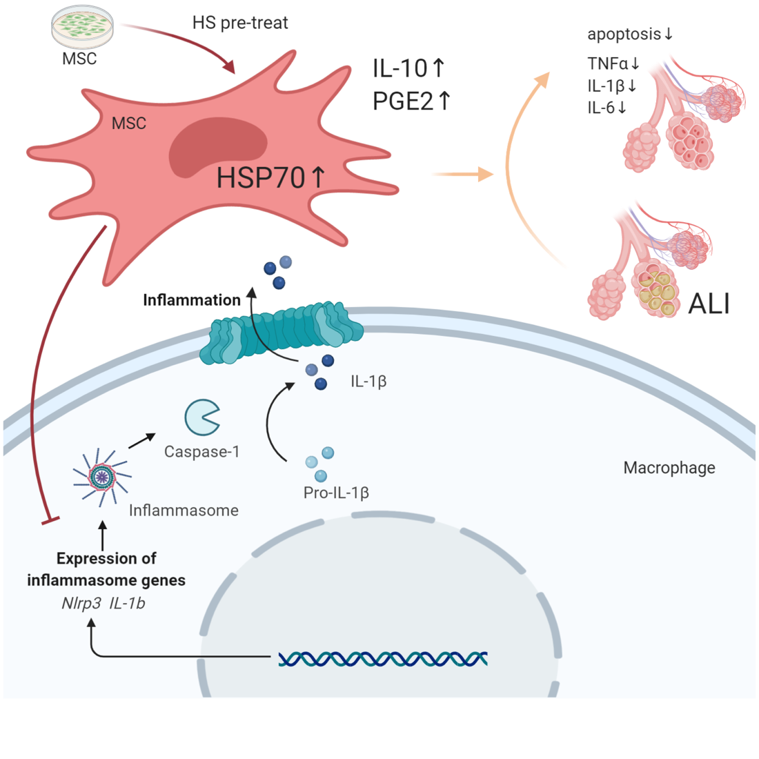
HS pretreatment enhanced the secretive abilities of UC-MSCs via increasing HSP70 expression
We further explore the potential mechanism that how HS-pretreated UC-MSCs improved ALI and reduced the NLRP3 inflammasome activation of macrophages. In the previous studies, both IL-10 and PGE2 could negatively regulate inflammasome activation [29, 30]. Therefore, in this setting, we detected the levels of IL-10, TGF-β and PGE2 in cultured supernatant to evaluate the secreted abilities of UC-MSCs that presented or absent HS. ELISA results shown that compared with the control group, HS led UC-MSCs to synthesize and secrete IL-10 and PGE2 remarkablely, but not TGF-β (Fig. 6A). In addition, as the evidence from Fan et al. demonstrated that HSP70 promoted PGE2 production via regulating COX-2 [31, 32], we, herein, targeted the expression of HSP70 in UC-MSCs in each group. As expect, the results from Western blot assay shown that the level of HSP70 was significantly higher in the HS-pretreated UC-MSC group than that in the control group (Fig. 6B). To further confirm this correlation, we added Apoptozole, an antagonist of HSP70, to treat HS-pretreated UC-MSCs, and the results showed that inhibiting HSP70 expression obviously reversed the increased PGE2 secretion of UC-MSCs induced by HS. Meanwhile, we used these cells to co-culture with macrophage and found from the results of ELISA and flow cytometry analysis that administration of Apoptozole significantly blocked the effect of HS on strengthening immunoregulative roles of UC-MSCs to modulate macrophage functions (Fig. 6C and 6D). Finally, we also performed Western blot assay to detect the inflammasome activation of macrophages in each group. The results indicated that Apoptozole might reversed the role of HS-pretreated UC-MSCs in reducing NLRP3 inflammasome activation of macrophages through downregulated PGE2 secretion (Fig. 6E).
Inhibition of HSP70 weakened the protective effect of HS-pretreated UC-MSCs on attenuating LPS-induced ALI
Furthermore, we also evaluated the degree of lung injury in each group to explore whether pretreatment of UC-MSCs with HS attenuate LPS-induced ALI model via regulating HSP70. The results showed that addition of Apoptozole to inhibit HSP70 abolished the protective effect of HS-pretreated UC-MSCs, as evidenced by the reversely aggravating pathological changes in lung architecture (H&E and injury score) and the increased lung wet/dry weight ratio (Fig. 7A, 7B and 7C). In addition, combined application with HS-pretreated UC-MSCs and Apoptozole showed that Apoptozole could reversely increase the reduced BALF inflammatory cell count treating by HS-pretreated UC-MSCs (Fig. 7D). And the results for investigating the TUNEL-positive cells indicated that the role of HS-pretreated UC-MSCs in alleviating alveolar epithelial cells was weakened by utilization of Apoptozole (Fig. 7E). In addition, we also performed ELISA assay to evaluate the inflammatory response, and the results showed that the significantly decreased pro-inflammatory cytokines, including TNF-α, IFN-γ, IL-1β, IL-6 and IL-18, by HS-pretreated UC-MSCs in this ALI model were remarkably reversed after union used with Apoptozole (Fig. 7F).