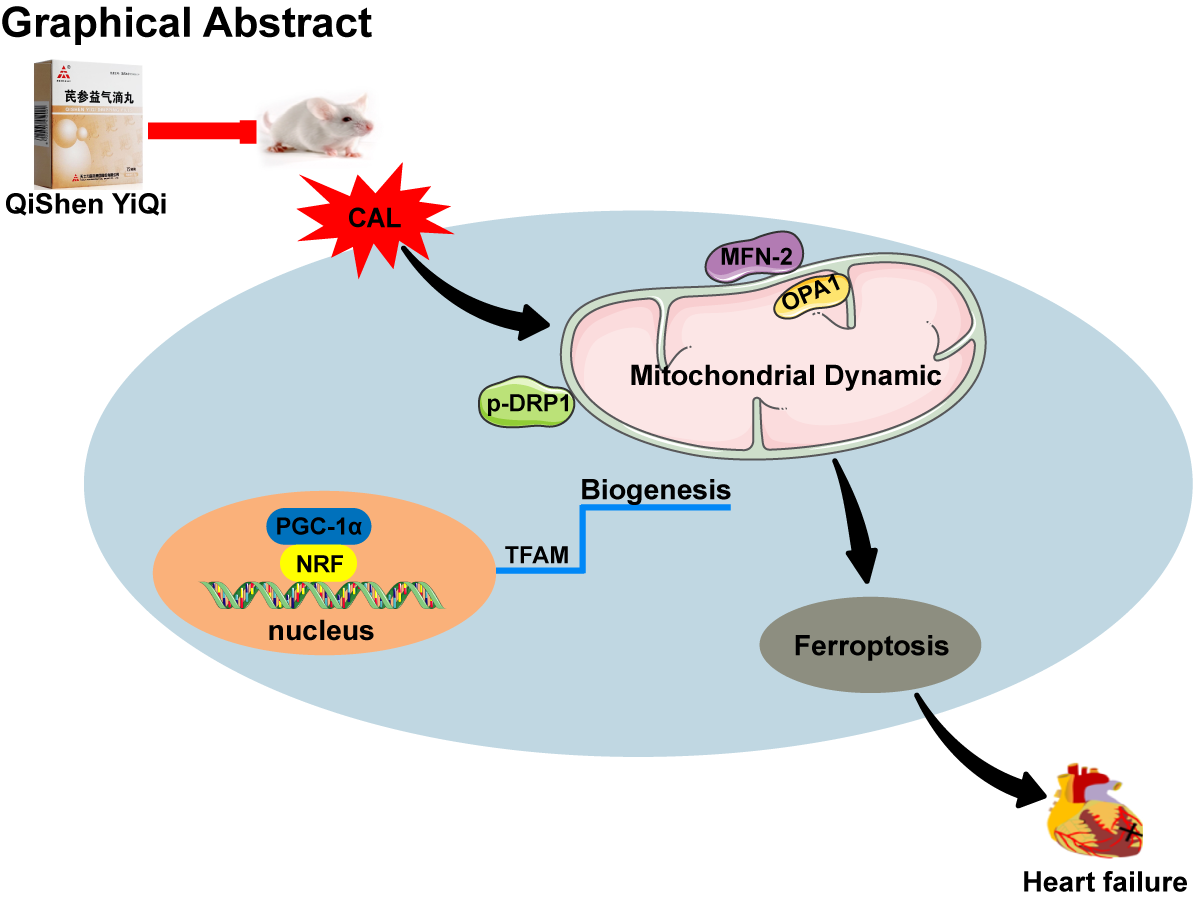
Drugs and Reagents
QSYQ were provided by Tasly Pharmaceutical Co., Ltd. (Tianjin, China) with the production batch number of 200402. The biochemical kits for detection of lactate dehydrogenase (LDH), malondialdehyde (MDA), glutathione (GSH) and tissue iron determination were obtained from Nanjing Jian Cheng Biotechnology Company (Jiangsu, China). The ELISA kits for detection of precursor brain natriuretic peptide (NT-pro BNP) and 4-hydroxynonenal (4-HNE) were obtained from Shanghai Heng Yuan Biotechnology Company (Shanghai, China). RIPA lysis buffer, protease inhibitor, and enhanced chemiluminescence (ECL) reagent were purchased from Vazyme Biotech Co., Ltd. (Jiangsu, China). Hoechst33342 staining kit was provided by Beyotime (Nanjing, China). Antibody against β-actin were provided by Proteintech Group (IL, USA) and antibody against PGC-1α, TFAM, Nrf1 were purchased from Santa Cruz (CA, USA). Antibody against FHC, GPX4, DRP1, phospho-DRP1, MFN-2 and OPA1 were obtained from Proteintech Group (IL, USA) and antibody against ACSL4 were obtained from Abclonal Proteinch (Wuhan, China).
Animal Experiments
ICR male mice (20–25 g) were purchased from the Comparative Medical Center of Yangzhou University (Jiangsu, China). Experimental animal production license number: SYXK 2021-0011. The animals were maintained on food and water in standard cages, at controlled temperature (22 ± 2°C), relative humidity (40%-80%), and 12 h light/dark cycles.
Animals And Cal Model
Mice were anesthetized with intraperitoneal injection of pentobarbital. Then, the thoracotomy was operated at the left third and fourth ribs intercostal space, and the heart was exposed followed by making a slipknot (6 − 0 silk) around the left anterior descending coronary. After ligation, the heart was promptly placed back in the intrathoracic cavity with the expulsion of air.
The surviving mice were randomly divided into six groups according to the experimental requirements: the sham group, the model group (CAL 14 days), the QSYQ low-dose group (12 mg/kg, ig), the QSYQ medium-dose group (35 mg/kg, ig), the QSYQ high-dose group (105 mg/kg, ig) and the Enalapril (EP) group (2.6 mg/kg, ig). The sham and model groups received an equal volume of physiological saline intraperitoneally. Mice in the administration group were treated for 14 days according to the experimental protocol.
Echocardiography
Mice were anesthetized with 2% isoflurane and then subjected to transthoracic echocardiography with the Vevo3100 imaging system to assess cardiac structure and function. Body temperatures were maintained between 36.9°C and 37.3°C. The heart rates were maintained at 400–550 bpm. At the target heart rate, the position and direction of ultrasonic probe were adjusted to find the maximum of the opening and closing apical value, and the left ventricular echocardiography was obtained. M-mode images were used to assess parameters of left ventricular function.
Histopathologic Examination
Mice were sacrificed, the hearts were harvested and fixed in 4% paraformaldehyde solution for at least 24 h at 4 °C. The samples were dehydrated and embedded in paraffin for slicing, the thick sections (4-5 μm) were stained with hematoxylin-eosin (HE) and Masson according to the manufacturer’s instructions (Servicebio Company, Wuhan, China). Then, histopathological changes were examined via an optical microscope (DX45 microscope; Olympus), quantification was performed by Image J software.
Serum Biochemical Indicators Detection
After echocardiography measurements, blood samples were collected by extracting the eyeball, rested for 60 min at room temperature then centrifuged at 3500 rpm for 10 min at 4°C, and separated to get serum. The levels of LDH, NT-pro BNP, MDA, 4-HNE and GSH were measured with kits according to the manufacturer's protocols.
Transmission Electron Microscopy
To observe the cardiac mitochondrial morphology, the fresh heart was removed rapidly fixed with 4% paraformaldehyde solution including 2.5% glutaraldehyde for 24 h, and cut into 1 mm 3 pieces (Leica, EMUC7, Germany). The ultrastructure was detected by using transmission electron microscopy (FEI Tecnai G220 TWIV, United States). All the samples were examined under the transmissive electron microscope. Mitochondrial lengths, mitochondrial numbers and mitochondrial volume was performed using Image J software.
Immunofluorescence Analysis
The fresh heart tissues removed from mice each group were collected and immediately cut into 5 µm thick frozen sections. The cardiac tissues were incubated with Mito-SOX Red dye in dark humidified chamber at 37°C for 30 min. Then the tissues were stained with Hoechst 33342 for 5 min in the dark. The images were acquired by a confocal laser scanning microscope (LSM700, Zeiss, Jena, Germany).
Quantitative Real-time Pcr
Total RNA was isolated from the heart tissues via using Trizol lysis buffer (Vazyme Biotech Co., Ltd, China). RNA concentration was measured by Nanodrop from Thermo Fisher Scientific (San Jose, CA, USA). Following the manufacture instructions, reverse transcription of RNA was performed with the cDNA Reverse Transcription kit (Vazyme Biotech Co., Ltd, China). The mRNA expression of prostaglandin-endoperoxide synthase 2(PTGS2), solute carrier family 7 member 11(SLC7A11) and glutathione peroxidase 4(GPX4) were measured by real-time fluorescence quantitative PCR. The sequences of the primer pairs are as Tabel1.
Western Blotting
Heart tissues were lysed in RIPA lysis buffer (Beyotime, Jiangsu, China) with 1 mM PMSF. The lysates were centrifuged at 12000×g for 10 min at 4°C and the supernatants were collected. The protein concentrations were measured utilizing a BCA protein assay kit (Beyotime, P0011). The same amount of protein (30 µg) was separated by SDS-PAGE and transferred to PVDF membranes (Millipore, Bedford, MA, USA). After blocked with 5% BSA for 2 h at room temperature, the membranes were incubated with indicated primary antibody overnight at 4°C. The primary antibodies against PGC-1α, TFAM, Nrf1, DRP1, p-DRP1, Mfn-2, OPA1, FHC, ACSL4, GPX4, and β-actin were mixed with the corresponding dilutions (1:1000, 1:1000, 1:1000, 1:2000, 1:2000, 1:2000, 1:2000, 1:2000, 1:2000, 1:8000). The membrane was washed and incubated with secondary antibody. Later, the membrane was submerged in an ECL reagent developing solution and the data were quantified using Image Lab™ Software (version 4.1; Bio-Rad).
Statistical Analysis
All statistical analyses were analyzed using GraphPad Prism version 8.0. All values in the figures were expressed as mean ± SD. Comparisons were performed with one-way analysis of variance (ANOVA). Statistical significance was considered to be present at p < 0.05.