5-HT induced autophagic response in hepatoma cells.
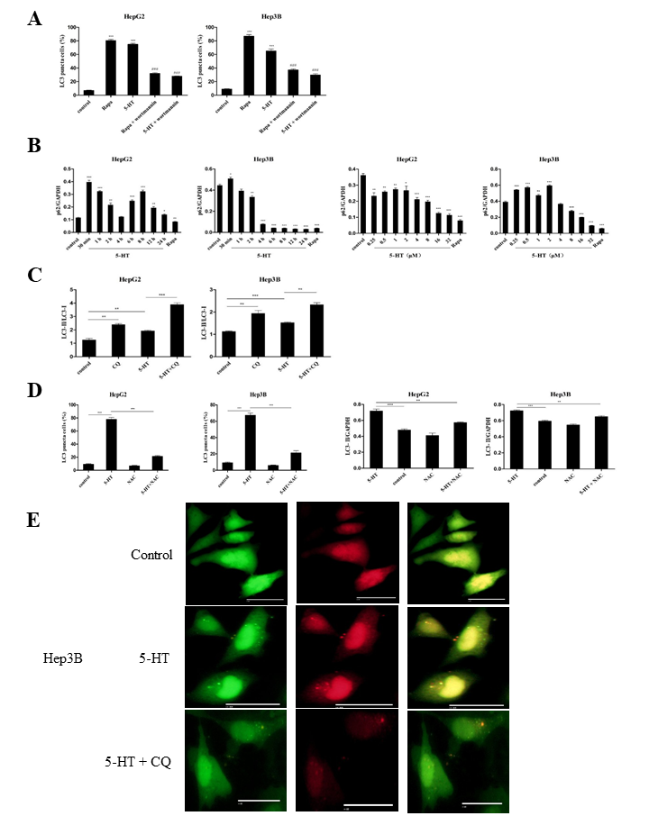
Expression of LC3-II in autophagy is considered to be an important sign of autophagy [25]. To determine whether 5-HT could induce autophagy, the HepG2 and Hep3B cells were transfected with GFP-LC3 plasmid. Following successful transfection, the HepG2 and Hep3B cells were treated with 5-HT at a concentration of 1 μM for 2 h. The rapamycin (an autophagy inducer) and wortmannin (an autophagy inhibitor) were also used to treat the transfected cells. The results showed that the expression of LC3 puncta were significantly increased in HepG2 and Hep3B cells when treatment with rapamycin or 5-HT, whereas, wortmannin (100 nM) could significantly inhibit rapamycin- or 5-HT-induced LC3 puncta (Fig. 1A and Fig. S1A). These date indicated that 5-HT might induce autophagy in hepatoma cells.
To further confirm the autophagy induced by 5-HT. The LC3 levels of 5-HT-treated hepatoma cells (HepG2 and Hep3B) were determined by western blotting. The HepG2 and Hep3B cells were treated with 1 μM 5-HT for 8 time points (0.5 h to 24 h) respectively. Rapamycin was used as a positive control. The results showed that the ratio of LC3-II/GAPDH was significantly increased in 5-TH-treated HepG2 and Hep3B cells in a time-dependent manner compared with that of untreated cells, and the expression of LC3 reached its peak in 2 h and 4 h in HepG2 and Hep3B cells, respectively (Fig. 1B). Then, the HepG2 and Hep3B cells were treated with 5-HT at a concentration gradient of 0.25 to 32 μM for 2 h or 4 h, respectively. We found that the ratio of LC3-II/GAPDH were increased in a dose-dependent manner and the expressions of LC3-II reached their peak in 1 μM and 8 μM in HepG2 and Hep3B cells, respectively (Fig. 1C). In summary, these data confirmed that 5-HT could induce autophagy in hepatoma cells.
5-HT induced a complete autophagic process in hepatoma cells.
The polyubiquitin-binding protein p62/SQSTM1 is a marker of complete autophagy, it was selectively incorporated into autophagosomes through indirect binding to LC3 and is efficiently degraded by autophagy [26]. So, we explored if 5-HT could induce degradation of p62 in hepatoma cells. After treatment of HepG2 and Hep3B cells with 5-HT for 0.5 to 24 h, the expression of p62 protein was detected by western blot. The results showed that the p62 protein was significantly degraded in 2 h and 4 h after treatment with 5-HT in HepG2 cells and in Hep3B cells, respectively (Fig. 2A and Fig. S1B). Then western blot were used to detect the effect of 5-HT concentration (0.25 to 32 μM) on the p62 protein in HepG2 and Hep3B cells for 2 h and 4 h, respectively. The results showed that the degradation of p62 reached its peak when cells were treated with 5-HT at 32 μM in HepG2 cells and 8 μΜ in Hep3B cells (Fig. 2B and Fig. S1B).
To future examine the occur of complete autophagic, HepG2 and Hep3B cells were transfected with RFP-GFP-LC3 plasmid, and then the HepG2 and Hep3B cells were treated with 5-HT alone or pretreated with CQ and then treated with 5-HT, the number of RFP-GFP-LC3 puncta in HepG2 and Hep3B cells was observed under fluorescence microscope. Chloroquine (CQ), an agent that impairs lysosomal acidification, which could block autophagy by block LC-II turnover, thus CQ not only increases the number of GFP-LC3 puncta, but also allows LC3-II to accumulate in cells. Our results showed that the 5-HT-treated groups contained more green and red LC3 puncta than the control groups. Interestingly, the number of red puncta was significantly increased than the green puncta (Fig. 2C and Fig. S1E), however, the number of GFP-LC3 puncta in CQ pretreated groups was significantly increased than 5-HT alone group (Fig. 2C and Fig. S1E), this data indicating that some green puncta had vanished during the autophagic process and suggesting that 5-HT induces a complete process.
We also measured the accumulation of the LC3-II used western blot. Our results showed that LC3-II accumulated upon CQ treatment (Fig. 2D and Fig. S1C). Most importantly, the level of LC3-II was obviously higher after treatment with the combination of 5-HT and CQ compared with that of 5-HT treatment alone (Fig. 2D and Fig. S1C), demonstrating that autophagy induced by 5-HT included the process of the fusion of autophagosomes with lysosomes. Taken together, these data suggested that 5-HT induced a complete autophagic process in HepG2 and Hep3B cells.
5-HT activated autophagy through the AMPK/ULK1 pathway instead of the mTOR-dependent pathway in hepatoma cells
Previous studies reported that many autophagy occurs by inhibiting the phosphorylation of mTOR, such as rapamycin [27]. To verify whether the autophagy induced by 5-HT in HepG2 and Hep3B cells was involved in mTOR pathway, the HepG2 and Hep3B cells were treated with 5-HT for 0.5 to 24 h, respectively, and the phosphorylation of mTOR pathway proteins were detected by western blot. The results showed that 5-HT did not inhibit the phosphorylation of mTOR and its related proteins (p70S6K and 4E-BP1) during the treatment period (Fig. 3A). Then HepG2 and Hep3B cells were treated with 5-HT in a concentration gradient from 0.25 μM to 32 μM for 2 h and 4 h, respectively. Western blot assays showed that the concentrations of 5-HT did not inhibit the expressions of mTOR and its associated proteins (Fig. 3B). Therefore, 5-HT activated autophagy through the mTOR-independent pathway in hepatoma cells.
ULK1 is an important protein in autophagy initiation process regulated by mTOR and AMPK [28,29,8] then we investigate whether 5-HT could induced the expression of ULK1 and AMPK. 5-HT were used to treat the HepG2 and Hep3B cells for 0.5 h to 24 h. Western blot results showed that the expressions of p-AMPK and ULK1 proteins first were increased and then decreased with elongated treated time in HepG2 and Hep3B cells (Fig. 3A). Furthermore, the western blot showed that 5-HT unregulated the phosphorylation of AMPK and ULK1 in HepG2 and Hep3B cells treated with 5-HT at concentrations from 0.25 to 32 mΜ for 2 h or 4 h, respectively (Fig. 3B).
In order to further explore the relationship between ULK1 and AMPK pathway related proteins and autophagy, ULK1 inhibitor (SBI-0206965) and AMPK inhibitor (Compound C) were used to detect the change of autophagy marker protein LC3. Our results showed that compared with 5-HT treatment alone, pretreated with SBI-0206965 or Compound C significantly reduced LC3-II expression in hepatoma cells (Fig. 3C). Therefore, 5-HT induced autophagy through AMPK/ULK1 pathway activation.
5-HT stimulated autophagy through JNK/p38/ERK pathway activation
It has been confirmed that MAPK-related signal pathways including ERK, JNK and p38 play important roles in autophagy [30], and then we speculated whether 5-HT induced autophagy through ERK/JNK/p38 pathway. The HepG2 and Hep3B cells were treated with 5-HT at 8 time points (0.5 h to 24 h). The western blot results showed that 5-HT could induce the phosphorylation of JNK, ERK and p38 and presented a trend that increased at first and then fell with elongated treated time, and the proteins expression reached its peak in 2 h and 4 h (Fig. 3A). After treatment with 5-HT at different concentrations range from 0.25 to 32 μM, it was found that the levels of phosphorylation of proteins were increased in a dose-dependent manner, then decreased (Fig. 3B).
In order to explore the relationship among ERK, JNK and p38 in 5-HT-induced autophagy. We used PD98059 (ERK Inhibitors, 20 μM), SP600125 (JNK Inhibitors, 10 μM) and SB203580 (p38 inhibitor, 10 μM) to pretreat HepG2 and Hep3B cells for 1 h, and then added 5-HT to treat HepG2 and Hep3B cells for 2 h and 4 h, respectively. The results showed that PD98059 could inhibit the phosphorylation of ERK but not the phosphorylation of JNK and p38. Moreover, SB203580 could inhibit the phosphorylation of p38 and ERK but not the phosphorylation of JNK. Nevertheless, SP600125 could inhibit the phosphorylation of ERK, JNK and p38. Furthermore, the treatment of all of inhibitors significantly inhibited the expression levels of LC3-II (Fig. 3D). Thus, we concluded that 5-HT-induced autophagy in HepG2 and Hep3B cells is dependent on the JNK/p38/ERK pathway, ERK is downstream of JNK and p38, and the ERK phosphorylation was regulated by JNK and p38, the phosphorylation of p38 was regulated by JNK.
ROS participated in 5-HT-induced autophagy in hepatoma cells.
The HepG2 and Hep3B cells were treated with 5-HT for 0.5 h to 24 h, respectively. And the hepatoma cells treated by 16 μM PMA for 2 h was used as a positive control for ROS induction. Fluorescence microplate reader was used to detect fluorescence intensity. The results showed that the ROS were significantly increased in HepG2 and in Hep3B in 2 h and in 4 h after treated with 5-HT, respectively (Fig. 4A). Then, HepG2 and Hep3B cells were treated with 5-HT or rapamycin at concentrations of 0.25 to 32 μM for 2 h and 4 h, respectively. And ROS production were significantly increased in 5-HT-treated HepG2 and Hep3B cells compared with that of untreated cells in a dose-dependent manner, and the production of ROS reached its peak when HepG2 and Hep3B cells were treated with 5-HT for 1 μM and 8 μM, respectively (Fig. 4B). In addition, The ROS scavenger NAC effectively decreased the production of 5-HT-induced ROS (Fig. 4C).
To further verify the relationship between ROS and autophagy, HepG2 and Hep3B cells were transfected with GFP-LC3 plasmid and treated with 5-HT or 5-HT pretreated with NAC. The GFP-LC3 puncta was observed under a fluorescencemicroscope. Compared with the 5-HT group, the GFP-LC3 puncta formation was significantly reduced in the NAC pretreated group (Fig. 4D and Fig. S1D). Further, western blot was used to assay the LC3 levels of 5-HT-treated hepatoma cells (HepG2 and Hep3B). The results showed that the level of protein LC3 was significantly increased in the 5-HT-treated group compared with that of the NAC-pretreated group and control group (Fig. 4E and Fig. S1D). All the data suggested that ROS was involved in 5-HT-induced autophagy of HepG2 and Hep3B cells.