2.1 Materials
AgNPs (5 nm and 50 nm) were purchased from Xi'an Ruixi Biological Technology Co., Ltd (Xi’an City, China). Triton X100, sodium lauryl sulfate, and soy peptone were purchased from Sinopharm Chemical Reagent Co., Ltd (China). Agar, pepsin, and Masson staining reagents were purchased from Solarbio Science and Technology Co., Ltd (Beijing, China). Tryptone was purchased from OXOID (Shanghai, China), calcein was purchased from Yeasen Biotech Co., Ltd. (Shanghai, China), and Cell Counting Kit-8 (CCK8) was obtained from APExBIO Technology LLC (USA). Trypsin was purchased from Sigma-Aldrich (USA). The 2,2-Diphenyl-1-picrylhydrazyl(DPPH) reagent was purchased from GlpBio (USA).
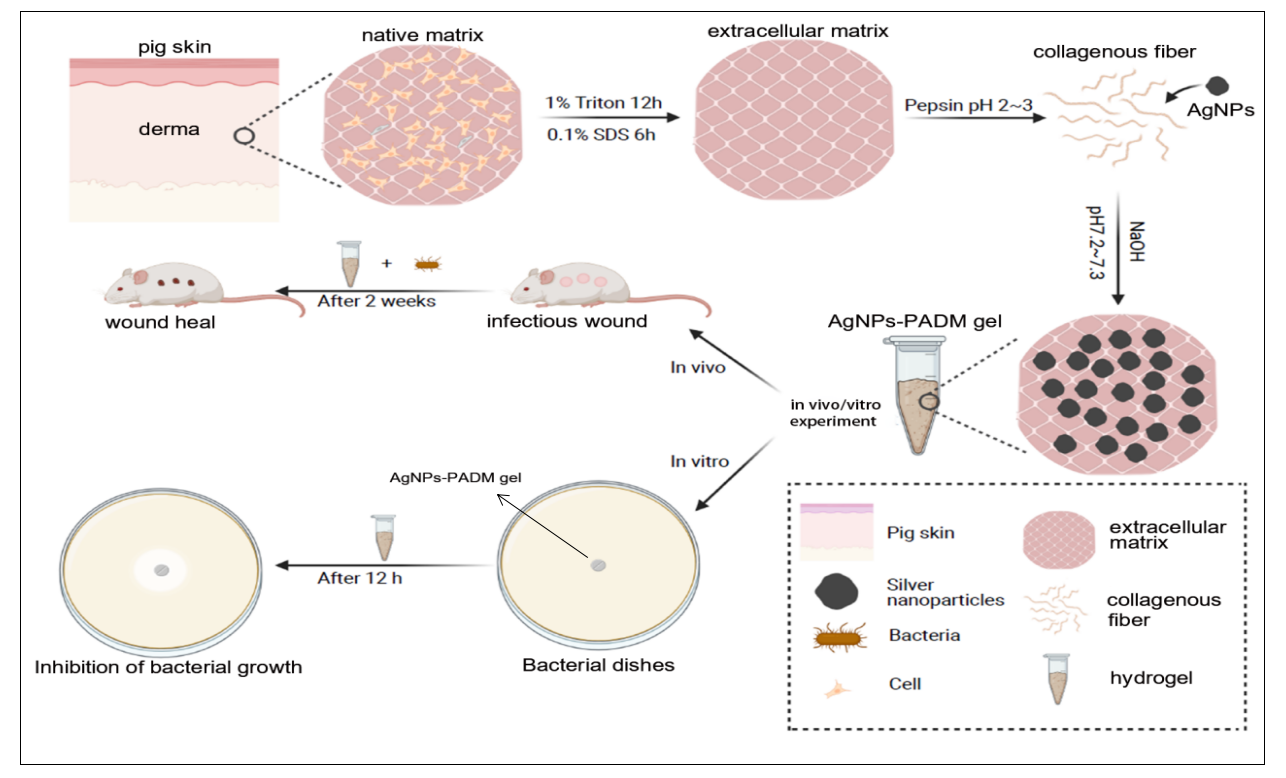
2.2 Synthesis of AgNPs-PADM hydrogel
Pig skin was harvested from fresh skin tissue from adult pigs and collected from local slaughterhouses. Then, the porcine skin tissue was rinsed using sterile water for 3 h, followed by three consecutive cycles of freezing and thawing (− 80–37 oC). The tissue was cut into 1×1 cm pieces, and the subcutaneous tissue was removed with scissors, shaken at 120 rpm at a constant temperature (25 oC)). It was then treated with 1% Triton X-100 solution for 12 h, 0.1% sodium dodecyl sulfate for 6 h. It was rinsed extensively in phosphate buffered saline (PBS) and lyophilized. The sample was then ground into powder. Then, 20 mg/mL of powder was digested with pepsin powder in dilute hydrochloric acid solution (pH 2–3) for 10 min(21), and a 5 nm Ag NPs solution was added dropwise to the solution and stirred rapidly. Subsequently, the above mixture continued to digest for 2 h until the gel was translucent and viscous, and it was stored in a refrigerator at 4℃. PBS solution was added to adjust the osmotic pressure, and pre-cooled 10 M NaOH was added to adjust the pH to 7–8. After maintaining the gel at 37℃ for 20 min, and a AgNPs-PADM hydrogel was prepared.
2.3 Characterization of a AgNPs-PADM hydrogel
Cell- and nuclear-removal performances were assessed using hematoxylin and eosin (H&E) and 406-diamino-2-phenylindole (DAPI) staining. The absorption of AgNPs, PADM hydrogel, and AgNPs-PADM hydrogel was investigated using UV-Vis spectrophotometry. The particle size and distribution of the 5 nm AgNPs were observed using transmission electron microscopy (TEM) before its addition to the hydrogel and after its release from the AgNPs-PADM hydrogel. The spatial structures of the PADM and AgNPs-PADM hydrogels containing 20, 50, and 80 µg/mL of AgNPs were studied using scanning electron microscopy (SEM). Briefly, after the fixation of the hydrogels in 2.5% (w/v) glutaraldehyde for 1 h, the samples were washed three times with PBS solution and then dehydrated sequentially with 30%, 40%, 50%, 70%, 80%, 90%, 95%, and 100% CH3CH2OH. Subsequently, the samples were dehydrated with liquid carbon dioxide in a critical point dryer and observed via scanning electron microscope (SEM) (JSM-6360LV, JEOL, Tokyo, Japan) to determine the structural characteristics of the PADM and AgNPs-PADM hydrogels. An energy-dispersive spectrometer (EDS, X-act, OXFORD, England) was used to study the composition and distribution of different elements.
The porosities of the PADM and AgNPs-PADM hydrogels were investigated by using two methods. The first is the medium-immersion method(22). First, a volume of the hydrogel was pre-frozen at -80 oC for 1 h and then placed in a freeze dryer for 12 h under negative pressure. The mass of the solid gel (W1) was measured, and the hydrogel was immersed in Dulbecco's modified Eagle’s medium (DMEM, Hyclone) until saturation. The beaker weight (W2) of the DMEM medium and that (W3) of the DMEM medium containing the hydrogel was measured. The hydrogel was removed, excess media was gently wiped off the gel surface with gauze, and the hydrogel was weighed after soaking (W4). Porosity was calculated as follows: Porosity= (W4-W1)/(W3-W2). For the second method, the cross-section of the sample was scanned via SEM, and the porosity of the hydrogel was calculated by using Image J (National Institutes of Health, USA).
2.4 Water-retention performance
The same mass of PADM and AgNPs-PADM hydrogels was placed in a 37 oC incubator, and their masses were measured at different time points until their masses no longer changed. The formula used to calculate the water-retention performance of the hydrogels is as follows: water retention rate = W2/W1×100%. W2 represents the weight of the gel measured at each time point, and W1 represents the initial weight of the hydrogel.
2.5 In vitro degradation performance
To measure the degradation performance under different conditions, we placed the PADM and AgNPs-PADM hydrogels in a centrifuge tube and then placed them in an incubator at 25, 37, and 42 oC and observed regularly. The dissolution of the PADM and AgNPs-PADM hydrogels was checked, the liquid dissolved in the centrifuge tube was promptly aspirated, and the remaining samples were weighed.
In addition, we prepared 500 µL of the PADM and AgNPs-PADM hydrogels into a solid state and placed them in a centrifuge tube containing 1 mL of PBS. Then, 7.5 mg of trypsin was added to the experimental group (trypsin group) at 37 oC. After the hydrogels were immersed for seven days, the two groups of solutions were changed once a day. The PBS and trypsin solutions in the centrifuge tube were removed at the same time every day, and the hydrogels were rinsed with PBS. The water on the surface of the hydrogel was wiped off, the sample was weighed, and the above solution was added in the same configuration after weighing.
2.6 Release of AgNPs from the AgNPs-PADM hydrogel
To determine the release performance of AgNPs in the AgNPs-PADM hydrogel under different conditions, we initially prepared a series of AgNP solutions, measured the absorbance values of different concentrations of AgNP solution via ultraviolet (UV) spectrophotometry, and created a standard curve. The release of AgNPs from the AgNPs-PADM hydrogel was then measured at different temperatures. The PADM hydrogels (500 µL of the PADM and AgNPs-PADM hydrogels) were placed in a centrifuge tube, which was placed in an incubator at 25, 37, and 42 oC in particular intervals. The dissolved solution was removed for a period of time, and its optical density(OD) value at a wavelength of ཞ400 nm was measured. The above operation was repeated until the OD value of the solution no longer increased.
Trypsin is a commercial food-grade enzyme with increased proteolytic activity, used to break down collagen(23).The AgNPs-release performances of the PADM and AgNPs-PADM hydrogels in trypsin-containing PBS and PBS alone were also measured. A group of 500 µL solid samples prepared from the PADM and AgNPs-PADM hydrogels were placed in centrifuge tubes, and trypsin (7.5 mg) and PBS solution (1 mL) were added to the tubes. Another set of centrifuge tubes containing two samples only were added with PBS solution (1 mL) respectively. A 1 ml solution was taken every day, and its OD value was measured at a wavelength of ཞ400 nm. Then, 1 ml of the corresponding solution was added to the centrifugal tube after measurement, and the above operation was repeated until the OD value of the removed solution no longer increased.
Finally, we measured the release of AgNPs from the PADM and AgNPs-PADM hydrogels using an ultrasonic crushing instrument. The solid samples prepared by using 500 µL of the PADM and AgNPs-PADM hydrogels were cut into small pieces and placed in a centrifuge tube. Deionized water (400 µL) was added, and an ultrasonic shatterer was used to vibrate the samples at frequencies of 25 Hz and 30 Hz. The samples were vibrated for 30 s and rested for 20 s in a cycle to prevent an excessively high solution temperature. After 24 cycles, the samples were centrifuged at 4000 g for 3 min, and then 200 µL of the supernatant was removed. The OD value was measured at a wavelength of ཞ400 nm. The supernatant was added to the sample after the test, and the above operations were repeated until the OD value of the solution did not increase.
2.7 Antibacterial properties in vitro
The in vitro antibacterial potential of the AgNPs-PADM hydrogel was investigated using the Oxford cup method (OCM) and colony count method (CCM). Gram-negative bacteria (Escherichia coli) and gram-positive bacteria (Staphylococcus aureus and Enterococcus faecalis) were used for the OCM experiments. First, single colonies of Escherichia coli, Enterococcus, and Staphylococcus aureus were added to the test tubes containing the medium, and the tubes were placed on a shaking table at 150 rpm and 37 oC overnight. Bacteria in the logarithmic growth stage were selected for the experiment. An appropriate number of bacteria were selected, and their OD value was adjusted to 0.1 (the number of bacteria was 0–1.5x10^8/L). The number of bacteria was diluted to 10^5/L. Then, 1/10 of the culture-medium volume was added to the uncoagulated culture medium containing Agar, the medium was fully mixed and added to a Petri dish. After cooling, the PADM and AgNPs-PADM hydrogels were added. The Petri dish was placed in an incubator at 37 oC. After culturing for 12 h, the diameter of the bacteriostatic zone around the samples was measured using a scale. Each experiment was repeated thrice.
Subsequently, we selected Staphylococcus aureus for CCM detection. First, the PADM and AgNPs-PADM hydrogels were cut into 3 mm pieces and sterilized for 2 h under UV light. Then, 2 mL of Staphylococcus aureus at a concentration of 1x10^8/mL was put into a test tube. The test tube was placed on a shaking table at 150 rpm and 37 oC to reproduce the bacteria. Bacterial suspension (100 µL) and EP tubes were centrifuged at 2, 4, and 12 h. After discarding the supernatant, the bacteria were suspended in normal saline.20ul bacterial suspension was evenly coated on nutrient agar plates, and bacteria were counted after 24 h of culture at 37 oC.
The hydrogels prepared with 80 g/mL AgNPs was placed in a centrifuge tube, and ultrasonic shock at a working frequency of 25 Hz was carried out for 30s and then for 20s. The AgNP release solution (100 µL) at 60, 100, and 140 min was obtained and added to an Oxford cup that was prepared in advance. The Oxford cup was prepared as follows. First, an appropriate amount of Escherichia coli solution was used, its OD value was adjusted to 0.1, and it was diluted to 10^6/L. Then, 1/10 of the volume of the culture medium was mixed into the unsolidified Agar medium. The culture medium was poured into the Petri dish, and after it cooled, the Oxford cup was placed, and an appropriate amount of the unsolidified Agar medium was added again. After the upper medium solidified, the prepared AgNPs release solution was added, and the Oxford cup was removed 12 h later to observe the bacterial-removal status at the bottom.
2.8 Cytotoxicity test
The hydrogel (100 µL) was added to a 24-well plate and sterilized using irradiation (25 kGy γ radiation). HeLa cervical epithelioid carcinoma cells were seeded at 4x10⁴ per well in blank Petri dishes and PADM and AgNPs-PADM hydrogels(24). DMEM (Hyclone) was added to 10% (v/v) fetal bovine serum, 100 µg/mL penicillin, and 100 µg/mL streptomycin. Cells were cultured in a humid environment containing 5% carbon dioxide at 37 oC After 48 h of co-culture, the cells were stained with a calcium lutein staining kit and observed under a 515 nm fluorescence microscope.
Cytotoxicity was investigated by using CCK8. PADM and AgNPs-PADM hydrogels were immersed in DMEM (hydrogel volume/medium volume 1:5). The supernatant was collected as a 100% leaching solution and then mixed with a particular proportion of DMEM medium to obtain culture solutions containing 0%, 20%, 50%, and 100% leaching solutions. For further experiments, HeLa cells were seeded in 96-well plates at a density of 2000 cells/well. After culturing at 37 oC for 24 h, the supernatant medium was removed from each well, and 100 µL of each medium containing different proportions of leaching solution were added to the DMEM medium without the leaching solution as the control group. Then, 10 µL CCK8 reagent was added to each well on the day 1, day 3 and day 5. Meanwhile, the cells were incubated at 37 oC for 1 h. Absorbance at ཞ450 nm was measured using a spectrophotometer.
2.9 Oxidation resistance
The free-radical scavenging capacities of the PADM and AgNPs-PADM hydrogels were measured using the DPPH free-radical scavenging assay (9), in which the clearance rate of ascorbic acid was used as the reference standard. Then, 80 µg/ mL ascorbic acid and PADM and AgNPs-PADM hydrogels were added to 0.4 mM DPPH anhydrous ethanol solution, and the solution was placed away from light for 10 min. An appropriate amount of supernatant was obtained from each sample, and its absorbance at 517 nm was measured. The content of the test sample in the anhydrous ethanol solution containing DPPH was adjusted until the absorbance of the DPPH anhydrous ethanol solution corresponding to the AgNPs-PADM hydrogel sample did not change. The scavenging ability of radicals in the samples at a particular concentration was calculated using the following formula: Inhibition (%) =(C1-C2)/C1*100%. C1 represents the difference between the maximum and minimum absorbance values of the DPPH solution containing ascorbic acid, and C2 represents the difference between the absorbance value of the DPPH solution at this sample concentration and that of the DPPH solution alone.
2.10 In vivo experiment
The rats used in this study were purchased from the Zhejiang Academy of Medical Sciences. They were six months old and weighed 200–300 g. They were reared in separate cages, fed standard feed and tap water, and the cages were maintained at a controlled temperature of 25°C and humidity of 55%, alternating day and night for 12 h. According to the National Institute of Health publication No. 18–23, 1985, which is typically referenced for the care and use of laboratory animals, great care should be taken with rats. The rats were anesthetized with ketamine (30.0 mg/kg). Their dorsal hair was removed, and their skin was rinsed with ethanol (70%). Three 1 cm square skin wounds were created on the midline side with a scalpel and tweezers, and 30 µL Staphylococcus aureus (bacterial concentration: 1x10^8 CFU/L) was dropped into each wound. Then, 2 h was given to allow the bacteria to fully infiltrate the wound. The wounds were divided into three groups. Group I was treated as the control group and did not receive any treatment (naked wound); group II was treated with PADM gel; group III was treated with the AgNPs-PADM hydrogel and covered with gauze, and the PADM and AgNPs-PADM hydrogels were replaced with new ones on the 3rd, 5th, 7th, 9th, and 11th days.
Reduction in the wound area (wound contraction) was used as an indicator of the therapeutic outcome. Wound contractions were recorded on day 0, 7, and 14. The wound healing area was expressed as a percentage. The wound-shrinkage percentage was estimated using the following formula: wound-healing ratio (%) =(C1-C2)/C1x100%, where C1 represents the initial wound area, and C2 represents the wound area at each measurement time.
On the 7th and 14th days, half of the rats in group I, in group II and group III were randomly anesthetized and sacrificed, and non-wound skin approximately 5 mm around the wound was biopsied. Skin tissue was immobilized in buffered formalin (4%) for 2–3 days before tissue processing and paraffin embedding. Tissue sections with a thickness of 5 µm were made using a sectioning mechanism and stained with hematoxylin and eosin (H&E). The neovascularization, epidermis, scarring, and granulation tissue were observed and photographed.
Masson’s trichrome staining can be used to dye early collagen in light blue and mature collagen in dark blue to assess wound healing. On the 7th and 14th days, the paraffin sections of the wound were stained with Masson’s trichrome staining, and the collagen content in the tissue sections was analyzed under a microscope. Masson's trichrome stain was designed to distinguish smooth muscle cells from collagen. Additionally, paraffin sections were stained using immunohistochemistry to measure the expression of CD68 and CD34 in the samples to evaluate the changes in macrophage content and tissue micro-vessel content during wound healing, respectively.
Statistical analysis was performed using GraphPad Prism 8 (GraphPad Software Inc., USA). The results are expressed as mean ± standard deviation. A one-way ANOVA and two-way ANOVA were used to determine whether there was a significant difference in the results, and a P-value ≤ 0.05 was considered acceptable.