Existing pesticide formulation solvents emit VOCs, are flammable, and are classed as hazardous air pollutants (HAP), which means they are harmful to users and phytotoxic to crops. Regulations, health, and environmental concerns have created the need for green solvents in formulations. The "green chemistry" trend has resulted in the use of less toxic solvents in emulsifiable concentration (EC) formulations. The least dangerous carrier solvent among four commonly utilised organic solvents [dimethyl sulfoxide (DMSO), dimethylformamide (DMF), aromatic hydrocarbon (C9), and methyl oleate] was chosen in this study after a comprehensive and comparative fungal growth inhibition assessment. We used methyl cis-9-octadecenoate (60%) as the bio-based green reserver for the effective creation of bioinspired EC formulations (30%) of Pongamia pinnata L extract using emulsifier blends based on the established toxicity order (DMF > DMSO > C9 > methyl oleate) (10 percent). In terms of emulsion stability, cold test, accelerated storage stability, flash point, and other parameters, EC1 beat the other thirteen formulations (EC1-EC13), confirming its appropriateness for commercial manufacturing. In-vitro fungicidal activities against Alternaria solani and Phytophthora spp. were investigated using four therapeutically relevant dosages of agricultural use. At the highest dose (1%), A. solani (EC50 = 0.08 percent) demonstrated the greatest growth suppression (87.4 percent), followed by Phytophthora sp. (71.1 percent) (EC50 = 0.49 percent). The study demonstrated its relevance in the development of eco-friendly green solvent-based herbal formulations as a sustainable crop protection alternative to hazardous chemical pesticides.
Research Article
Selection of green carrier solvent for the effective creation of bio-inspired liquid formulation and anti-fungal potency evaluation
https://doi.org/10.21203/rs.3.rs-1549486/v1
This work is licensed under a CC BY 4.0 License
You are reading this latest preprint version
Pongamia pinnata extract
Herbal formulation
Emulsifiable concentrate
Emulsion stability
Alternaria solani.
Phytophthora sp.
Chemical fungicides used for plant protection might leave hazardous residues, endangering human and environmental health as well as non-target creatures (Bhandari et al., 2020; Rani et al., 2020). (Barbieri et al., 2020). Furthermore, due to rising crop protection costs as a result of fungicide resistance, the use of some chemical pesticide classes for plant disease control has been curtailed (Gao et al., 2020). As a result, developing novel mechanisms for alternative fungicides for sustainable agriculture is a top goal (Campos et al., 2018; Lengai et al., 2019).
The use of natural antifungal chemicals for plant protection is receiving a lot of attention these days (Jamiolkowska and Kopacki, 2020). The seeds of the Pongamia pinnata (L.) plant (Family: Leguminaceae) contain a wide range of naturally occurring biologically active compounds, including flavonoids, terpenoids, phenols, saponins, alkaloids, and fatty acids (Badole et al., 2020; Purkait et al., 2019), as well as antifungal properties (Badole et al (Raja and Sreenivasulu, 2016; Sharma et al., 2012; Shaheen et al., 2017). Without suitable formulations, bioactive chemicals in plant extracts can quickly deteriorate and volatilize in the field (Borges et al., 2018). As a result, improving the efficiency of these natural phytochemicals in farmers' fields may necessitate the development of industrial formulations (Mugao et al., 2020). Many botanicals have been created using standard aromatic solvents, according to researchers (toluene, xylene, etc.).
These volatile organic solvents, on the other hand, are poisonous to users and have been demonstrated to have unacceptable phytotoxicities (Kewei et al., 2006), as well as negative environmental consequences due to their non-biodegradability (Donglu et al., 2012). Concerns about food safety and the environment, as well as growing crude oil prices and the harmful effects of organic solvents, have driven interest in producing easily degradable, repeatable, and non-petroleum solvents in recent years (Morya et al., 2020). Vegetable oil derivatives, such as the methyl oleate group of fatty acid methyl esters (FAME), are being studied to see if they can mimic the characteristics and performance of petroleum-based solvents (Purkait and Hazra, 2019). FAME is a user-friendly, environmentally friendly solvent that is said to be biodegradable and environmentally benign (Ping et al., 2016).
The goal of this research was to determine the inherent toxicity profiles of routinely used organic solvents against two plant pathogenic fungus, A. solani and Phytophthora sp. The goal of the research was to create herbal antifungal formulations using Pongamia pinnata seed oil and the least toxic solvent possible. In-vitro bio-efficacy testing was also carried out against Alternaria solani and Phytophthora spp., the pathogens that cause early and late blight in tomatoes and potatoes, resulting in severe damage and high production losses of up to 95.8% (Lees et al., 2019; Mugao et al., 2020) and yield losses of up to 79 percent in vegetable crops (Lees et al., 2019). (Kalpashree and Raveesha, 2016; Lal et al., 2016).
2.1. Botanicals, chemicals and reagents
Pongamia pinnata L. (Fabaceae) seeds were collected and taxonomically recognized from the local market. E. Merck India provided emulsifiers (such as Tween 20, Tween 80, nonylphenol ethoxylates, calcium alkylbenzene sulphonates, and dodecylbenzene sulphonate) and carrier solvents (such as aromatic hydrocarbon (C-9), dimethyl sulfoxide (DMSO), N,N-dimethylformamide (DMF), and methyl oleate). Without additional purification, the reagents were employed. As standard hard water of 342 ppm strength with an electrical conductivity (EC) value of 0.69 dSm-1, a solution of anhydrous CaCl2 (2.74 mM) and MgCl2.6H2O (0.68 mM) was produced in double-distilled water (1 liter) (CIPAC MT 18, 1995).
2.2. Hexane extract preparation
Pongamia pinnata seeds were washed carefully under a gentle flow of tap water to eliminate dust and other pollutants before being dried in the shade at room temperature. As reported in our earlier research paper (Purkait et al., 2019), air-dried seeds (1 kg) were ground to powder in a household grinder (Bajaj, Bravo Dlx 500) and extracted with hexane (4 liters) in a Soxhlet apparatus for 6 hours at 60 C. (Figure 1). The hexane extract was filtered and concentrated in a rotary vacuum evaporator (Buchi (R-3), Switzerland) at 40°C under decreased pressure (370 mbar) to yield (28.37 percent) of the desired extract, which was then stored at 4°C for further use.
2.3 Toxicity assessment of organic solvents
2.3.1. Isolation and maintenance of a pure culture
Phytophthora sp. and A. solani were isolated from infected potatoes using a single spore isolation technique, and identified by scientists from the Department of Plant Pathology, Bidhan Chandra Krishi Viswavidyalaya (BCKV), Mohanpur, West Bengal, India, based on colony morphology, morphometric characteristics of acervuli, seta, conidia, and conidiophores, as described by Prittesh (2016). Potatoes infected with the pathogen were chopped into little pieces along the edges of lesions (5 mm in diameter). The pieces were surface sterilized in a 0.1 percent w/v aqueous mercuric chloride solution, rinsed five times, dipped in streptomycin, and placed to a potato dextrose agar (PDA) growth plate. Mycelial bids were moved from culture plates to PDA slants afterwards and allowed to sporulate for 8–10 days.
The single spore isolation procedure developed by Alexopoulos et al. was used to establish pure cultures of Phytophthora sp. and A. solani (2002). At 4°C, a pure fungal culture was maintained on PDA media. Conidia from 10-day-old cultures grown on PDA on Petri dishes were combined to make pathogen inocula. Flooding the plates with sterile distilled water and gentle scraping with a sterile slide were used to remove conidia from the surface of the media. To eliminate mycelial pieces, the suspensions were filtered through a thin layer of absorbent cotton wool and the spore number in suspension was adjusted to 106 conidia-mL-1 using a hemocytometer. In in-vitro pathogenicity testing, this spore suspension was utilized to inoculate banana fingers and chili.
2.3.2. Bioassay in-vitro
Following the poison food technique, an in-vitro bioassay was used to screen out the toxicities of several organic carrier solvents (Jang and Kulk, 2018). According to the Clinical and Laboratory Standards Institute (CLSI) standard recommendations, the concentration of DMSO, C9, DMF, and methyl oleate in the final test solutions was 1% (v/v) (Hazen, 2013). At the moment of full radial growth (9 cm) of each control plate, i.e. 6 days following the incubation period, colony diameters of treatment plates were measured. All treatments and controls were duplicated three times and incubated at 28 1°C for the duration of the experiment. The following formula was used to compute the percentage inhibition of mycelial growth (Dutta et al. 2019):

Where dc is the test fungus's average radial growth (cm) on control plates, and dt is the test fungus's average radial growth (cm) in treatment plates. A statistically significant difference was defined as a difference of p 0.05.
2.4. Preparation of emulsifiable concentrate
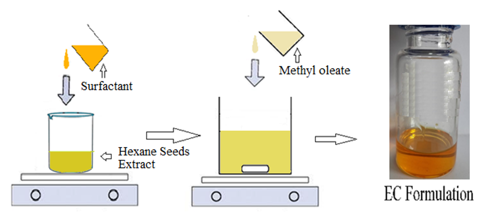
With various adjustments, different emulsifiable concentrate (EC) formulations were created using the approach outlined in our earlier article (Purkait et al., 2019). From the toxicity assessment of organic solvents, seed extract (30 percent w/w) was dissolved in the least harmful solvent (Methyl oleate) (60 percent w/w) (Section 2.3). To prepare EC formulations, different emulsifiers in a blend (10%) were added to the extract solution and thoroughly swirled on a magnetic stirrer at 200 rpm at 50-60 C. (Supplementary Figure 1).
2.5. Physico-chemical characteristics
P. pinnata seed extracts were produced in thirteen distinct EC (30% w/w) formulations (EC-1 to EC-13), as indicated in Table 2. The generated formulations' physico-chemical properties were tested in triplicates according to CIPAC and Indian Standard (IS) requirements (BIS, 1997).
2.5.1. Emulsion stability
In a clean, transparent beaker, the prepared sample (2 mL) was poured (250 mL). At 32 degrees Celsius, standard hard water was poured on the sample at a rate of 15 to 20 mL min-1 to get the volume up to 100 mL and swirled constantly with a glass rod. The diluted emulsion was transferred to a clean and dry graduated cylinder (100 mL) with a stopper and left undisturbed for one hour to check for the formation of any creamy layer on top or deposition on the bottom (CIPAC MT 36.3, 2003).
2.5.2. Cold test
In ice-cold water, the formed sample (50 mL) was placed in a glass container with a stopper (-10 C). For 1 hour, the container was swirled at brief intervals and looked for turbidity, an oily layer, or both.
2.5.3. Flashpoint
Abel's equipment was used to test the flashpoints of the produced EC formulations (Scavini, IP0170-110). Each formulation was carefully placed in the cup and slowly heated. At regular intervals, an external flame was directed at the cup, and the temperature at which the formulation lit was recorded. The formulation's flashpoint should be above 24.5 C, according to CIPAC MT12 (1995).
2.5.4. Storage stability evaluation
The formulations were stored at increased temperatures (4, 25, and 54 2C) for 14 days in three duplicated sets, equating to a two-year shelf life at ambient temperature (27 2C) (CIPAC MT46.3, 2000). After fourteen days of storage, the EC formulations were visually examined for phase separation or the creation of a creamy layer.
2.5.5. pH and specific gravity
A pH meter pre-calibrated at 25 1 C was used to measure the pH of the formed samples (1 percent aqueous solution) (Systronics, Model 335; Gujarat, India). Using a calibrated hydrometer (Fisher Scientific, 11-603-4F & 11-603-4G), the specific gravity of the produced formulations was also determined (CIPAC, 2000).
2.6. Bioassay of an emulsifiable concentrate (30 EC) formulation in vitro
The antifungal activity of the produced EC was assessed using an in-vitro bioassay based on the inhibition of A. solani and Phytophthora sp. mycelial radial growth. In conical flasks containing previously sterilized and cooled PDA medium, four doses (0.1, 0.25, 0.5, and 1.0 percent) of the selected formulation (EC-1) were created alongside a control (without formulation). 15 milliliters of medium were placed into disinfected petri dishes after thorough mixing (9 cm in diameter).
Five-day-old mycelial discs (7 mm in diameter) were removed and deposited separately in the center of PDA plates using aseptic techniques. All treatments and control plates were triple-duplicated and incubated at 28°C. At the moment of full radial growth (9 cm) of each control plate, i.e. 6 days following the incubation period, the colony diameters of the treatment plates were measured. Section 2.3.2 shows how to compute the percentage inhibition of mycelial development using equation (1). (Dutta et al. 2019). The logarithm of each concentration and the accompanying probit value for each inhibition percentage were used to compute the EC50 values of various concentrations.
3.1. Toxicity of the solvents
The effects of organic solvents on the growth of two plant pathogenic fungus, A. solani and Phytophthora sp., were studied (Figure 2). The solvent DMF had the most potent inhibitory effect on A. solani fungal growth (Figure 2: A4), while DMSO had the most potent inhibitory effect on Phytophthora sp (Figure 2: P3).
Table 1 shows the results of the inhibition of fungal growth. Table 1 shows that DMF (18.2-22.4%) had the greatest inhibitory effect on fungal growth, followed by DMSO (16.7-18.7%). DMSO has previously been shown to be harmful to Botrytis cinerea at concentrations of 1.0 percent (Randhawa, 2006) and 0.5 percent (Randhawa, 2006). (Petruccelli et al., 2020).
When methyl oleate was utilized as a solvent (3.3–5.6 percent), the least inhibitory impact was detected, followed by C-9 (4.7–13.3). (Table 2). As a result, the observed toxicity of the solvents on fungal growth can be arranged as Methyl oleate C-9 DMSO DMF. The findings match those previously published (Dyrda et al., 2019; Okumura et al., 2001). As a result, the carrier solvent for the production of the herbal fungicidal formulation from P. pinnata seed oil was found as methyl oleate, which has the least toxic effect on the fungi.
Table 1. Inhibition of radial growth of two fungi using organic solvents @ 1.0% dilution
|
Solvents |
% inhibition of radial* growth of the fungi |
|
|
Alternaria solani |
Phytophthora sp. |
|
|
Methyl oleate |
3.34 |
5.61 |
|
Aromatic hydrocarbon (C-9) |
4.73 |
13.35 |
|
Dimethyl sulfoxide (DMSO) |
18.68 |
16.72 |
|
N,N-dimethyl formamide (DMF) |
18.24 |
22.38 |
* Data are average of three observations
3.2. Development of EC formulations of P. pinnata extract
Emulsifiable Concentrate (EC) formulations were created utilizing P. pinnata seed extract (30%) and methyl oleate (60%) as the least hazardous suitable solvent. To prepare the EC formulations (EC-1 to EC-13), different combinations of emulsifiers (such as NP-13, CABS, DBS, Triton–X–100, Tween 20, Tween 80, Span 60, and Span 40) were utilized as blends (10%). The blend emulsifiers' hydrophilic-lypophilic balance (HLB) value met the 10-17 HLB requirement for EC formulation (Fernandes et al., 2013). Table 2 lists the physicochemical characteristics of the developed EC formulations.
3.2.1. Physico-chemical properties
Because of the natural colour of P. pinnata seed extract, the produced formulations appeared translucent and light brown. The formulations' pH (6.4–6.8), specific gravity (0.978–0.984), and flash point (66–78 oC) are all satisfactory (Table 2). (Purkait et al., 2019). EC-3 and EC-10, on the other hand, produced unstable emulsions among the EC formulations. The cold test also demonstrated phase separation in the formulations EC-3, EC-6, EC-8, EC-10, EC-11, and EC-13 (Table 3), indicating that they are unsuitable for further development.
3.2.2. Stability of storage
EC-1, EC-5, and EC-9 were shown to be stable after 14 days of accelerated storage at 4, 25, and 54 degrees Celsius and passed all physico-chemical tests (Table 2). These formulations resulted in milky white emulsions with good blooming phenomena during emulsification. After a one-hour dilution in ordinary hard water, no flocculation or creamy layer separation occurred.
Table 2. Results of physico-chemical properties of EC formulations of P. pinnata seed extract (30%) using methyl oleate (FAME, 60%) as solvent
|
Formulation code |
Composition of Emulsifiers (10%, w/w) |
Emulsion stability |
Cold test |
pH# |
Sp. Gravity |
Flash point (ºC) |
Storage stability after 14 days at |
|||||
|
Name |
Ratio |
HLB value |
HLB (blend) |
|||||||||
|
4ºC±1 |
25ºC±1 |
54ºC±1 |
||||||||||
|
EC-1 |
NP-13 CABS |
1:1 |
14.57 10.50 |
12.53 |
Dense milky |
Pass |
6.6 |
0.980 |
72 |
Pass |
Pass |
Pass |
|
EC-2 |
DBSa DBSb |
4:1 |
10.50 10.50 |
10.50 |
Dense milky |
Pass |
6.5 |
0.984 |
74 |
Fail |
Pass |
Pass |
|
EC-3 |
Tween 80 Tween 20 |
1:1 |
15.0 16.72 |
15.86 |
Not stable |
Phase separation |
6.6 |
0.978 |
70 |
Pass |
Pass |
Fail |
|
EC-4 |
Tween 80 Span 60 |
1:2 |
15.0 4.7 |
8.13 |
Dense milky |
Pass |
6.6 |
0.983 |
72 |
Pass |
Fail |
Pass |
|
EC-5 |
Tween 20 Span 40 |
1:1.5 |
16.72 6.7 |
10.71 |
Dense milky |
Pass |
6.6 |
0.980 |
71 |
Pass |
Pass |
Pass |
|
EC-6 |
DBSa CABS |
1:1 |
14.57 10.50 |
12.53 |
Milky |
Phase separation |
6.8 |
0.982 |
68 |
Pass |
Fail |
Fail |
|
EC-7 |
NP-13 DBSb |
2:3 |
14.57 10.50 |
12.13 |
Dense milky |
Pass |
6.4 |
0.981 |
76 |
Fail |
Fail |
Pass |
|
EC-8 |
DBSa Triton-x-100 |
4:1 |
10.50 13.4 |
11.08 |
Milky |
Phase separation |
6.5 |
0.984 |
72 |
Fail |
Pass |
Fail |
|
EC-9 |
CABS Tween 20 |
1.5:1 |
15.0 16.72 |
15.69 |
Milky |
Pass |
6.6 |
0.978 |
74 |
Pass |
Pass |
Pass |
|
EC-10 |
Tween 80 Span 60 |
1:0 |
15.0 16.72 |
15.0 |
Not stable |
Phase separation |
6.6 |
0.983 |
72 |
Fail |
Fail |
Fail |
|
EC-11 |
Tween 20Span 40 |
1:1.5 |
16.72 6.7 |
10.71 |
Milky |
Phase separation |
6.6 |
0.980 |
73 |
Fail |
Pass |
Pass |
|
EC-12 |
DBSb CABS |
1:1 |
14.57 10.50 |
12.53 |
Dense milky |
Pass |
6.8 |
0.982 |
78 |
Pass |
Fail |
Pass |
|
EC-13 |
NP-13 Triton-x-100 |
2:3 |
14.57 13.4 |
13.87 |
Milky |
Phase separation |
6.4 |
0.981 |
66 |
Fail |
Fail |
Fail |
*FAME: methyl oleate group of fatty acid methyl esters (FAME); NP-13 = nonylphenol ethoxylates (NP-13); CABS = calcium alkyl benzene sulphonates (CABS); DBS = dodecyl benzene sulphonate (DBS) [a = active matter 60%; b = active matter 70%]; HLB = hydrophilic-lypophilic balance (HLB);
EC-1 was chosen because it has the best HLB value (12.53) of the surfactant mix for long-term emulsion stability. Because of differences in the hydrophile-lipophile balance of the emulsifier system from lot to lot of solvent or batch to batch of pesticidal chemical, modifications in the hydrophile-lipophile balance of the emulsifier system may be necessary to achieve consistent emulsification in the completed product. The use of the paired-emulsifier system substantially facilitates adjustment. Moreover, these formulations, including EC-1, are transparent, indicating that the seed oil was dissolved uniformly in the solvent. Unlike most typical liquid formulations, the solution stayed homogeneous and consistent during application, with no sedimentation or crystallisation issues. Thakur et al. (2014) used 10–13 percent emulsifiers to create a 5–20 percent (w/w) EC of turmeric oil. The authors have created an EC formulation of botanicals (30% w/w) with only 10% emulsifier mixes. The content of seed extract formulations was also improved. Because of the higher active components, there was a lower solvent requirement and a lower amount of emulsifiers, resulting in lower toxicity.
3.3. Results
3.3.1. In-vitro effectiveness against Alternaria solani and Phytophthora sp.
The selected formulation (EC-1 of P. pinnata seed extract) was tested in an in-vitro bioassay against two post-harvest plant diseases, A. solani and Phytophthorasp. Figures 3 and 4 clearly show the inhibitory effect of the chosen formulation (EC1) on plant pathogen radial growth. When the concentration levels of the EC-1 formulation were increased, the radial growth of the fungus was reduced compared to control. Both fungi showed the greatest reduction of fungal growth when given a 1.0 percent dose of EC-1.
After 6 days of incubation, the diameter of the fungal colony in the treated plates was measured at the time of full radial growth (9 cm) in each control plate, and the results are reported in Table 3. The EC-1 formulation's inhibitory efficacy differed significantly between the two fungal species. The radial growth of A. solani (3.87 cm) was significantly suppressed (57 percent) at T1 (0.1 percent), compared to Phytophthora sp. (6.60 cm), which was only decreased by 26.7 percent (Table 3). The fungal growth was gradually reduced as the EC-1 formulation concentrations were increased from 0.1 percent to 1.0 percent. The EC-1 formulation inhibited the growth of A. solani by 87.4 percent at T4 (1.0 percent), but only 71.1 percent in the case of Phytophthora sp (Table 3).
Table 3. Effect of P. pinnata seed extract formulation(EC-1) on the growth of Alternaria solani and Phytophthora sp.
|
Applied concentration of EC-1 |
Mean radial growth* (cm)(±s.d.) along with growth inhibition (%) of the fungi |
|
|
Alternaria solani |
Phytophthora sp. |
|
|
T1 (0.1%) |
3.87±0.01 [57.0%] |
6.60±0.01 [26.7%] |
|
T2 (0.25%) |
3.13±0.01 [65.2%] |
5.70±0.022 [36.7%] |
|
T3 (0.50%) |
2.60±0.015 [71.1%] |
5.30±0.023 [41.1%] |
|
T4 (1.0%) |
1.13±0.03 [87.4%] |
2.60±0.014 [71.1%] |
|
T5(Control without EC-1 Formulation) |
9.0 |
9.0 |
|
SEm (±) |
0.017 |
0.016 |
|
CD (p < 0.05) |
0.052 |
0.05 |
|
EC50 value |
0.08% |
0.49% |
* The data are an average of three observations on colony diameter of treated plates recorded [at the time of full radial growth of each control plate (9 cm) after 6 days of incubation]; and % inhibition was calculated compared to control; s.d. = standard deviation; SEm = standard error of mean; CD = Critical difference,
The EC-1 formulation's determined median effective concentration (EC50) value for A. solani was 0.08 percent and 0.49 percent for Phytophthora sp (Table 3). The antifungal activity of aqueous, alcoholic, and ethyl acetate crude seed extracts (10–50 percent) of P. pinnata against A. solani was previously described (Kalpashree and Raveesha, 2016; Latha et al., 2009; More and Baig, 2013). Phytophthora infestans (L) zoospore germination was inhibited by acetone leaf extract of P. pinnata by more than 50%, according to Rani et al. (2017). In comparison to an earlier report of only 30% radial growth inhibition by 0.05–2.5 percent crude extract, the effectiveness of P. pinnata extract formulation against Phytophthora sp. in the current study is significantly superior (Rani et al., 2015; Sarpong, 2016).
The first stage in developing an emulsifiable concentration is to locate a suitable solvent for the toxicant to be created. The emulsifier that produces the greatest results in terms of dispersibility and emulsion performance is then chosen. Following that, storage was made to determine the emulsifier's solubility and compatibility with the toxicant-solvent system. Surfactant systems are normally miscible in formulation, especially when aromatic-type solvents are utilised. Solvents can change the human skin penetration of pesticides, influence the composition's toxicity to the human eye, and so on.
As a result, the findings of comprehensive safety reviews must be integrated into labelling. suitable material safety data sheets to ensure the safety of all those who come into contact with the content. Solvents also have an impact on the phytotoxicity of plant protection formulations. Hydrocarbon solvents are usually more phytotoxic than other types of solvents. Higher boiling hydrocarbons are more harmful to plants than lighter solvents. In this study, methyl oleate was found to be the least hazardous organic solvent against both fungi, outperforming C-9, DMSO, and DMF (Figure 2 and Table 1). Solvents having log P values of 1 to 5 are generally regarded highly hazardous to microorganisms (Dyrda et al., 2019). As a result, methyl oleate's reduced fungal toxicity may be partly explained by its higher log P value of 7.45 (Table 4). According to hazard identification and ecotoxicity values, methyl oleate (commonly known as biodiesel) is regarded non-hazardous and less harmful than other hazardous solvents such as DMF (Pan et al., 2020). (Table 4).
Other exceptional solvent features of methyl oleate include a high flash point, a high boiling range, inflammability, and mid polarity, in addition to its reasonable price, non-hazardous identity, and low toxicity (Table 4). Furthermore, biodiesel (methyl oleate) is a good alternative solvent for liquid pesticide formulations due to its simple biodegradability and low production of volatile organic compounds (VOC). It is easily reproduced from waste cooking oil, waste edible oil, animal fat, and other sources (Bitonto and Pastore, 2019; Jume et al., 2020). As a result, for the synthesis of herbal fungicidal formulations from P. pinnata seed oil, methyl oleate was regarded a green solvent.
Table 4. Solvency and hazardous properties of different organic solvents used in pesticide formulation
|
Property |
Methyl oleate |
C-9 |
DMSO |
DMF |
|
Viscosity (cp) (300C) |
3.3–5.2 |
0.88 |
2 |
0.79 |
|
Specific gravity (300C) |
0.917 |
0.875 |
1.0904 |
0.9445 |
|
Flash point (0C) |
157–182 |
42 |
89 |
60 |
|
Industrial Safety |
Inflammable |
Flammable |
Inflammable |
Inflammable |
|
Hazard(s) identification# |
Not considered hazardous |
Hazardous |
Hazardous |
Hazardous |
|
log P |
7.45 |
3.27 |
4.96 |
4.38 |
|
Eco-toxicity$ |
Acute risks to aquatic life is minimal |
Very toxic to aquatic life |
Harmful to aquatic life |
Due to high volatilization, it is low toxic to aquatic life. |
|
Toxicity |
Non irritant |
Serious eye irritation |
Mild irritant |
Severe irritant |
|
Price (Rs/Kg) |
83 |
52 |
125 |
76 |
# OSHA Hazard Communication Standard, 2012; $WHO, 2001; Pubchem, 2020; P =partition coefficient of a given solvent in an equimolar mixture of octanol and water.
Using biodegradable surfactant blends (10%), seed extract (30%) from the plant P. pinnata was made into a transparent and stable emulsifiable concentrate (EC) that passed the quality requirements set out by CIPAC and IS (BIS, 1997). Plant extract-based formulations have a difficult time preserving the bioactive chemicals found in the extracts against fast degradation and volatilization under field circumstances (Borges et al., 2018). Methyl oleate is a mid-polar fatty acid that is well compatible with P. pinnata seed extract and surfactants, and it has a long shelf life (54 20C for 14 days).
Purkait and colleagues (2019). The flash point was around 700 °C, which was higher than our prior study (about 500 °C), which used C-9 as a carrier solvent in the EC formulation and produced better results (Purkait et al., 2019). Liquid products have a high flash point, which allows for safer storage, transit, and application (Chin et al., 2012a; 2012b). Methyl oleate also improved the distribution of oil droplets over the applied surface by lowering interfacial tension and contact angle. The low surface tension indicates that P. pinnata elements adsorb well, uniformly, and easily on the fungal surface, resulting in effective growth inhibition (N.A et al., 2019).
The concentration of the EC-1 formulation (from 0.1 percent to 1.0 percent) considerably enhanced the inhibition of A. solani and Phytophthora sp. fungal growth. At the highest concentration level of 1%, the maximum growth inhibition of two fungi (71.1-87.4%) was discovered (Table 3). The sensitivity to fungicides of different species of fungi, or even isolates of the same fungal species, might vary (Panja et al., 2013). Furthermore, the effectiveness of P. pinnata seed extracts is greatly reliant on the kind, portion, and maturity stage of the extracted plant, as well as the pathogen's mechanism (Pizzolitto et al., 2015). Antifungal components (17-pentacontene; 2-[5 (2-methyl-benzo-oxazole-7-yl)-1-H-pyrazole-3-yl]-phenol) in P. pinnata seed extract (Purkait et al., 2020) may also be responsible for the antimicrobial action found.
The solvent utilised in the formulation has a significant impact on the toxicity of fungicides, according to this study. EC-1 outperformed the other thirteen EC formulations developed from the hexane extract of P. pinnata in terms of emulsion stability, cold test, accelerated storage, and flash point, demonstrating commercial viability. The use of a biodegradable carrier solvent (methyl oleate) in botanical formulation development can make the product more environmentally and user-friendly. The study revealed its significance in the development of herbal fungicide formulations as an alternative to damaging synthetic chemicals, potentially expanding the number of environmentally friendly plant disease control choices.
hazardous air pollutants (HAP)
emulsifiable concentration (EC)
dimethyl sulfoxide (DMSO)
dimethylformamide (DMF)
fatty acid methyl esters (FAME)
Bidhan Chandra Krishi Viswavidyalaya (BCKV)
potato dextrose agar (PDA)
volatile organic compounds (VOC)
calcium alkyl benzene sulphonates (CABS)
dodecyl benzene sulphonate (DBS)
hydrophilic-lypophilic balance (HLB)
- Alexopoulos, C.J., Mims, C.W., Blackwell, M.M., 1996. Introductory Mycology, John Wiley and Sons, Inc., New York, NY, USA.
- BIS Specification, 1997. Indian standardmethods of test for pesticides and their formulations, IS: 6940-1982, Reaffirmed 21-26.
- Borges, D.F., Lopes, E.A., Moraes, A.R.F., Soares, M.S., Visotto, L.E., Oliveira, C.R., Valente, V.M.M., 2018. Formulation of botanicals for the control of plant-pathogens: A review. Crop Protec. 110, 135-140.https://doi.org/10.1016/j.cropro.2018.04.003.
- Campos, E.V.R., Proenca, P.L.F., Oliveira, J.L., Bakshi, M., Abhilash, P.C., Fraceto, L.F., 2018. Use of botanical insecticides for sustainable agriculture: Future perspectives.Ecol. Indic. https://doi.org/10.1016/j.ecolind.2018.04.038.
- CIBRC, 2019.Majoruses of pesticides (Registered under the Insecticides Act, 1968),http://ppqs.gov.in/divisions/cib-rc/registered-products (Accessed on March, 2019).
- CIPAC MT12. 1995. Flash point. In: Dobrat W, Martijn A, editors. CIPAC handbook F. Physico chemical methods for technical and formulated pesticides.Harpenden, England: Collaborative International Pesticides Analytical Council Ltd. 1.
- CIPAC MT 18. 1995. Preparation of standard waters A and D. In: Dobrat 255 W, Martijn A, editors. CIPAC handbook F. Physico-chemical methods for technical and formulated pesticides.Harpenden, England: Collaborative International Pesticides AnalyticalCouncil Ltd.59-62.
- CIPAC MT 36.3. 2003. Emulsion stability and re-emulsification in: Dobrat W, Martijn A, editors. CIPAC handbook K. Physico-chemical methods for technical and formulated pesticides. Harpenden, England: Collaborative International Pesticides Analytical Council Ltd.137.
- CIPAC MT 46.3. 2000. Accelerated storage procedure. In: Dobrat W, Martijn A, editors.CIPAC handbook J. Physico-chemical methods for technical and formulated pesticides.Harpenden, England: Collaborative International Pesticides Analytical Council Ltd. 128.
- CIPAC MT 75.3. 2000. Determination of pH. In: Dobrat W, Martijn A, editors. CIPAC handbook J. Physico-chemical methods for technical and formulated pesticides. Harpenden, England: Collaborative International Pesticides Analytical Council Ltd. 131.
- Fernandes, C.P., Mascarenhas, M.P., Zibetti, F.M., Lima, B.G., Oliveira, R.P.R.F., Rocha, L.,Falcao., D, Q., 2013.HLB value, an important parameter for the development of essential oil phytopharmaceuticals. Braz. J. Pharmacog. 23, 108-114.https://doi.org/10.1590/S0102-695X2012005000127.
- Pubchem, NIH, US, National Library of Medicine, National central for biotechnology information. https://pubchem.ncbi.nlm.nih.gov/(Accessed on March, 2020).
- Thakur, L.K., Roy, S., Prajapati, R., Singh, M.K., Raza, S.K., Mangave, B.D., Singh, A., Jha, S., 2014. Development and evaluation of environment & user friendly turmeric oil emulsifiable concentrate (EC) formulations for postharvest quality and life in rose CV. Poison.Int. J. Recent Sci. Res. 5, 178-185.
- More, D.R., Baig, M.M.V., 2013, Fungitoxic properties of Pongamia pinnata (L) Pierre extracts against pathogenic fungi. International Journal of Advanced Biotechnology and Research, 4, 560-567.
- Rani, A., Singh, R., Kumar, P., Shukla, G., Singh, C., 2017. Ecofriendly management of late blight of potato caused by Phytophthora infestans (L). International Journal of Pharmacy Research and Technology. 1: 21-27.
- Hazen, K.C., 2013. Influence of DMSO on antifungal activity during susceptibility testing in vitro. 75(1) 60-63.Diagnostic Microbiology and Infectious Disease.doi:10.1016/j.diagmicrobio.2012.09.002.
- Randhawa, M,A., 2006. The effect of dimethyl sulfoxide (DMSO) on the growth of dermatophytes. Nippon Ishinkin Gakkai Zasshi, 47(4), 313–318.doi:10.3314/jjmm.47.313.
- Petruccelli, V.; Brasili, E.; Varone, L.; Valletta, A.; Pasqua, G. (2020). Antifungal activity of dimethyl sulfoxide against Botrytis cinerea and phytotoxicity on tomato and lettuce plants. Plant Biosystems - An International Journal Dealing with all Aspects of Plant Biology, 154(4), 455–462.doi:10.1080/11263504.2020.1779846.
- Purkait, A., Biswas, S., Saha, S., Hazra, D.K., Roy, K., Biswas, P.K., Ghosh, S.K., Kole, R.K., 2019. Formulation of plant based insecticides, their bio-efficacy evaluation and chemical characterization. Crop Prot. 125, 104907.https://doi.org/10.1016/j.cropro.2019.104907.
- Chin CP, Lan CW, Wu HS. 2012a. Application of biodiesel as carrier for insecticide emulsifiable concentrate formulation. J Taiwan Inst Chem Eng. 43:78–584.
- Chin CP, Lan CW, Wu HS. 2012b. Study on the performanceof lambda cyhalothrin microemulsion with biodiesel as an alternative solvent. Ind Eng Chem Res. 51(12):4710–4718.
- N. A., M. Mukhtar; Abd Aziz, Abd Rashid; Hagos, Ftwi Y.; Noor, M. M.; Kadirgama, Kumaran; Mamat, Rizalman; Abdullah, A. Adam (2019). The influence of formulation ratio and emulsifying settings on tri-fuel (diesel–ethanol–biodiesel) emulsion properties. Energies, 12(9), 1708–.doi:10.3390/en12091708.
- Y. Okumura; J. Koyama; H. Takaku; H. Satoh (2001). Influence of organic solvents on the growth of marine microalgae, 41(2), 123–128.doi:10.1007/s002440010229.
- Dyrda, G., Boniewska-Bernacka, E., Man, D., Barchiewicz, K., Słota, R., 2019. The effect of organic solvents on selected microorganisms and model liposome membrane. Molecular Biology Reports, 46, 3225-3232,doi:10.1007/s11033-019-04782-y.
- Kewei, L., Zhang, Y., Liping, Z., Tao, Y., Anhui, H.,2006, Pesticide missible oil preparation containing biological diesel oil solvent, patent no: CN 101194606.
- Donglu, X., Haiyan, Y., Puchao, L., Guangze, L., Jian, K., Fuming, C., Mingzhang, C., Min,L., 2012, Pesticide solvent using jatropha curcas source, and preparation method and application for pesticide solvent, patent no: CN 102907418A.
- Morya, S.,Amoah, A.E.DD., Snaebjornsson, S.O.,(2020). Microorganisms for Sustainable Environment and Health, Food poisoning hazards and their consequences over food safety.383–400.doi:10.1016/B978-0-12-819001-2.00019-X.
- Ping, H., Hongmei, Y., Donghui, W., Jianhua, L., Guohua, Z., Jianfeng, Y., Chunlin, W., Baojiang, D., 2016, Dimethyl dichloroviny phosphate emulsifiable concentrate and preparation method thereof, patent no: CN 105613566.
- Bhandari, G., Atreya, K., Scheepers, P.T.J.; Geissen, V., (2020). Concentration and distribution of pesticide residues in soil: Non-dietary human health risk assessment. Chemosphere, 253, 126594–.doi:10.1016/j.chemosphere.2020.126594.
- Rani, L., Thapa, K., Kanojia, N., Sharma, N., Singh, Sukhbir; G., Ajmer S., Srivastav, A.L., Kaushal, J., (2020). An extensive review on the consequences of chemical pesticides on human health and environment. Journal of Cleaner Production,124657,doi:10.1016/j.jclepro.2020.124657.
- Jamiolkowska, A., Kopacki, M., 2020. Natural Remedies for Pest, Disease and Weed Control, Natural compounds against plant pests and pathogens. 55–63.doi:10.1016/B978-0-12-819304-4.00005-1.
- Barbieri, M, V., Peris, A., Postigo, C., Moya-Garces, A., Monllor-Alcaraz, L.S., Rambla-Alegre, M., Eljarrat, E., de Alda, M,L., (2020). Evaluation of the occurrence and fate of pesticides in a typical Mediterranean delta ecosystem (EBRO RIVER delta) and risk assessment for aquatic organisms. Environmental Pollution, 115813.doi:10.1016/j.envpol.2020.115813.
- Gao, J., Wang, F., Jiang, W., Miao, J., Wang, P., Zhou, Z., Liu, D., (2020). A full evaluation of chiral phenylpyrazole pesticide flufiprole and the metabolites to non-target organism in paddy field. Environmental Pollution, 264(), 114808–.doi:10.1016/j.envpol.2020.114808.
- Badole, S.L., Zanwar, A.A., Khopade, A.N., Bodhankar, S.L., (2011). In vitro antioxidant and antimicrobial activity cycloart–23–ene–3β,-25–diol (B2) isolated fromPongamia pinnata(L. Pierre). , 4(11),910-916. doi:10.1016/s1995-7645(11)60217-4.
- Lengai, G.M.W.; Muthomi, J.W.; Mbega, E,R., (2019). Phytochemical activity and role of botanical pesticides in pest management for sustainable agricultural crop production. Scientific African, 7, e00239–.doi:10.1016/j.sciaf.2019.e00239.
- OSHA Hazard Communication Standard, 2012, https://www.osha.gov/dsg/hazcom/ (Accessed on 10th December 2020).
- WHO, 2001, Concise International Chemical Assessment Document 31https://www.who.int/ipcs/publications/cicad/en/cicad31.pdf?ua=1 (Accessed on 10th December 2020).
- Latha; P., Anand; T., Ragupathi; N., Prakasam; V., Samiyappan R., (2009). Antimicrobial activity of plant extracts and induction of systemic resistance in tomato plants by mixtures of PGPR strains and Zimmu leaf extract againstAlternaria solani,50(2), 85–93.doi:10.1016/j.biocontrol.2009.03.002.
- Sharma, KK; Kotoky, J; Kalita, JC; Barthakur, R (2012). Evaluation of antidermatophytic activity of Ranunculus sceleratus and Pongamia pinnata available in North Eastern Region of India. Asian Pacific Journal of Tropical Biomedicine, 2(2), S808–S811.doi:10.1016/S2221-1691(12)60317-7.
- Shaheen I, Parveen S, Parveen Z (2017) Evaluation of Pongamia pinnata products against the Sclerotium rolfsii extracted from chickpea. Adv Crop Sci Tech 5: 291. Doi: 10.4172/2329-8863.1000291.
- Vigya Kesari; Archana Das; Latha Rangan (2010). Physico-chemical characterization and antimicrobial activity from seed oil of Pongamia pinnata, a potential biofuel crop.Biomass and Bioenergy, 34(1), 108–115.doi:10.1016/j.biombioe.2009.10.006.
- More, D.R., Baig, M.M.V., 2013, Fungitoxic properties of Pongamia pinnata (L) Pierre extracts against pathogenic fungi, International Journal of Advanced Biotechnology and Research, 4(4), 560-567.
- Raja, R.R.,Sreenivasulu, M., 2016,Pongamia Pinnata - Phytotherapeutic review, World Journal of Pharmaceutical Research, 5(4), 505-511.
- Mugao, L.G.; Muturi, P.W.; Gichimu, B.M.; Njoroge, E.K. (2020)In Vitro Control of Phytophthora infestans and Alternaria solani using crude extracts and essential oils from selected plants International Journal of Agronomy, 2020, 1–10.doi:10.1155/2020/8845692.
- Lees, A.K., Roberts, D,M.; Lynott, J; Sullivan, L; Brierley, J.L., (2019).Real-Time PCR and LAMP assays for the detection of spores of Alternaria solani and sporangia of Phytophthora infestans to inform disease risk forecasting.Plant Disease, 103(12), 3172-3180,doi:10.1094/PDIS-04-19-0765-RE.
- Mehi Lal, Saurabh Yadav, Vivek Singh and M Nagesh (November 16th 2016). the use of bio‐agents for management of potato diseases, plant growth, Everlon Cid Rigobelo, IntechOpen, DOI: 10.5772/64853.
- Rani, A., Shukla, G., Sing, R., Kumar, A., Girdharwal, V., 2015, Antifungal Activity of Plant Extracts Against Phytophthora in festans, International Journal of Scientific Research. 4(8), 666-668.
- Sarpong, M.T., In vitro evaluation of the effect of selected plant extracts on the Phytophthorafungus causing disease in MD2 variety of pineapple in the central region of Ghana. EC Microbiology 4.1 (2016): 623-632.
- Jume, B,H., Gabris, M.A., Nodeh, H,R., Rezania, S., Cho, J., (2020). Biodiesel production from waste cooking oil using a novel heterogeneous catalyst based on graphene oxide doped metal oxide nanoparticles. Renewable Energy, 162, 2182–2189.doi:10.1016/j.renene.2020.10.046.
- Bitonto, L., Pastore, C., (2019). Metal hydrated-salts as efficient and reusable catalysts for pre-treating waste cooking oils and animal fats for an effective production of biodiesel. Renewable Energy, 143, 1193-1200,doi:10.1016/j.renene.2019.05.100.
- Pan, L., Chang, P., Jin, J., Yang, Q., Xing, F., (2020). Dimethylformamide inhibits fungal growth and aflatoxin b1 biosynthesis in Aspergillus flavus by down-regulating glucose metabolism and amino acid biosynthesis. Toxins, 12(11), 683–.doi:10.3390/toxins12110683
No competing interests reported.
- Supplementaryfigure1.png
Supplementary Figure 1. Schematic process of EC formulation