Bone tissue engineering aims to create materials that spontaneously enter irreparable bone tissue lesions and subsequently regenerate through resident cells. Biomaterials can mimic the ECM's function, resulting in cellular and vascular infiltration and structure of the bone matrix in injured tissue(3, 13, 15). Numerous research has been conducted to determine the best ability of nanofiber scaffolds incorporating nanoparticles, such as bioceramic, metal, Etc., to repair bone defects. Even in the absence of bone growth agents, nanostructured materials operate as an effective signal in the mechanism of osteoblastic differentiation(8, 16, 17). On the other hand, ideas such as bone protein uptake, topography, and surface calcification are proposed for assessing the ossification function of chemicals that can cause it(8, 18).
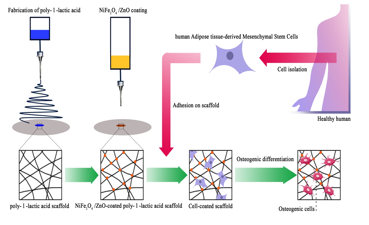
The ability of hAMSCs to differentiate into osteogenic cells was evaluated in this work using NiFe2O4/ZnO-coated PLLA as a scaffold to target bone tissue engineering. According to this, the mechanical properties of the PLLA scaffold improved following plasma treatment and nanocomposites coating. This method revealed evidence of additional tensile strength. Plasma therapy leads to carboxyl and hydroxyl groups forming electrostatic bonds with soluble ions such as calcium and growth hormones (19). Due to the scaffolds' high stiffness, osteogenic proteins (Osteonectin and Osteopontin) and vascularization are promoted, resulting in a significant correlation between vascular growth, bone formation, and bone matrix deposition(18, 19).
Additionally, the nanocomposites applied to the scaffold surface did not obstruct the porosity space of the scaffold. As shown in Fig. 4.a, nanocomposites coated on the scaffold surface have no inhibitory effect on adhesion cell proliferation and survival but rather enhance cell proliferation. Because connected pore networks can facilitate the movement of nutrients, oxygen, and waste products and thus angiogenesis, the porous area of the scaffold receives the majority of cell infiltration. Roughness on the surface and other topological properties can enhance cell adhesion and fate(21).
On day 7, DAPI staining revealed that the number of cells in the NiFe2O4/ZnO-coated PLLA scaffold was much more significant (Fig. 5.d). The presence of magnetic nanoparticles to control and proliferate stem cells is responsible for this action(10). These nanocomposites with magnetic properties, i.e., nickel and ferrite, affect cell proliferation and survival, achieved with surface modifiers(22). The broad porous nanofibers contain anchors that promote cell attachment and proliferation while boosting cell cohesion and diffusion (23).
Cell growth and proliferation were increased on both scaffolds throughout the 21-day monitoring of MSC differentiation into the osteogenic lineage (Fig. 4.b) but were significantly more on the nanocomposites-coated scaffold, which also had more cell layers on the surface. The presence of hydroxyapatite granules on the surface of the NiFe2O4/ZnO-coated PLLA demonstrates that it can promote differentiation into the osteogenic lineage.Carbonate hydroxyapatite is a type of bone mineral necessary for the formation of phosphate and calcium. It is found between collagen fibers. These compounds are responsible for bone's structural, hierarchical, and mechanical properties. Microporous scaffolds have been shown to promote MSC adhesion and osteogenic differentiation(3). Through intracellular and intercellular signaling, the material's topographic and biochemical properties can alter the microenvironment of the desired location. These microenvironmental modifications, such as regulating enzymes, cells, and ions containing radical species, affect cell differentiation (21).
ALP activity was assessed based on our findings from the process of osteoblastic differentiation of MSC on PLLA, NiFe2O4/ZnO-coated PLLA, and TCP. The maximum activity of the alkaline phosphatase enzyme (Fig. 5, b) was seen in nanofibers of NiFe2O4/ZnO-coated PLLA throughout this procedure. This is due to the presence of zinc in this composite, which acts as a cofactor in ALP activity and promotes the development of osteoblastic activity(13). ALP, active in osteoblasts, is the primary marker of osteogenesis and hard tissue(24).
As a significant factor, calcium functions in creating osteogenesis(25). On the NiFe2O4/ZnO-coated PLLA scaffold (Fig. 5), calcium levels rise during the differentiation phase from day 7 to day 21 due to the support of ALP activity, which initiates the mineralization process. By releasing signaling ions, biodegradable biomaterials can alter the environment. Calcium can excite calcium sensory receptors, essential for cell proliferation, differentiation, and chemotaxis. In addition, releasing ions from the phosphate, calcium, and biomaterials bases can stimulate endogenous cells, causing them to develop into the osteogenic lineage (21).
On day 21, the highest amount of calcium mineralization occurred in the NiFe2O4/ZnO-coated PLLA, as shown in Fig. 4.c of Alizarin red staining, which serves as a seal of approval for the process of osteoblast cell mineralization and maturation during the differentiation process into the osteogenic lineage. The magnetic mechanism of action is mediated through the calcium ion transport channel in the cell membrane. Additionally, the magnetic field affects the structure and crystallization of biomineral products, alters the spatial organization of proteins in the cytoskeleton, and has an effect on the biomineralization behavior during the early stages of gene expression and protein synthesis (10). Cell receptors, proteins, and peptides interact to enable the cell to store minerals and ECM proteins (3).
During the 21-day observation of osteogenic lineage development, bone-related mRNAs (Osteonectin, Osteocalcin, ALP, Runx2, and Collagen type I) were detected during the molecular behavior of h-AMSC. The nanocomposites used on the scaffold surface boosted and maintained runx2 expression until day 21. (Fig. 6). The Runx2 gene regulates the expression of phenotypic markers such as osteocalcin and ALP during the differentiation of MSCs into pre-osteoblasts (26). The highest level of ALP mRNA expression on day 14 indicated the nanocomposites' osteoblastic activity, which can be attributed to the activity of the essential element zinc, which had a significant effect on the amount of ALP, as well as the magnetic field produced by nickel ferrite, which is effective at interacting with cells and their ossifying role (12, 26).Karimi et al. investigated the osteogenic development of MSCs on bone PLLA nanofibers using Baghdadite bioceramic nanoparticles. The expression of the Runx2 gene was shown in this study to increase Bmp9 expression (19). Osteonectin is a calcium-binding glycoprotein that plays a function in the crystallization of osteoblasts during their early stages of development (27). Maximum expression of this gene was seen on day 21 on the NiFe2O4/ZnO-coated PLLA scaffold, which corresponds to the mineralization stage (two weeks of differentiation) in previous studies (27, 23). The osteocalcin gene encodes a bone-growth protein (BGP) that osteoblasts generate and release during their maturation (three weeks of differentiation) (27, 28). Although all groups had an increase in gene expression, the complete transcription of this gene was seen on day 21, which correlates with the magnetic field of nanocomposites (10).Collagen type I is the most abundant protein in the bone matrix, as it aids in the mineralization process during bone formation (11). Collagen type I expression increased in this study, while there was no significant difference between the groups on days 7 and 14 of osteoconductive culture. As a result, they provide surfaces capable of transmitting biological signals by mimicking the structure of the ECM. Moving electrons provide magnetic properties on stem cells, which aid in their cohesion, survival, migration, proliferation, and differentiation, and their porosity space can result in the formation of arteries, angiogenesis, and a bone-like environment, which enables bone tissue regeneration(29, 30, 31).Our results indicate that NiFe2O4/ZnO-coated PLLA and PLLA nanofibers prolong the osteoblastic obligation process in hAMSC. Combining PLLA nanofibers with nanocomposites (NiFe2O4/ZnO) promotes cell interaction and ossification in an osteogenic environment. The NiFe2O4/ZnO-coated PLLA scaffold may be used in animal models of bone injury to confirm this work's findings, consistent with the link between osteogenic function in vitro and in vivo.