Histopathological Findings
Out of 65 colon biopsy specimens 35 showed adenocarcinoma of colon (Figure 10 & 11) 25 specimens were with carcinoma and 10 with squamous cell carcinoma. In ascending colon samples 15 were detected with adenocarcinoma, 7 with carcinoma (Figure 13) 3 with adenoma while in descending colon specimens 13 were with adenocarcinoma (Figure 12) 4 with metaplasia (Figure 14) and 3 with carcinoma. Rectal polyp specimens showed 8 specimens with adenoma, 4 with tubulovillous adenoma with severe dysplasia and 3 with carcinoma. In rectal specimens 4 were detected with adenocarcinoma, 3 with squamous cell carcinoma (Figure15) and 3 with metaplasia as shown in table I.
Figure 10
Figure 11
Figure 12
Figure 13
Figure 14
Figure 15
GeneXpert System
The GeneXpert Dx system uses real-time polymerase chain reaction to automate sample preparation, nucleic acid amplification, and target sequence detection in simple and complicated samples (PCR). The system is designed for in vitro diagnostic applications that need hand-off processing of patient samples (specimens), and it provides data in tabular and graphic representations for both summarised and comprehensive test findings. The samples are prepared and processed in GeneXpert cartridges that are designed for single-use assays. The sample and reagents are placed in a cartridge, which is subsequently put into the instrument module.
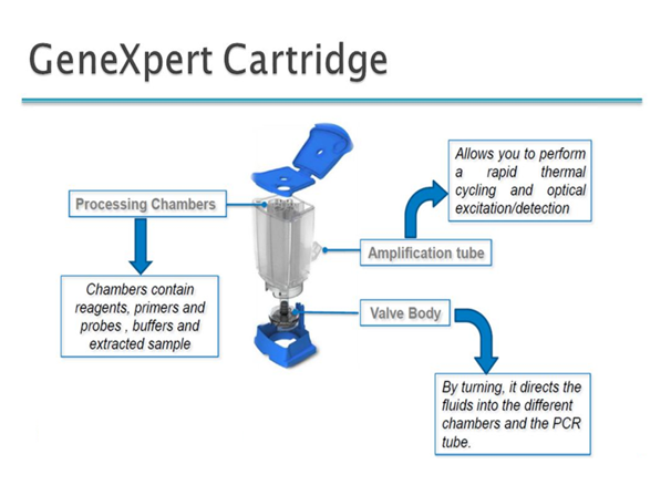
GeneXpert Cartridge
Each cartridge consists of 1) Processing chamber 2) Optical window 3) Valve and Reaction tube Total of 135 histopathologically confirmed colorectal paraffin embedded biopsy specimens were collected from histopathology department of Ayub Medical College (Abbottabad) and histopathology department of Abbott Laboratory and other hospitals of Hazara Division. The experimental work was carried out in Abbot Lab Histopathology and PCR departments. After melting the specimens to remove the wax, they were treated with xylol (MERCK, Germany), then washed with 100 percent ethanol. After removing wax tissues were cut in to pieces and treated with liquid nitrogen and by using mortar and pestle/tissue grinder tissues were crushed in to fine powder. Now each sample powder form was dissolved in preserved cyst solution provided and vortex for 10 to 15 seconds. After vortexing 1ml solution was loaded into HPV cartridge (Figure16) and after scanning bar code of cartridge it was lodged into PCR system and after 1 hour 50 minutes results were obtained (Figure17).
Figure 16
Figure 17
Out of 135 specimens 85 (62%) were detected with HPV DNA, 37 (43%) with HPV-16, 25(29%) with HPV-18 and 16(19%) with HPV-45 genotype (Table I). Seven specimens were detected with low risk genotypes and 43 samples were negative for HPV DNA there was no detection in normal control specimens as shown in Table II. Our results showed that HPV plays a definite role in carcinogenesis of colon and rectum so it should be considered in colorectal cancer cases like cervical cancer.
Table I
Table II