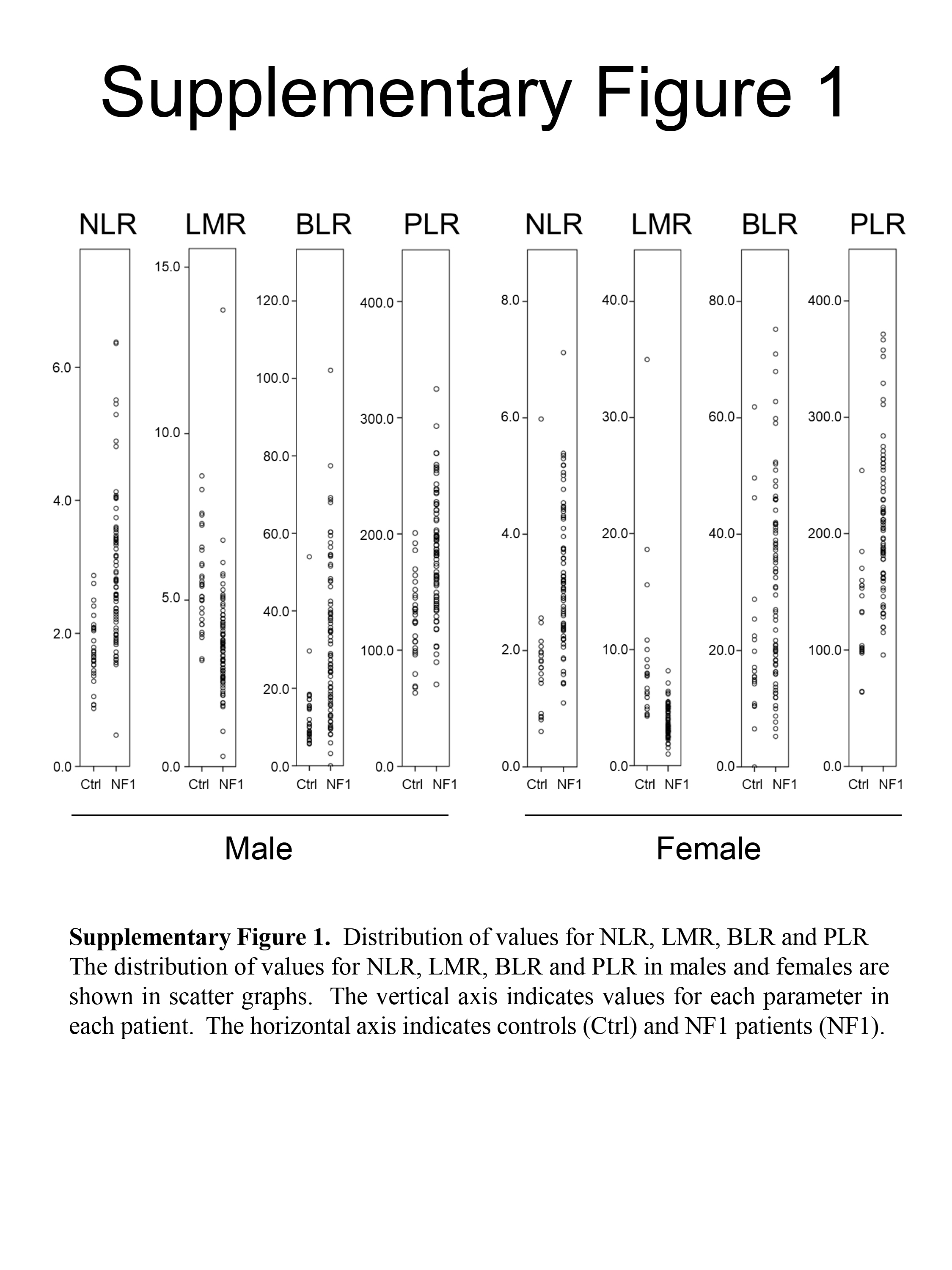
This study offers the first evidence that NLR, PLR and BLR are higher, and lymphocyte count and LMR are lower in NF1 patients than in non-NF1 individuals.
Many reports in the literature have described that high NLR and PLR are associated with the pathogenesis and clinical course of cancers 9,10,18,22, compatible with several reports that neutrophils play various oncogenic roles, including tumor growth, angiogenesis, metastasis and T-cell suppression 22,23, while lymphocytes potentially act as tumor-suppressive immune responders 24. Considering that high NLR and PLR also indicate inflammation 8,16, the high ratios in NF1 are suggested to represent tumorigenesis of neurofibroma through inflammation, mainly associated with mast cells located in neurofibroma 5–7. Because the inflammation localized in neurofibromas may not sufficiently stimulate signals for the production of common inflammation markers such as C-reactive protein, the assessment of inflammation by CBC testing in NF1 may have rarely been a focus before now.
We also focused on BLR for NF1, because: i) mast cells infiltrate into neurofibromas; and ii) mast cells and basophils share functional similarities, including long-term inflammatory or immunologic responses as well as immediate hypersensitivity reactions 25. This study clearly showed that BLR was higher in NF1 patients than in controls. In contrast, Yang et al. demonstrated a significantly low BLR in systemic autoimmune rheumatic diseases 19. These facts led to three considerations. First, BLR should not be regarded as a universal inflammatory marker. Second, a high BLR may be somewhat specific for NF1. Third, reference to BLR may be difficult in NF1 patients with complicating systemic autoimmune rheumatic diseases. On the other hand, basophils and mast cells exhibit fundamental differences 25. Basophils do not proliferate after terminal differentiation and the cells circulate in the peripheral blood. In contrast, mast cells reside in peripheral tissue and retain the ability to proliferate. In NF1, the association between basophils and mast cells should be further investigated.
The application of these parameters to clinical practice may be premature because the results obtained did not reveal relationships with clinical symptoms, including the tumor burden, skeletal abnormalities, vasculopathy, and learning difficulties. Therefore, further studies are warranted to identify the symptoms that affect these parameters and also the extent of their effects.
NF1 is an RASopathy, a comprehensive pathological concept covering genetic syndromes caused by germline mutations in the genes that encode the molecules associated with the RAS/MAPK pathway 26. In addition to NF1, RASopathy includes Noonan syndrome, Noonan syndrome with multiple lentigines, Costello syndrome, cardio-facio-cutaneous syndrome, and Legius syndrome. 26. Whether these other RASopathies also show high NLR, PLR and BLR and low lymphocyte count and LMR is an interesting question worth exploring.
Lee et al. calculated mean NLR, LMR and PLR across all ages from 12160 blood samples collected from healthy patients in South Korea 27. Our data may be comparable with the data described by Lee et al., because patients included in our data were uniformly Japanese, representing an East Asian ethnicity similar to South Koreans. NLR for our lipoma patients compared to the data from Lee et al. showed 1.76 vs. 1.63 for males and 1.84 vs. 1.66 for females. Similarly, our data compared to theirs indicated LMR of 5.52 vs. 5.05 for males and 9.36 vs. 5.60 for females, and PLR of 127.66 vs. 122.75 for males and 128.82 vs. 142.76 for females. All parameters calculated from our lipoma patients were broadly compatible with those calculated from healthy South Koreans, with the exception of LMR in females. This information supports the notion that lipoma patients can largely be regarded as healthy controls in terms of these comparisons.
Some limitations to this study need to be considered. First, data were lacking for other inflammatory markers such as C-reactive protein and erythrocyte sedimentation rate. Therefore, whether the NF1 patient group included NF1 patients with acute inflammatory diseases causing high NLR and/or PLR remains unclear. Second, an assessment of the clinical severity of NF1 was not performed. It currently remains unclear whether the parameters examined in the present study are associated with the clinical severity of NF1, including the tumor burden. Third, data were lacking in terms of complications in NF1 patients and control patients, such as systemic inflammatory diseases, cardiovascular diseases, diabetes mellitus and malignancies. NLR, LMR, PLR and/or BLR may be affected by patient status in terms of systemic inflammatory disease, risk of cardiovascular events, control of type 2 diabetes mellitus or the presence of malignancy 9,12,19,22,28,29. Fourth, the present results lead only to speculations that the mechanisms of inflammation may be associated with NF1. Therefore, further studies are needed to show direct evidence for a relationship between the mechanisms of inflammation and NF1.
In conclusion, this study demonstrated that NF1 patients show high NLR, PLR and BLR and low lymphocyte count and LMR. These parameters may represent the inflammation associated with neurofibroma tumorigenesis in NF1.