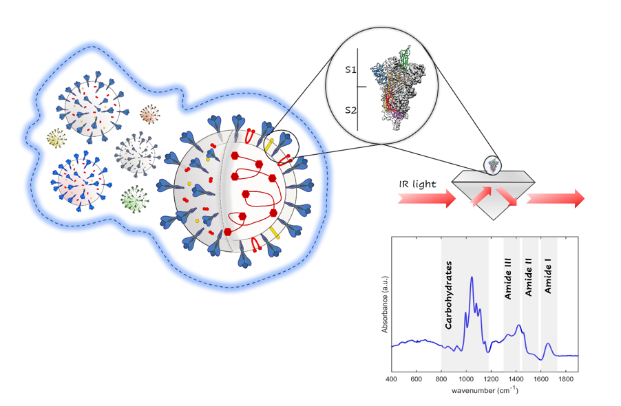
Amide I: Protein secondary structure
Figure 2 shows the absorbances A(ω) vs. frequency (ω) of the S1 glycoprotein of MERS-CoV (B), SARS-CoV (C) and SARS-CoV-2 (D) in the Amide I band, between 1580 and 1750 cm− 1, measured at pH 7.4 and at a concentration of 0.25 mg/ml. Similar data have been obtained for other concentrations (see Fig. S1 in supporting information). A first qualitative comparison can be made looking at Fig. 2A, where the absorption spectra of the three S1 proteins are shown. While MERS-CoV (blue line) and SARS-CoV (red line) show a quite similar broad absorption band centered at about 1660 cm− 1, in SARS-CoV-2 (yellow line) the band has a maximum around 1650 cm− 1. This red-shift can be further quantified by calculating the differences A(ω)(SARS−CoV−2)-A(ω)(MERS−CoV), and A(ω)(SARS−CoV−2)-A(ω)(SARS−CoV) (blue and red line in the inset of Fig. 2A, respectively) and comparing them with the reproducibility of the A(ω)(SARS−CoV−2) absorption measurements. The reproducibility has been estimated by the difference A(ω)(SARS−CoV−2)-A(ω)(SARS−CoV−2) for two different measurement runs (yellow line in inset of Fig. 2A). The SARS-CoV-2 absorption reproducibility fluctuates less than 2% in the whole Amide I spectral range. Instead, a sizeable difference (actually far out of the reproducibility of absorption spectra), can be observed when comparing SARS-CoV-2 with MERS-CoV and SARS-CoV absorptions. In particular, for both MERS-CoV and SARS-CoV there is an absorption reduction between 1600–1650 cm− 1 (in agreement with the main panel of Fig. 2A), and a smaller enhancement at higher frequency.
In order to identify the secondary structure for MERS-CoV, SARS-CoV and SARS-CoV-2, a global fitting approach (40–42, 47) have been used for deconvoluting in Gaussian spectral components the Amide I band. The total fit (empty circles), and the spectral decomposition (colored Gaussians), of MERS-CoV, SARS-CoV and SARS-CoV-2 Amide I band (black lines), are reported in Fig. 2B, C and D, respectively. The area of each component can be used to estimate the secondary structure content of the S1 protein.
Table 1 summarizes the vibrational frequencies of the different Gaussian-components and their relative intensities, together with their assignment to specific secondary conformation structures (40, 44, 47–49). In particular, we notice in all A(ω) an intense peak around 1658 cm− 1, associated with the α-helix structure (40,47–49). The β-sheet components are instead observed between 1620–1640 cm− 1 and around 1690 cm− 1 (49). In particular, the bands near 1630 cm− 1 and 1690 cm− 1 are typically related to an antiparallel arrangement of the β-sheet. The bands in the 1665–1690 cm− 1 range can be assigned instead to the β-turn structure. The broad absorption band centered at 1643 cm− 1 corresponds to random coils. The absorption band at 1619 cm− 1, present only in SARS-CoV-2 S1 unit, is finally assigned to side chains and intermolecular anti-parallel β-sheets (40,44,47–49). The actual secondary structure of S1 unit in MERS-CoV, SARS-CoV and SARS-CoV-2 viruses at pH 7.4 can be estimated through the ratios among the intensity of each component of the Amide I band over the total intensity (also reported in Table 1) as obtained by the previously mentioned fitting procedure. From these data, one can observe that SARS-CoV and SARS-CoV-2 show similar α-helix (27.7% and 29.4%) and random coil contents (13.5% and 12.3%). The larger difference in the secondary structures of the S1 proteins can be instead observed in the arrangement of β-sheet and β-turn. Notably, a significant increase is revealed in the β-sheets content passing from MERS-CoV (20.6%) to SARS-CoV (26.8%) and SARS-CoV-2 (30.6%). This is mainly due to the strong increase of the antiparallel β-sheet structure observed at 1619 cm− 1 for the SARS-CoV-2 S1 unit, which corresponds to nearly 5% of the total protein secondary structures. An opposite trend is shown by the β-turn (1665–1687 cm− 1): MERS-CoV and SARS-CoV S1 proteins exhibit approximatively the same β-turn content (30.6 and 32 ), compared to ~ 28 of the SARS-CoV-2 S1 unit. Observing the results concerning MERS-CoV and SARS-CoV S1 proteins, we notice a similarity in the secondary conformational structure (α-helix and β-turn), attributable to the aminoacid sequence identity of their S1 unit (3, 50). Instead, although both SARS-CoV-2 and SARS-CoV S1 proteins interact with the human hACE2 receptor and show a sequence similarity (between 73–75 ) as determined by Cryo-EM (3), they exhibit, on the basis of our vibrational absorption measurements, a robust secondary conformation difference.
Table 1
Secondary structure assignment for MERS-CoV, SARS-CoV and SARS-CoV-2 S1 units derived from the Gaussian decomposition of the absorption spectra. For each S1-protein, we show (first column) the characteristic absorption frequency and (second column) the peak relative intensity. In the third column the observed absorption peaks are assigned to specific secondary conformations (40, 49).
| MERS-CoV peaks [cm− 1] | MERS-CoV peak relative intensity [%] | SARS-CoV peaks [cm− 1] | SARS-CoV peak relative intensity [%] | SARS-CoV-2 peaks [cm− 1] | SARS-CoV-2 peak relative intensity [%] | Assignment |
| - | - | - | - | 1619 | 5.2 | β-sheet (intermolecular) |
| 1628 | 14.0 | 1625 | 7.9 | 1628 | 11.4 | β-sheet |
| 1632 | 2.4 | 1633 | 12.7 | 1633 | 9.8 | β-sheet |
| 1641 | 16.5 | 1642 | 13.5 | 1643 | 12.3 | Random coils |
| 1650 | 13.7 | 1650 | 12.9 | 1650 | 13.6 | α-helix |
| 1658 | 18.6 | 1658 | 14.9 | 1658 | 15.9 | α-helix |
| 1666 | 10.6 | 1666 | 12.2 | 1666 | 5.6 | β-turn |
| 1673 | 8.3 | - | - | 1673 | 6.7 | β-turn |
| - | - | 1675 | 10.4 | - | - | β-turn |
| 1680 | 11.8 | 1681 | 9.3 | 1678 | 15.2 | β-turn |
| 1693 | 4.2 | 1690 | 6.1 | 1693 | 4.2 | β-sheet |
Being the receptor-protein recognition conformation-dependent, the observed differences can be related to their differential receptor-binding affinities. Indeed, recent Cryo-EM and computational studies have shed light on this behavior (19, 51–54), investigating the binding affinity of the RBD sites for the hACE2-peptidase domain. Experimentally, Wrapp et al. (54) and Tai et al. (19) have reported that the SARS-CoV-2 RBD has a higher binding affinity for hACE2-peptidase domain than the SARS-CoV RBD.
Amide I: Changes In Secondary Structure Induced By Ph Variation
The pH-dependent conformation changes in proteins play a key role in virus replication, pathogenesis, and transmissibility. In particular, the local-environment pH can strongly influence the protonation state in folded proteins by promoting changes in interchain interactions. Consequently, the stability of proteins is pH-dependent, favoring conformational flexibility, protein activation and/or inactivation. In this framework, we have studied the secondary conformation of SARS-CoV-2 S1 protein at different pH values (pH = 4.55, 5.5, 7.4, 8.8 and 11.2), by measuring the absorbance A(ω) of the Amide I band (Fig. 3A). The absorption behavior vs. pH is not monotonic; while SARS-CoV-2 S1 at serological (yellow line) and 8.8 (purple line) pH values show a similar broad absorption band centered at about 1650 cm− 1, the spectra relative to mild low pHs (blue line at 4.55 pH, and red line at 5.5 pH) and alkaline pH (green line at 11.2 pH) are red-shifted of about 10 cm− 1 and exhibit a maximum around 1640 cm− 1. This red-shifting can be further observed in Fig. 3B, where the calculated absorption differences A(ω)(pH 7.4)-A(ω)(pH x), are compared with the reproducibility of A(ω)(pH 7.4). This last quantity has been estimated by the difference A(ω)(pH=7.4)-A(ω)(pH=7.4) for two different measurement runs (yellow line in Fig. 3B). Let us notice that a similar reproducibility can be observed at any pH. The absorption reproducibility fluctuates less than 2% in the whole Amide I spectral range. Significant differences, actually far out the reproducibility, emerge when the pH moves from mild low values to alkaline ones. Indeed, an absorption frequency redistribution is observed around an isosbestic point at about 1647 ± 1 cm− 1. As the S1 protein concentration and all physical parameters, e.g., temperature, pressure and relative humidity, are kept constant during experiments, the occurrence of the isosbestic point can be associated to the conformational changes in the protein structure induced by the pH.
The influence of pH on the S1 protein conformation can be quantified by studying the change in peak position and intensity of the spectral components of the Amide I band through a global Gaussian fitting. The global fit (empty circles), and the spectral decomposition (colored Gaussians), of the Amide I band for different pHs (4.55, 5.5, 8.8 and 11.2), are compared in Fig. 3C, D, E and F, respectively. Absorption at pH = 7.4 has been already reported in Fig. 2D. The area of each spectral component has been used to estimate the secondary structure content and then its variation with pH. In Table S1, the peak frequencies for the Amide I S1 protein vs. pH and associated to specific secondary conformation structures are shown while their intensities vs. pH are represented in Fig. 4 which will be discussed in the following (40, 44, 47, 49, 55).
The main effect of pH concerns, firstly, the α-structure (see Table S1 in supporting information), which give rise to a main absorption band at 1658 cm− 1 and to a shoulder at lower frequencies around 1650 cm− 1 at 7.4 pH. While the main α-structure component remains nearly constant in frequency (1658–1659 cm− 1) for pH moving from 7.4 to 5.5, this band shifts near to 1663 cm− 1 at pH = 4.55 and at alkaline levels. Usually, a blue-shift is associated to the formation of a short α-helix. Moreover, for acid pHs, the whole α-helix absorption reduces its intensity of about 50% (see Fig. 4). The band at 1643 cm− 1 for 7.4 pH associated to random coils, is also influenced by pH variation, blue-shifting to 1646 cm− 1 for acid and alkaline pHs. Its intensity is maximized at pH = 5.5 (see Fig. 4). The intermolecular β-sheet located around 1619 cm− 1 at serological and alkaline pHs, instead red-shifts for acid conditions. The other β-sheet components are located between 1620–1640 cm− 1 and around 1690 cm− 1 (see Table S1 in supporting information) (44) at pH = 7.4. The band at 1629 is red-shifted at 1628 cm− 1 at acid pH levels suggesting the aggregation of sheets with a large number of strands (40, 49), and blue-shifted at 1633 cm− 1 for alkaline pHs. The absorption of β-sheet for pH = 4.55, 5.5 and 11.2 pH is related to a new band at 1637 cm− 1. Finally, the absorption at 1693 cm− 1 disappears for extreme pH values. From Table S1, the total intensity of β-sheet presents a minimum at serological pH, being maximized at acid pHs. The bands in 1666–1678 cm− 1 range, assigned to β-turn structures (40, 44, 47,49, 55) are blue-shifted compared to their spectral positions at 7.4 pH, while a new band appears at 1686 cm− 1 for pH values of 4.55, 8.8 and 11.2. The total intensity of β-turn structures, which shows a maximum at pH = 7.4 reduces both at acid and alkaline pHs.
For a quantitative evaluation of secondary conformation changes, we show in Fig. 4 the ratio between the secondary structure peak areas and the total area vs. pH. At the serological pH 7.4, the α-helix and β-turn structures show their maxima, reducing their intensity both in acid and alkaline conditions. Conversely, the random coil and the β-sheet present an opposite behavior with minima at pH 7.4 while increasing towards the acid and alkaline pH values. Moving from serological to alkaline environment, at pH 8.8 the contents of random coil and α-helix are almost unchanged, differently from β-sheet and β-turn structures which increase and decrease, respectively. Thus, a slight unfolding of β-structures appears at pH 8.8, that is the typical environmental pH observed in bat caves where high ammonia concentrations build up. Moreover, at pH 11.2 the decrease of the β-turn content by an average of ~ 10% and the increase of β-sheet structure of 12% are observed. This behavior suggests a transition from β-turn the to the β-sheet structure in the strong alkaline medium (pH = 11.2), while a slight rearrangement of the α-structure is observed under mild alkaline conditions. This is also consistent with a loss of secondary structure, perhaps due to a partial protein unfolding. The reduction of the β-turn content is also observed for acidic pHs. In addition, the β-sheet content increases from its value at 7.4 pH (~ 30.6%) moving from neutral to mild low pH at 47% of all Amide I band. The content of side chains and intermolecular β-sheets (~ 1619 cm− 1), associated to a low frequency band, exhibits a pH-dependent behavior, with a maximum value of ~ 19% at 5.5 pH, to be compared with the serological value (5%).
The strong changes of the protein secondary conformation at pH 5.5 is associated with the rearrangement and modification of the RBD sites. Some investigations have shown that several regions of the SARS-CoV-2 S protein are susceptible to mutations, with the RBD site particularly vulnerable to a conformational change (56, 57). The RBD undergoes indeed a hinge-like movement that brings up or down the specific amino-acid sequence responsible for the binding to the hACE2 receptor (54) inducing a change in the secondary conformation. In our work, we observe a large variation of intermolecular β-sheets content for the SARS-CoV-2 S1 vs. pH (see Fig. 4), with a maximum value at pH = 5.5 (nearly 20%). This behavior can be related to a structural transition from a closed over a locked form of S1 as pH is increased from mild acidic to neutral as observed by Cryo-EM microscopy studies (58, 59), with an optimal condition for the pre-fusion configuration at pH 5.5.
The overall strong conformational changes of SARS-CoV-2 S1 unit vs. pH by passing from the alkaline ones, characteristic of the ecological niche bats ambient, to the human serological environment, suggests a high capability of S1 SARS-CoV-2 glycoprotein to adapt to different environments, further indicating the existence of a correlation between the S1 protein secondary conformation and the virus transmissibility.