3.1 Preparation and characterization of HLaPc-BNNSs
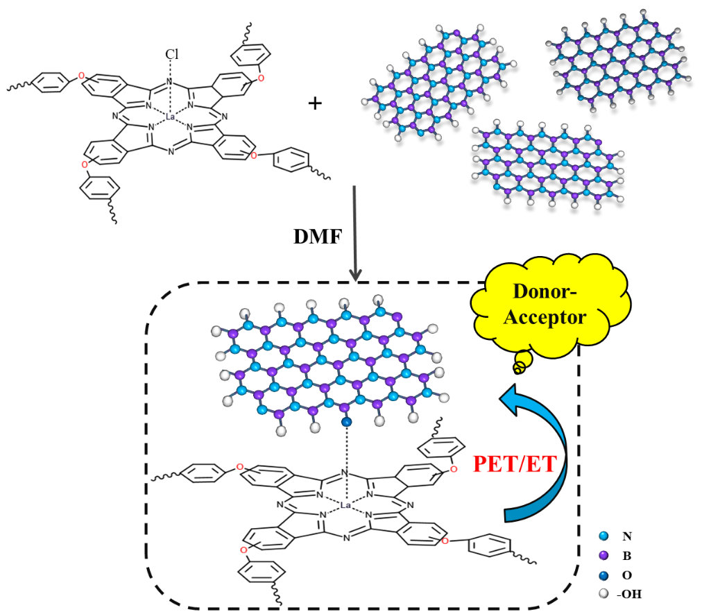
The synthesis procedure of HLaPc-BNNSs was shown in Scheme 1. By heating a mixture of silver trifluoromethanesulfonic acid, OH-BNNSs and HLaPc in DMF, the corresponding axially covalent connected boron nitride to phthalocyanine can be obtained.The changes of some functional groups in the material structure were verified by analyzing infrared spectra. The infrared spectra of h-BN and OH-BNNSs are shown in Fig S2.The strong absorption peak at 1417 cm-1 is assigned to the in-plane bending vibration of BN. The peak at 804 cm-1cans be attributed to the out-of-plane bending vibration of BN. This is consistent with the previously reported spectrum[20]. However, there is a new peak at 1190 cm-1 in the OH-BNNSs spectrum, and this additional absorption peak results from the deformation of the B-O bonds of OH-BNNSs. A significant hydroxyl absorption peak appears at 3360 cm-1,indicating that the hydroxyl group has been successfully linked to the BN[21, 22]. The infrared spectra of OH-BNNSs, HLaPc and HLaPc-BNNSs are shown in Fig. 1. The characteristic peaks have good correspondence, which indicates that the synthesis is successful. 1456.16cm-1, 1519.80cm-1 correspond to the stretching vibration absorption peak of C = N bond and C = C bond respectively.AnAr-O-Ar vibration absorption peak of hyperbudgetedphthalocyanine was observed at 1252 cm-1, as well as the vibration absorption peak of phthalocyanine ring skeleton is at 729 cm-1, 1149 cm-1.The weak peak at 1090cm-1 is associated with the C-H bonds of phthalocyanine. The peak near 660-900cm-1 corresponds to the out-of-plane bending vibration of the phenyl C-C bond[23]. The peak at 1386 cm-1 may be attributed to the vibration of La-Cl bond, which disappeared after bonding with OH-BNNSs.
The composition of the sample phase can be qualitatively analyzed by XRD. The XRD patterns of h-BN, OH-BNNSs, HLaPc and HLAPC-BNNSs are shown in Fig. 2(a). h-BN has a strong and sharp diffraction peak at 2θ = 26.7° and belongs to a layer structure with a distance of 0.1091 nm in the direction of (002)[24]. The functionalization of BN usually leads to changes in lamellar structure and space. The introduction of hydroxyl groups broadens the peak shape and increases the peak intensity significantly, which can be used to judge that the particle size of the crystal becomes smaller. It also can be seen that HLaPc-BNNSs has the peak shape of both h-BN and HLaPc, and the HLaPc-BNNSs hybrid shows a wide (002) peak at 2θ = 27.14 °, and the corresponding layer spacing is 0.2243 nm, which is lower than the diffraction peak of the original BN[25]. It can be explained that the introduction of hyperbranched phthalocyanine reduces the regularity of BN lamellar, increases the layer spacing, decreases the intensity of diffraction peak and widens the peak type. The widening of the lamellar distance is often beneficial to improve the dispersion of the hybrid, thus further improving the nonlinear optical effect.
Raman spectroscopy is a common and effective method to characterize materials. The Raman spectra of h-BN and HLaPc-BNNSs hybrid materials are shown in Fig. 2(b). Under 532 nm wavelength excitation source, the characteristic spectra of all hybrid materials appear in the range of 1000 cm-1 to 2000 cm-1. Raman spectrometric measurements show that there is a G band at ≈ 1366cm-1(E2g mode), which is caused by the base plane defect. The sharper the peak, the better the crystallinity. The G band is used to evaluate the distribution of functionalized modifications [26]. The G band transfers to a lower frequency when hybridized with the electron donor component, indicating that charge is transferred from HLaPc to BNNSs.
The XPS survey spectra of the OH-BNNSs, HLaPc and HLaPc-BNNSs are shown in Fig. 2(c-e). There are four peaks around 532.5eV, 398 eV, 288.8eV and 190.7eV, corresponding to O1s, N1s, C1s and B1s, respectively, and the peak of La 4d at 97.6eV.It can be seen that compared with HLaPc, the Cl 2p peak near 198.9eV in HLaPc-BNNSs is obviously disappeared, which fully indicates that the Cl atom is replaced by the BNNSs group. Figure S4 is the XPS peak spectrum.It can be seen that the B1s level of OH-BNNSs can be divided into two peaks. The binding energy of B-O is 191eV, and B-N is 190.2 eV[27, 28]. The XPS spectrum of the C 1s level of HLaPc-BNNSs is divided into three peaks, involving C–C at 284.4 eV, C–N/C = O at 285.9 eV and C-O at 285.7 eV [29, 30]. Two peaks were observed at 835.9 eV and 853 eV, corresponding to the 3d5/2 and 3d3/2 orbits of La, respectively[31].
3.2 Photophysical properties
Absorption spectra can be used to study the photophysical properties of substances. As shown in Fig. 4(a), the spectrum of HLaPc contains a clear Q-band peak at approximately 678 nm, which can be assigned to the π–π* transition,and B-band peak was observed in the nearUV region (345nm)[32]. In addition, there are shoulder peaks in HLaPc due to the aggregation of phthalocyanine rings,the aggregation will increase the relaxation channel, shorten the excited state lifetime, and reduce the effective nonlinear absorption. The Q band acromion of HLaPc-BNNSs weakened or even disappeared due to the decrease of aggregation. Compared with HLaPc, the Q-band absorption peak intensity of HLaPc-BNNSs showed a decreasing trend a certain degree of redshift. This is because the bonding of BNNSs increases the π electron conjugation system of hyperbudgeted lanthanide phthalocyanine, which makes the phthalocyanine compound have lower S1 state energy level[33, 34],as well as the peak pattern widened to a certain extent, showing the effect of combination with BN absorption spectrum. These results also provide evidence for covalent functionalization between HLaPc and BN nanosheets.
The electron transfer process between BNNSs and HLaPc was studied by fluorescence emission spectroscopy. As shown in Fig. 4(b), HLaPc showed a fluorescence emission peak at 706 nm at the excitation wavelength of 698 nm. The peak position is slightly larger than the wavelength of the maximum absorption peak of Q band in the UV-vis absorption spectrum, indicating that HLaPc is excited to the singlet S1 level at about 698 nm and thus fluoresces at 700–720 nm. HLaPc-BNNSs has a certain degree of redshift compared with HLaPc, which is because the introduction of BN expands the degree of conjugation of phthalocyanine molecules and increases the electron cloud density[2]. In addition, the fluorescence intensity has obvious quenching phenomenon, which is because BN acts as electron acceptor in the molecule, while the phthalocyanine has abundant π conjugated electrons as electron donors.Therefore, the fluorescence quenching of HLaPc-BNNSs is due to an effective charge transfer process from phthalocyanine to BNNSs unit[35, 36]. In addition, as a rare earth element, lanthanum can effectively promote the electron transition from singlet state to triplet state, which has a positive effect on the efficiency of PET/ET process between hyperbudgetedphthalocyanine and BNNSs.
3.4 Non-linear optical absorption studies
3.4.1 nonlinear optical properties of solution
To determine the non-linear optical properties of the functionalised hybrid materials, the OA Z-Scan measurements were carried out under nanosecond pulse excitation at 532 nm and 1064nm. The non-linear optical absorption characteristics of h-BN, HLaPc and HLaPc-BNNSs in DMF are presented in Fig. 5. The modified compound (HLaPc-BNNSs) not only ensures the effective nonlinear optical response at 532nm, but also shows a significant improvement in nonlinear optical response at 1064nm, indicating that the limiting region of hyperbranched lanthanum phthalocyanine grafted BNNSs is effectively broadened to near infrared region, which is of guiding significance for the development of new optical limiting materials with wide limiting region. Due to the strong absorption of phthalocyanine compounds in the linear absorption region, the nonlinear optical absorption mainly occurs in the visible region. Therefore, the limiting region of phthalocyanine compounds is narrow and there is no linear absorption at 1064nm (as shown in Fig. 5b). The strong absorption of the modified compound at 1064nm was mainly due to the synergistic effect of the desaturation of h-BN and the effective PET/ET process between the constructed donor and acceptor [37, 38]. The effective nonlinear absorption coefficient βeff (cm/GW) and the third–order nonlinear susceptibility Im[χ(3)] were obtained by fitting the numerical data are given in Table 1. The βeff values of HLaPc functionalized boronitroides were better than those of other individual com-ponents, showing better NLO properties. The third-order nonlinear susceptibility Im[χ(3)] values of HLaPc-BNNSs at 532 nm and 1064 nm are 9.46×10⁻¹⁴esu and 7.44×10⁻¹⁴esu, respectively, showing that the perturbation caused by laser pulse is robust. Eq. (1)[39] can be used to calculate the nonlinear absorption coefficient βeff (cm/GW).
$${ T}_{OA}={\sum }_{m=0}^{{\infty }}\frac{[-{\beta }_{\text{eff}}{I}_{0}{L}_{eff}/(1+{\left(z/{z}_{0}\right)}^{2}){]}^{m}}{(m+1{)}^{3/2}}$$
1
TOA is the normalized transmittance, and Leff (Leff =[1–exp(–αL)]/α) is the effective thickness of the sample. I0 and z are the axial light intensity and the position of the sample. z0 (z0 = πω02/λ) is the Rayleigh range, where ω0 is the beam waist at focal spot and λ is the wavelength of input light. The third–order nonlinear sensitivity values (Imχ(3)) can be figured out from Eq. (2)[40] :
$${ Im}[{\chi }^{\left(3\right)}]={n}_{0}^{2}{\epsilon }_{0}c\lambda {\beta }_{eff}/2\pi$$
2
Where n0 and ε0 are the linear refractive index and the permittivity of a vacuum, respectively, c is the speed of light and λ is the wavelength of the laser. Based on the definition of Im[χ(3)], it can be found that the value of Im[χ(3)] is positively correlated with the nonlinear response. The Im[χ(3)] of HLaPc-BNNSs is larger than that of HLaPc and h-BN, which is fully consistent with the electron transfer rule.
3.4.2 nonlinear optical properties of thin films
The nonlinear optical properties of solid film materials are of great significance for the practical application of optical limiter materials. For this reason, the nonlinear optical properties of h-BN/PPSU, HLaPc/PPSU and HLaPc-BNNSs/PPSU were studied by Z-scan technique (Fig. 6).The film sample is placed vertically and clamped to the mold, and then movethe mold uniformly in the Z-axis direction. At 532nm, when Z = 0, the linear transmission of pure PPSU, h-BN/PPSU, HLaPc /PPSU and HLaPc-BNNSs/PPSU are 69%, 54%, 52% and 46%, respectively. By fitting, the βeff value of HLaPc-BNNSs/PPSU reaches 1800(cm/GW) at 532nm and 1400(cm/GW) at 1064nm, respectively. The NLO performance of HLaPc-BNNSs/PPSU is greatly improved compared with that of the solution. The remarkable improvement of NLO performance is due to the excellent chemical and structural stability of PPSU as a special engineering plastic and the large number of benzene ring conjugated electrons in PPSU structure. And it reflects that the HLaPc-BNNSs/PPSU film accelerates the effective charge transfer between individual BNNSs and HLaPc [41].
Table 1
Nonlinear optical parameters of h-BN,HLaPc and HLaPc-BNNSs.Nonlinear optical parameters of PPSU,h-BN/PPSU,HLaPc/PPSU and HLaPc-BNNSs/PPSU.
| Sample | λ | I0 (µJ) | βeff (cm/GW) | Im[χ(3)](esu) |
| h-BN | 532 1064 | 158 150 | 5.5 4.3 | 5.84×10− 14 4.57×10− 14 |
| HLaPc | 532 1064 | 158 150 | 8.7 / | 9.24×10− 14 / |
| HLaPc-BNNSs | 532 1064 | 158 150 | 10.8 7 | 9.46×10− 14 7.44×10− 14 |
| PPSU | 532 | 15 | 650 | 6.91×10− 12 |
| 1064 | 20 | 250 | 2.78×10− 12 |
| h-BN/PPSU | 532 | 15 | 1100 | 1.91×10− 11 |
| 1064 | 20 | 850 | 1.38×10− 11 |
| HLaPc/PPSU | 532 | 15 | 1500 | 2.03×10− 11 |
| 1064 | 20 | / | - |
| HLaPc-BNNSs/PPSU | 532 | 15 | 1800 | 2.43×10− 11 |
| 1064 | 20 | 1400 | 1.69×10− 11 |