Given the nature of the experimental ingredients and the research that has already been done previously in shrimp [5-7, 10, 16] we were rather confident that the by-products tested here would meet the profile to partially replace FM in the feed for carnivorous fish. It resulted true in the case of shrimp head (SD) and Pen shell viscera meals (PD). However, from the growth results reported before [9] we knew that almaco jack fed CD and SCPD had very low specific growth rate, 1.1 and 1.5% a day, respectively, half that of the RD (2.8 %) or the other two experimental diets, SD (3.2 %) and PD (3.4 %). Previous analyses of other batches of CD showed low concentrations of essential fatty acids, which are necessary for a rapid growth of S. rivoliana. Catarina scallop meal did have much less HUFA than any of the other by-products, with values of HUFA around 1.2 g/kg dw, while shrimp head meal had 11.2 g/kg dw, Pen shell meal had 29.6 and FM had 17.3 g/kg dw. The requirements of HUFA n-3 for Seriola species, range between 5.0-20.0 g/kg of EPA + DHA in the diet [17, 18]. Here we found EPA + DHA in the feed ranged from 12.4 to 14.5 g/kg of the diet for RD, SD, and PD, but were below 10 g/kg for CD and SCPD, so even these diets had sufficient HUFA n-3 according to literature. We assumed that the triple diet would be the best for growth since nutrients that were lacking in one by-product could be provided by another by-product. For example, scallops in general are low in cholesterol and rich in phytosterols not present in FM. In contrast, shrimp contains three-fold the cholesterol of scallops (Table 1). However, the growth results of fish fed the SCPD (117.1 g) were similar to that of the CD (95.2 g), lower than the other treatments: RD (258.9 g), SD (328.9 g) and PD (365.1 g) [9]. We then supposed that Catarina scallop viscera meal could have been contaminated: Here, we found that Catarina scallop viscera meal did have low but nonetheless detectable levels of 18:5n-3 (0.02 g/kg). This fatty acid, 18:5n-3 is found in marine dinoflagellates, such as Gymnodinium sp. or Prorecentrum sp. [19]. The presence of this fatty acid could indicate that Catarina scallops were in contact with dinoflagellates, probably in the form of an initiating red tide, that they were ingested and that the toxins were accumulated in the viscera of the Catarina scallops. In situ, when the viscera of the Catarina scallops were collected, we noticed no evidence of contamination. However, a slightly higher mortality than usual for the season was reported, attributed to the higher water temperature during the summer (fishermen of the community 2014, pers. comm.). There have been reports of the presence of dinoflagellates (Gambierdiscus toxicus, Prorocentrum mexicanum and P. rhathymum) producers of diarrheic toxins such as okadaic acid in the Magdalena-Almejas lagoon system, B.C.S, Mexico in the periods 1980-1989 and 2005-2006 [20] and again in the Laguna Ojo de Liebre, B.C. S., from May to June 2014 [21, 22]. These types of dinoflagellates produce toxins, particularly okadaic acid that are accumulated by bivalve mollusks, such as oysters and clams in their tissues after consuming these dinoflagellates [23]. After detecting 18:5n-3, we tested all meals for toxins using immunochromatography test of lateral flow and found that the Catarina meal used here did contain low levels (0.02 g/kg) of okadaic acid (Nuñez-Vázquez 2018, pers. comm.). When diets (CD and SCPD) containing Catarina scallop viscera meal were offered to fish, they initially ate them at the same rate as the fish in other treatments, indicating that palatability was not affected and fish initially probably did not detect an off-flavor in the feed. However, after some days, fish were observed to actively reject the CD and SCPD feeds, by nipping the pellets as they sank in the water column and then spitting them out. This is consistent with a learned discomfort, probably in the digestive tract. The feeding intake was similar at the beginning of the experiment (3.8 and 4.1 g/day for each fish, for CD and SCPD, respectively), with a slight decrease at 15 days, but by day 30 it had reduced to half, 1.6 and 2.0 g/day in fish fed CD and SCPD, and it decreased even further after 45 and 60 days, while in the others treatments, feeding intake was of 5.4 g/day for the RD, 5.9 g/day for SD and 6.3 g/day for PD for each organism. Decreased feeding in CD and SCPD is in accordance with a lack of growth in these two treatments. By the end of the trial weight decreased 10.9% and 5.2 % in CD and SCPD fed juveniles, compared to their weights at day 30 [9]. In agreement, total lipids in muscle and liver of fish fed CD and SCPD were significantly lower than fish sampled at the beginning of the experiment, indicating that the fat fish started with had been exhausted, instead of accumulated, as was the case in the other treatments. Interestingly, total lipids in brain did not decrease in CD and SCPD fed fish and remained fairly similar to initial levels, denoting a differential use of fat from different tissues.
The effect of the long-held non-intentional fasting in the juveniles fed CD and SCPD on the fatty acid accumulation are also interesting. These diets had similar levels of lipid, ARA and DHA compared to the RD. However, the concentration of DHA (Fig. 1) and ARA (Fig. 2) differed among diets. The concentration of DHA was significantly lower in muscle of the CD and SCPD fed juveniles compared the initial values, RD and SD, even if the proportion of DHA in muscle was significantly higher, particularly for the SCPD fed juveniles, indicating a selective conservation of DHA in muscle for CD and SCPD during the imposed fast. Clearly, juveniles struggled to maintain some essential fatty acids necessary for survival, and DHA is essential for neural tissue, sensory organs and skeletal system [24]. However, DHA concentration in mesenteric fat in juveniles fed CD and SCPD was similar to other treatments, indicating that even with this level of fasting, fat was not burned to cover for lipid necessities. These would indicate a much more regulated lipid metabolism in mesenteric fat that previously though, and not just a deposit of excess fatty acids from feed [25, 26]. DHA in brain had similar concentrations and proportions of DHA in all treatments. Interestingly, juveniles fed PD had much more DHA accumulated in the muscle compared to liver, in comparison to all other treatments, indicating that there might be other component in PD that help the transference from liver to other tissues. The concentration of ARA (Fig. 2) was fairly stable in all tissues despite differences in treatments, indicating a stronger conservation of this fatty acid compared to others during the forced fasting, even more so than DHA. ARA is the substrate of eicosanoids that are needed for immune response, maturation, growth, etc., so its levels are tightly regulated in cells [27].
Putting aside the effect of a possible contamination with dinoflagellates of the Catarina scallop viscera meal, substituting FM with shrimp head meal or Pen shell viscera meal gave very good results in juvenile S. rivoliana. Pen shell viscera meal in particular, had even higher levels of EPA (12.6 g/kg) and DHA (12.1 g/kg) than FM (Table 1) with much lower DHA levels in shrimp head meal (3.4 g/kg) compared to FM (11.5 g/kg), but similar values of EPA between shrimp head meal and FM (4.0 and 3.4 g/kg). ARA levels were also higher in Pen shell viscera meal and shrimp head meal compared to FM. These differences in the meals were reflected in the diets (Table 2): PD with slightly higher levels of DHA and EPA compared to SD and RD. The n-3/n-6 ratio was similar among the feeds, but the DHA/EPA ratio was higher for RD, even if the PD had more DHA, since it also had more EPA. Several studies have suggested that the particular diet DHA/EPA ratio is important for marine fish [28, 29]. In studies using feed with different ratios of DHA/EPA ranging from 0.8 to 1.7, the best results on growth of Seriola sp. [18, 30] were obtained using the highest ratio (DHA/EPA 1.5 to 1.7). Here, the three diets were equal or above this ratio (1.7-2.1), and we did obtain very good daily weight gain in all three diets (2.8 % for RD, 3.2 % for SD, and 3.4 % for PD) [9]. In tissues, the DHA/EPA increased compared to initial values and to diets; in muscle values ranged from 3.4 for RD to 3.5 in the PD (without considering the diets containing Catarina meal), indicating a stronger accumulation of DHA relative to EPA in the last. In liver, the ratio increased from 1.8 in the initial fish to 2.6 in the RD, and to 3.2 in SD. In mesenteric fat, from 0.97 to 1.7 in the RD. The only exception was the brain, where the initial values were 7.9, and they significantly decreased to 5.7 in the PD, mostly given by a greater increase of EPA in brain tissue of PD fed juveniles, with no significant differences with the other two treatments. PD fed juveniles also had the highest increase of EPA and DHA in mesenteric fat, suggesting an accumulation of these HUFA from diet. In contrast, levels of these two fatty acids in muscle of juveniles fed PD were lower compared to SD and RD, suggesting a differential transference and accumulation depending on the source of fatty acids. This could be a result of where these fatty acids are stored, i.e. acylglycerides or phospholipids, or even different kinds of phospholipids. In contrast to FM, marine by-products are rich in lipid reserves that are composed of triacylglycerides [10]. HUFA can be digested, absorbed, and accumulated differently when united to an acylglyceride, such as in fish oil, or to a phospholipid [31].
In this case, Pen shell viscera meal was obtained from viscera of Pen shell that had a developed gonad, which has a very high proportion of vitellin that is composed of phospholipids. Another explanation, is that gonad of Pen shell accumulates great amounts of carotenoids (Table 2) that can reduce the oxidation of lipids during the meal-production process [10]. Phospholipids in particular, can be very prone to peroxidation [32], and since Pen shell viscera meal had the highest carotenoid content, a higher quality of lipids in this meal can be expected.
One difference between SD and the other diets was the very high levels of cholesterol in the former. Most fish are able to synthetize cholesterol, so it is generally not actively included in the feed. We expected more cholesterol accumulation in the tissues of juveniles fed SD, but particularly in liver, levels were lower compared to the initial values or to the RD. The liver uses cholesterol to produce bile so it aids in digestion [33, 34], and an excess of cholesterol in the diet might reduce the need to accumulate cholesterol in this tissue. It is possible that the higher concentration of cholesterol in the diet stimulates the synthesis of bile salts in juveniles almaco jack. In liver, cholesterol is also used to produce lipoproteins and aid lipid transport in the blood [35], in accordance to a slight, also not significant, increase of total lipids in muscle of juveniles fed SD.
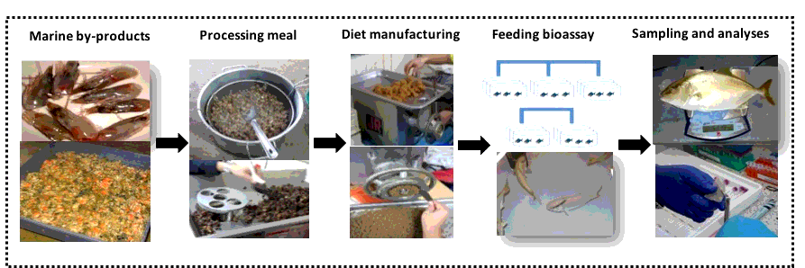
In all, it is concluded that the inclusion of some marine by-product meals, in this case, shrimp heads and Pen shell viscera, can reduce the use of fishmeal in the diet, allowing to maintain or even improve the fatty acid profile (HUFA) and the cholesterol content in the different tissues of S. rivoliana compared to the RD diet. From a human nutritional point of view, the almaco jack fillets (muscle) had levels of DHA and HUFA similar to RD with FM, when fed SD. However, as an experience derived from the present assay, it is important to perform a prior toxicity analysis to rule out any type of toxin in the marine by-products, particularly those that are prone to filtrate and accumulate lipidic toxins, as is the case of mollusks.