The coronavirus entry mechanism consists of viral spike glycoprotein interaction to host cell receptor ACE-2 and subsequent membrane fusion. Spike protein is a type- I membrane fusion protein consisting of two non-covalently associated subunits S1 (14–685 residues) and S2 (686–1273 residues). S1 binds with ACE2 receptor, whereas S2 subunit secures spike protein to the membrane and proceeds membrane fusion. The binding of S1 to the ACE2 receptor is the crucial step for SARS-CoV-2 entry. S1 subunits have four domains such as amino terminal domain, receptor binding domain (RBD) and two carboxy terminal domain. RBD (319–541 residues) consist of a loop termed receptor binding motif (RBM), which binds directly with ACE2. Currently available vaccines are developed based on the Spike protein, which is present in all SARS-CoV-2 variants. However, mutations in the Spike protein enable the CoV-2 variants to be increased transmissibility, pathogenicity, and minor to moderate antibody escape. Thus, the rapid mutation in spike protein may necessitate the development of new vaccines and therapeutic antivirals against SARS-CoV-2 [7, 16].
Accelerated targeted drug discovery is essential for identifying lead molecules against the SARS CoV-2 entry into mammalian cells. Multiple cell and cell free assays were developed for ACE2- Spike interaction modulators and their utility for neutralizing antibody testing is well established [14, 17]. Few of the approaches were also modified for the discovery of viral entry blockers [6, 18]. Most cell-based assays are influenced by the toxicity of the host cell and clinically useful viral entry blockers needs to be less toxic to mammalian cells necessitating sensitive assays for antiviral activity simultaneous with cell toxicity readout. The high-throughput adaptable assay system described here could precisely identify the molecules that could inhibit the interaction of SARS CoV-2 spike protein with host ACE2 receptors and also toxicity. SARS CoV-2 permissive assay system could be used for the screening of large compound libraries or synthetic molecules for antiviral activity against SARS CoV2. Moreover, the possibility of detecting H2B in SARS CoV-2 permissive cells could also be useful for segmentation and detection of toxicity of test compounds (Fig. 5). Histone2B also serves as the molecular marker of cellular toxicity. H2B phosphorylation occurs later in apoptosis during chromatin condensation and DNA fragmentation [19]. The developed SARS CoV-2 permissive assay system with H2B mCherry is a promising renewable, cost-effective, sensitive, rapid, BSL-2 compatible assay system for systematic identification of promising drug candidates against SARS CoV-2 with toxicity analysis.
The BSL-2 compatible replication-competent VSV-eGFP-SARS-CoV2 psuedovirion assay system performs equivalent to the clinical isolate of SARS CoV-2 [12], and facilitates the detection of syncytia formation. From our primary screening at 10 µM concentration, we have identified 24 natural products as primary hits (> 80% inhibition of infection) (Fig. 4.A,C) and dose response study revealed six natural product lead at minimum toxicity level. 17-Amino dimethoxy geldanamycin, Didemnin B, Streptonigrin, Scillaren A, Proscillaridin, and Acetoxycycloheximide have SARS CoV2 entry inhibition at 1 µM (> 80% inhibition of infection) (Fig. 4.D).
17-Amino dimethoxy geldanamycin is a geldanamycin analogue that inhibits heat shock protein-90 (HSP-90). HSP90 inhibitor SNX-5422 significantly inhibited SARS-CoV-2 replication and could be used as early therapy and which will reduce the disease severity, improve the clinical condition and reduce hospitalization of COVID-19 patients [20. Didemnin B cyclic depsipeptide antiviral marine natural product reported to have inhibitory activity against COVID-19 main protease (MPro) [21]. Didemnin B derivative plitidepsin (Aplidine) or dehyrodidemnin B were reported to have potent SARS-CoV-2 inhibition, targeting host eukaryotic translation elongation factor 1A [22]. Streptonigrin tetracyclic aminoquinoline-5,8 dione is an antitumor antibiotic and a previous screening against SARS-CoV-2 nsp15 endoribonuclease identified as positive hit [23]. Scillaren A, is a cardiac glycoside derived from Drimia species [24] and in silico studies reported to have SARS CoV-2 Mpro binding affinities [25]. Proscillaridin, is a cardiac glycoside isolated from Drimia maritima, an anticancer drug that inhibits DNA topoisomerase I and II [26]. Acetoxycycloheximide is an acetylated derivative of cycloheximide, which inhibits translation elongation by binding to the ribosomal E-site [27].
Inhibition of psuedoviral infection readout may be influenced by the toxicity of natural products and high-throughput screening may identify this as a false positive hit, if pseudoviral infection was the only evaluation parameter [10]. The present reporter assay system having integration of H2B mCherry also enabled the detection of toxicity of the molecule using the compatible imaging platform (Fig. 4.B). Among the six positive hits, Scillaren A and Proscillaridin showed slightly increased H2B mCherry intensity in high-throughput imaging and subsequent analysis (Fig. 8). Histones are chromatin structure proteins, function as damage-associated molecular pattern molecules (DAMPs) involved in multiple cellular processes, drug induced toxicity etc. [28, 29]. Thus, the observed increased H2B mCherry intensity could be due to the stress response of the natural products.
Spike mediated syncytia formation increases virus spread and disease complexity, urge the development of assay system and screening of drug that inhibit the syncytia formation [14, 17]. Spike protein alone can initiate syncytia formation in SARS permissive cells without any other viral protein [14]. Syncytia formation was observed after 12 hrs of post infection, which is line with previous observation of Zhang [30]. Syncytia formation generated by the VSV-eGFP-SARS-CoV2 psuedovirion in the SiHA ACE2 cerulean H2B mCherry cell proved as a promising imaging tool to study the fusion mechanism of the SARS virus and subsequent viral entry in real-time.
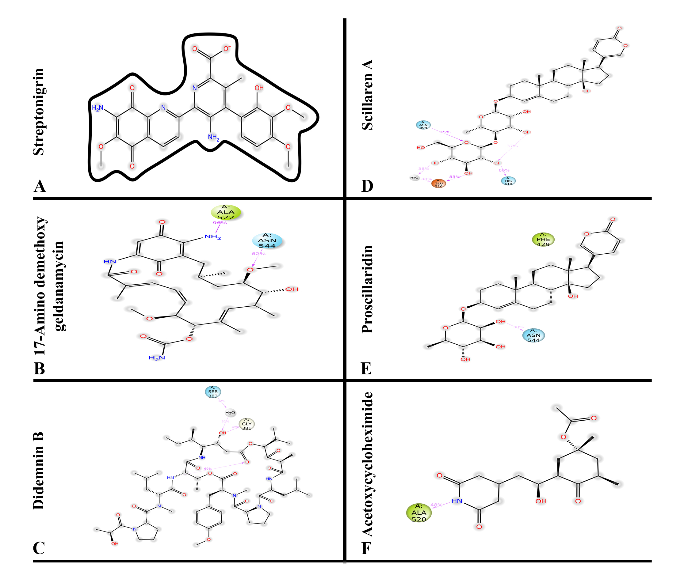
Molecular dynamic simulation analysis determined the stability of the molecules such as streptonigrin, 17-Aminodemethoxygeldanamycin, didemnin B, scillaren A, proscillaridin and acetoxycycloheximide complex with SARS-CoV-2 spike protein. Streptonigrin forms a hydrogen bond with ASN 331 which disappears during simulation and forms a water bridge with the same residue (Supp Fig. 1). Two residues, ALA 522 and ASN 544 are forming and maintaining hydrogen bonds with 17-Aminodemethoxygeldanamycin for 96% and 62% of the simulation time (Fig. 7). Didemnin B interact with spike through GLY 381 and Scillaren A maintains a hydrogen bond with ASN 394 for 95% of the simulation time and makes the complex stable by interacting with GLU 516 and HIS 519 (Fig. 7, Supp Fig. 1). Similarly, proscillaridin makes a hydrogen bond with ASN 544 and a hydrophobic interaction with PHE 429 and acetocycloheximide interacts with ALA 520 of spike protein and make a stable complex. All these molecules are forming interaction with at least one of the RBD residues which are essential for binding with hACE2 and can be potential inhibitors against SARS-CoV-2 spike protein [31].
Further studies are needed to assess the efficacy of the lead natural products against clinical isolate of SARS-CoV-2 and toxicity modification of identified molecules if required. Our present approach of simultaneous screening of pseudoviral inhibition, toxicity and ACE2-Spike mediated syncytia formation is advantageous for multiple applications. In order to validate the cell system, we have attempted the screening using only a small group of natural products that led to the identification of few leads. Interestingly most of the leads were previously reported to have antiviral activity confirming the sensitivity of the assay system. Since the cell system also supports real-time imaging of syncytia formation and cytopathic changes with no fixation or pre-processing, they will be an ideal tool for antiviral discovery using live virus.