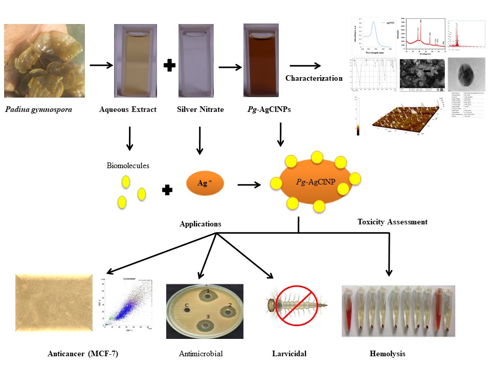
Initially, the formation of green synthesized Pg-AgClNPs was visually identified by the appearance of dark brown colour in 24 hrs of incubation (P. gymnospora with silver nitrate) (Fig. 1). The colour change from pale yellow to dark brown clearly indicating the formation of silver chloride nanoparticles. Then it was optically confirmed by UV-spectrophotometric analysis. A strong surface plasmon resonance peak was obtained around 377 nm (Fig. 2), which indicates the formation of AgClNPs [25, 26]. The maximum absorbance indicates the relative size of the nanoparticle, where the higher number corresponds to a larger particle size [27]. The chloride ions present in the seaweed extract converts AgNO3 to AgClNPs. Thus the chlorine content of the extracts could be the source for the formation of AgClNPs [28, 29]. The chlorine ions in the seaweed extracts were confirmed by EDAX Analysis. It reveals the presence of silver (56.6%) and chloride (22.9%) along with other elements such as B, C, O, Na, and Si was found to be contributed by the bioorganic materials present in the seaweed extract bound to the nanoparticle surface (Fig. 3).
The FTIR spectrum of P. gymnospora (Fig. 4a) shows that the strong bands observed at 1100, 1137 and 1416 cm− 1 are assigned to -C-O, -C = O and -C = C stretching of aromatic acids and esters present in the seaweed extract respectively. The band at 1643 cm− 1 is attributed to -C = O stretching of amides. The sharp bands at 2307, 3399 and 3727 cm− 1 correspond to NH stretching vibrations of primary and secondary amines in the crude extract. In the fingerprint region, the peaks observed at 601 and 676 cm− 1 correspond to the presence of C-Cl groups in the seaweed extract. These functional groups could act as biomolecules for stabilizing, capping and reducing agents for the formation of AgClNPs. On the other hand, the IR spectrum of Pg-AgClNPs shows the peaks of -C-O stretching shifted from 1100 cm− 1 to 1088 cm− 1 and the peak of N-H stretching shifted from 3399 cm− 1 to 3142 cm− 1 (Fig. 4b). It reveals that the carboxylic and amine groups are involved in the stabilization and immobilization of AgClNPs in the seaweed extract. The role of -C = C in the reduction of silver salt is evident by the peak observed at 1392 cm− 1 which was found at 1416 cm− 1 in seaweed extract respectively. Further, the presence of peaks at 1633 cm− 1 confirms the amide stretching group. The sharp peak observed at 594 cm− 1 confirms the presence of AgCl NPs stabilised by the seaweed P. gymnospora extract.
The powder XRD analysis of synthesized Pg-AgClNPs (Fig. 5) exhibits the crystalline structure with diffraction peaks obtained at 27.87°, 32.35°, 46.20°, 54.45°, 57.45° and 67.49° corresponds to the planes of (111), (200), (220), (311), (222) and (400) respectively which matches with the standard JCPDS File No. 85-1355. There is no other peak indicating that the high purity of prepared Pg-AgClNPs and the strong intensity of the peak shows a high crystallinity of the sample. The grain size was calculated as 30.39 nm using Scherrer’s formula. The absence of additional reflection indicates that Pg-AgClNP's lattice was not affected by other molecules in seaweed extract. SEM micrographs showed that the synthesized nanoparticles were observed with fine spherical crystals (Fig. 6) and the morphological features obtained from TEM reveal that the Pg-AgClNPs are mono-dispersed spherical shape with the average particle size of around 33 nm (Fig. 7). AFM micrographs indicate that the formulated Pg-AgClNPs possess a spherical shape and the calculated sizes in the range of 15.5 to 21.38 nm (Fig. 8). It characterizes the interactions between the Pg-AgClNPs and the supported lipid bilayers in real-time. The particle size is varied from 10 to 28 nm.
The Pg-AgClNPs were evaluated for anticancer activity against breast cancer cell line (MCF-7 cell line), and the cytotoxicity to the normal (Vero) cell line was also studied. Pg-AgClNPs showed a cytotoxic effect on breast cancer cells in a dose-dependent manner. The IC50 values of Pg-AgClNPs against breast cancer cells were found 31.37 µg/mL for MCF-7 and 421.46 µg/mL for Vero Cells (non-tumor cells) (Fig. 9). In vitro cell viability assay clearly revealed that Pg-AgClNPs were severely toxic to human breast cancer cells even more than AgNPs (data not shown). Interestingly, the synthesized Pg-AgClNPs have lower cytotoxicity effect on Vero non-tumour derived cell line. Growth inhibition related to cell cycle arrest and apoptosis was determined in flow cytometry using propidium iodide. MCF7 cell lines were treated with IC50 concentration of compounds. MCF7 cells treated with IC50 concentration of compounds increased the percentage of apoptosis in G0-G1 phase (5.16% when compared with control of 1.39%). An increase in the percentage of apoptotic cells was measured by flow cytometry, clearly clarifying an induction of programmed cell death in the cancer cells after treatment with AgClNPs. The Pg-AgClNPs induce cell death through the mitochondrial pathway. Most of the studies reported that AgNP's mediated cell death is independent of p53 [30] [12, 13]. Previously, it was reported that the biosynthesis of AgClNPs induces the cytotoxicity and apoptosis effect on human breast cancer cells through the up-regulation of apoptotic factors [6].
The larval bioassays performed by aqueous extracts of P. gymnospora and Pg-AgClNPs against larvae of Aedes agypti are a dengue vector. For the extract of P. gymnospora showed LC50 of 34.92 µg/ml in 24 hrs and 21.61 µg/ml in 48 hrs and 11.82 µg/ml in 72 hrs. While the synthesized Pg-AgClNPs show LC50 and LC90 of 7.15 µg/ml in 24 hrs, 6.06 µg/ml, in 48 hrs and 3.61 µg/ml in 72 hrs (Table 1). Among them, the Pg-AgClNPs showed higher larvicidal activity against A. aegypti with low concentration compared to the aqueous extract of P. gymnospora. The toxicity mechanisms of mosquito mortality in the nanoparticle treatment were studied recently. It is hypothetically suggested that the mechanism of toxicity against mosquito larvae by the penetration of nanoparticles through the exoskeleton. In intracellular space, nanoparticle degrades the enzymes and organelles, and it leads to the loss of cellular function and cell death [31, 32]. The AgNPs from Artemisia vulgaris leaf extracts showed the mosquito larvicidal activity. By the accumulation of nanoparticles in the midgut region of mosquito, larva causes damage in the midgut, cortex region, and epithelial cells. Similar to seaweed mediated synthesis of nanoparticles, several plants mediated synthesized nanoparticles were shown in mosquito larval control [33].
Table 1
The larval bioassays performed by aqueous extract of P. gymnospora and green synthesized silver chloride nanoparticle
| Sample | Conc (µg/ml) | Exposure Time(hr) | LC50 95%LCL-UCL | LC90 95%LCL-UCL | Intercept | X2 Value | pValue |
| Crude Extract | 5 10 20 30 40 | 24 | 34.92 (29.69 ± 43.95) | 61.73 (50.46 ± 86.43) | -1.67 | 3.75 | 0.99 |
| 48 | 21.61 (17.48 ± 25.91) | 44.87 (37.98 ± 57.59) | -1.19 | 6.90 | 0.90 |
| 72 | 11.82 (7.19 ± 15.35) | 31.02 (26.18 ± 39.58) | -0.79 | 4.25 | 0.98 |
| Pg-AgClNPs | 2 4 6 8 10 | 24 | 7.15 (6.30 + 8.16) | 11.99 (10.44 + 14.86) | -1.89 | 2.84 | 0.99 |
| 48 | 6.06 (5.19 ± 6.95) | 10.88 (9.49 + 13.36) | -1.61 | 6.19 | 0.93 |
| 72 | 3.61 (2.49 ± 4.42) | 7.82 (6.81 ± 9.56) | -1.10 | 5.79 | 0.95 |
| LC – Lethal Concentration, X2 – Chi square value; p – significant (0.05) |
AgClNPs act as an alternative source for microbial pathogens. The green synthesized Pg-AgClNPs were assessed for the antimicrobial activity against the human pathogens. Pg-AgClNPs showed antimicrobial activity in a dose-dependent manner. They were reported in Table 2. Compared to pathogenic bacteria and fungi, it acts as a good antifungal activity (Fig. 11). Moreover, the MIC also investigated these microbes by using Pg-AgClNPs at the range of 1 mg/mL by the serial dilution method. They were reported in Table 3. Among all concentrations of the selected pathogens, Canadian Albicans showed the best minimum inhibition at the concentrations of 8 mg/L, followed by VREF 32 mg/L. AgNPs act as an alternative source for microbial pathogens. Due to different variations and great impact on antimicrobial properties, it was predominately used in pharmaceuticals and drug industries [34]. Pg-AgClNPs showed antimicrobial activity in a dose-dependent manner. Compared to pathogenic bacteria and fungi, it acts as a good antifungal activity. Microbial growth inhibition in AgNPs is due to the surface and particle size variance [35]. The possible mechanism of antimicrobial activity is because of the generation of free radicals. The AgNPs interact with bacteria and release the Ag+ ions inside the cell as leads to cells leaked out caused by protein denaturation [36, 37]. The small size of AgNPs has a larger surface area, facilitates the interaction with the bacterial cell membrane, and altered the primary function of the cell membrane, including permeability and cell respiration, causing cell apoptosis [38]. Previous studies reported that the bacterial cells treated with AgClNPs caused the morphological changes and that AgClNPs were distributed on the surface of the bacterial cells [29].
Table 2
Antimicrobial activity of Green synthesized silver chloride nanoparticle compound against human pathogens
| Concentration (mg/mL) | Zone of Inhibition (mm) |
| Gram-positive bacterium | Gram-negative bacterium | Fungal Pathogen |
| MRSA | VREF | E. coli | P. aeruginosa | C. albicans |
| 1 | 22 | 26 | 14 | 19 | 29 |
| 2 | 22 | 26 | 14 | 21 | 31 |
| 3 | 23 | 27 | 15 | 22 | 31 |
Table 3
Minimum Inhibitory Concentration of MIC Green synthesized silver chloride nanoparticle.
| Nano- Particles | Minimum inhibitory concentration (MIC) (mg/L) |
| Gram-positive bacterium | Gram-negative bacterium | Fungal Pathogen |
| MRSA | VREF | E. coli | P. aeruginosa | C. albicans |
| NP | PC | NP | PC | NP | PC | NP | PC | NP | PC |
| MIC value | 64 | 4 | 32 | 1 | 128 | 1 | 64 | 2 | 8 | NA |
| MRSA: Methicilline resistance Staphylococcus aureus; VREF: Vancomycine resistance Enterococcus feacalis |
| NP: Nanoparticles, PC: Positive control, NA: Not Appearing |
Hemolytic activity of synthesized silver nanoparticles was screened at different concentrations among them. Pg-AgClNPs particles showed the hemolytic activity in 100 µg/mL 92% followed by 50 µg/mL 12% of hemolysis from the 25 µg/ml concentration showed no hemolysis to the human blood cells (Table 4). This result suggests that Pg-AgClNPs can be safe and also used for further investigation (Fig. 12). At the nanotoxicity level, the investigation on blood compatibility is essential because the blood cells are affected by nanoparticles directly or indirectly. The erythrocytes circulate to various organs through the cardiovascular system leading to DNA damage, cell membrane injury, and congenital malformation [7, 8]. In this condition, the biocompatibility of AgNPs rupturing and releasing of erythrocytes have more attention to analyzing the toxicity level of nanoproducts [34].