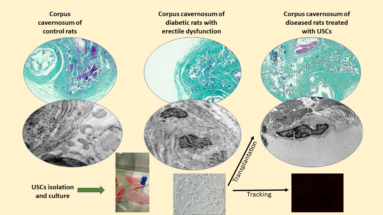
The present study compared the effect of transplantation of urine-derived stem cells (USCs) and their lysate (USCs-L) into the corpora cavernosa (CC) of rats with diabetic erectile dysfunction (DED). A rat model of DED was established via intraperitoneal injection of streptozotocin. After proving DED, USCs and USCs-L were transplanted into CC of two groups of rats. Our histopathological and morphometric results showed marked obliteration of the cavernous spaces with bands of collagen fibres, significant increase of collagen/smooth muscle ratio and decrease the mean area percentage of α-SMA in the CC of diabetic rats. The ultrastructure of the endothelium lining the cavernous spaces of the same group revealed irregular surface with villi-like projections along with thickening and splitting of their basal lamina. The diabetic rats exhibited deterioration of the all the parameters of copulatory function compared to the control rats. Transplantation of USCs and USCs-L significantly restored the structure and the ultrastructure of the CC and improved the sexual function compared to the diabetic rats without significant difference between them. The transplanted USCs were detected in a good number in the CC four weeks after transplantation, while their number decreased by the 8th week. As a conclusion, USCs and USCs-L can repair the structure and the ultrastructure of CC and improve the sexual function in a rat model of DED without significant different between them. however, the USCs-L could be better used as a treatment strategy in DED to avoid the undetected behaviour of USCs and decreased their survivability after transplantation.
Research Article
Urine-derived Stem Cells versus their lysate in Ameliorating Erectile Dysfunction in a Rat Model of Type II Diabetes
https://doi.org/10.21203/rs.3.rs-250173/v1
This work is licensed under a CC BY 4.0 License
Journal Publication
published 09 May, 2022
Version 1
posted
You are reading this latest preprint version
Urine-Derived Stem Cells
Diabetes
Erectile Dysfunction
Rat
Diabetic erectile dysfunction (DED) is a major health problem which has a strong impact on the quality of life of patients and their families. It was reported that prevalence of DED reached 78.7%, with 12.3% of patients had mild erectile dysfunction (ED), 30.9% had mild to moderate ED, 19.1% had moderate ED and 16.4% had severe ED. Duration of diabetes had a significant correlation with the severity of ED (1).
Chen et al., (2) reported a higher rate of DED that ranged from 35–90%. Moreover, men who have diabetes are thought to develop ED between 10 and 15 years earlier than men who do not suffer from diabetes. Additionally, ED in patients with diabetes mellitus is more severe and refractory to treatment compared with that in non-diabetic patients.
We can imagine the overall magnitude and burden of ED among diabetic and their families if we know that, the global diabetes prevalence in 2019 was estimated to be 9.3% (463 million people), rising to 10.2% (578 million) by 2030 and 10.9% (700 million) by 2045 with type 2 diabetes makes up about 90% of all diabetes cases (3).
The pathophysiology of ED in diabetes mellitus is likely multifactorial with vascular, neurological and hormonal alterations. Among these factors, endothelial dysfunction is recognized as a mainstay in the pathophysiology of DED. There are many modalities of current therapeutics for ED including a list of oral medications, intracavernosal injections, vacuum erection devices, and penile implants. Such therapeutic options provide only symptomatic improvement the ED but, none of these modalities, however, repair the pathogenesis or restore function (4).
Phosphodiesterase type 5 inhibitors (PDE5i), which is the most famous and successful first line oral medication for the treatment of ED was found to be less effective in treatment of DED. This is probably caused by the decrease in nitric oxide (NO) production resulting from endothelial dysfunction (5).
Therefore, new therapeutic strategies targeted towards repairing endothelial function, particularly in the early stage of this disorder, are needed. Stem cell (SC) therapies represent a promising new frontier for the treatment of DED. A growing body of research is demonstrating the therapeutic effects of numerous types of stem cells including mesenchymal stem cells (MSCs), such as rat bone marrow-derived mesenchymal stem cells (BMSCs), vascular endothelial growth factor (VEGF) transfected BMSCs and urine derived stem cells(4).
Urine derived stem cells (USCs), may be isolated from human urine samples using a cost-effective and simple method of isolation, incorporating not such complex reagents or invasive methods of collection compared with other stem cells. USCs are superior to conventional stem cell sources in terms of favourable gene profile and inherent multipotent potential. They also possess the features of a progenitor and are a convenient cell source. USCs display many characteristics of MSCs and can differentiate into multiple cell-lines including endothelial and smooth muscle cells (6).
Moreover, the intracavernous injection of SC in treating ED appears straightforward and logical, with proposed regenerative effect achieved by either secreting growth factors locally via a paracrine mechanism or by migration to the major pelvic ganglia to promote the propagation and differentiation of resident progenitor cell types. They also encourage the recovery of injured tissue via the production of antiapoptotic and proangiogenic factors, rather than trans differentiation into different cell types (7).
The first reported clinical trial of SC therapy in diabetic men with ED showed a reasonable increase in penile rigidity after a single intracavernous injection of umbilical cord blood SC. While penile rigidity was maintained for more than 6 months, the erection was not hard for sexual penetration, suggesting that the amount and a single administration of SC were likely insufficient for adequate penile rigidity (8).
In a different study on the use of SC in men with ED following radical prostatectomy, the intracavernous injection of bone marrow mononuclear cells appeared to be safe and improved the erectile function for a same period of 6 months (9).
Although previous studies confirmed that stem cell therapy is beneficial for treating DED, the optimal strategy has not been determined up to now. Therefore, new ideas for the treatment of DED are needed to design the most effective strategy for the future experimental research (4).
In this study, we simply try to investigate the underling mechanism of using Stem cells in improving DED either by their multipotentiality and homing capacity or their paracrine effect by comparing the effectiveness of urine-derived stem cells and their lysate in improving erectile dysfunction in type II diabetes rat model where, research work is still insufficient.
2.1. Animals:
The experiments were performed on fifty-five male Wistar albino rats of average 200–250 gm each. They were maintained under standard laboratory conditions with free access to standard rodent laboratory chow and water.
All experimental procedures and animal maintenance were conducted in accordance with the accepted standards of animal care of medical research centre, Ain Shams University.
The laboratory work was performed in the tissue culture lab of the centre of Pancreatic Cell Culture and Diabetic Research, Ain Shams Hospitals.2.2. Study design:
Ten Wistar male albino rats were served as donors for urine-derived stem cells. Ten rats were considered as control group (group I) and did not subjected to any intervention till the end of the study. The remaining thirty-fife rats were subjected to induction of diabetes; diabetes was established in 31 of them. Erectile dysfunction was tested and proved in 24 of them which were further divided randomly and equally into three groups (8 animal each):
Diabetic-induced erectile dysfunction group (group II): the animal of which was injected by phosphate buffer saline (PBS)(0.2ml) through intra-cavernous route Diabetic induced erectile dysfunction/ USCs-treated group (group III): the rats of which were subjected to intra-cavernous injection of USCs (2X10⁶) suspended in 0.2ml PBS. Diabetic induced erectile dysfunction/ USCs-L-treated group (group IV): the rats of which were subjected to intra-cavernous injection of USCs-L (400 µL of lysate/ each animal). Four and eight weeks after injection erectile function and sexual behaviour were assessed and the animals were euthanized at the end of the 8th week after transplantation. The penile tissue was harvested from all animals for further histopathological studies. The following illustration shows the steps and the duration of each one.

2.3. Preparation of a diabetic rat model and confirmation of diabetic erectile dysfunction:
Thirty-five rats were fed high fat diet 4 weeks prior to induction of diabetes. The diet was purchased from the medical research centre, Ain shams university. Its composition was 24%fat, 41% carbohydrates, 24% protein with total caloric value of 4.7 Kcal/g. Intraperitoneal injection of two dose of 30 mg STZ/kg/BW; dissolved in normal saline (Sigma Chemical Co., St. Louis, MO) with 7 days interval was performed to induce diabetes. The animals were considered to have diabetes if blood glucose was > 280 mg/dl for 2 consecutive days2. Body weight was assessed every week starting from the beginning of high fat diet till the end of the study. Insulin tolerance test was performed after 5 weeks of diabetic induction to test the insulin resistance of the animal according to Chen et al., (2); 1 IU/ml of bovine insulin was intraperitoneally injected in each rat followed by measuring blood glucose at 0,15, 30, 60, 90 and 120 min after insulin injection to attain blood glucose response curve. Confirmation of erectile dysfunction took place according to the method of Brein et-al., (10) by injection of Apomorphine hydrochloride (Sigma Chemical Co., St. Louis, MO) (APO); 80 µg/kg dissolved in a vehicle of 100 µg ascorbic acid in normal saline 0.9% (l ml/kg). the injection was subcutaneously in the back of the neck then erectile and yawning responses to APO were recorded for 30 min. the apomorphine screening test was performed to the animal after 4 weeks of diabetic induction and repeated to all the animal in all groups at 8 weeks after USCs and USCs-L transplantation.
2.4 Urine collection from the donner rats: the urine was collected from the donner rats after strict disinfection of the skin of the lower abdomen and the external genitalia. application of gentle trans-abdominal pressure over the bladder to overcome normal urethral pressure was performed to force the urine out into a sterile disposable plastic Petri dish and then aspirated and poured off into falcon tube; 50 ml. The mid and the last stream was collected (30 to 100 ml/ micturition) and immediately taken to the tissue culture lab (11).
2.5 Isolation, culture and passaging of urine-derived stem cells (USCs):
The collected urine samples were centrifuged (1800 rpm for 10 min) and the resulting cell pellets were washed with PBS (Lonza, Belgium) supplemented with 1% penicillin/ streptomycin (PS) (1:1) (Lonza, Belgium). Then cells were plated in 6-multi-well tissue culture plates at about 2000 cells per well with complete media (CM) composed of 89% DMEM (Lonza, Belgium), 10% fetal bovine serum (FBS) (Seralab, Brazil) and 1% PS. Cell monitoring and follow up took place daily to the cells of primary culture (P0). The first exchange of media was after 48 h of seeding and the subsequent exchanges were performed every 3 days. when reaching confluency (80–90%), the culture was trypsinized by 0.25% trypsin/EDTA (Gibco, Grand Island, NY) and expanded through 3 passages (P1-P3). Cells of P3 was used for characterization and transplantation (12).
2.6. Preparation of USCs-L:
Modified technique after Albersen et al., (13), was used for this purpose; After trypsinization and obtaining the USCs pellet of P3, the supernatant medium was discarded, and the cells were resuspended into 10 ml of deionized H2O for half an hour at room temperature to cause cell membrane rupture. Three Freeze-thaw cycles were performed to obtain cell free lysate followed by Centrifugation at 2000 rpm for 10 minutes to get rid of any insoluble fragments. 400µL of the lysate was frozen in a separate Eppendorf for each animal till the time of injection in – 80oC.
2.7. Characterization of USCs:
Flowcytometric analysis of the isolated USCs of P3 took place to show the incidence of expression of the following cell markers; CD73 and CD 44 and CD 45 in the flowcytometry unite, Poisoning Treatment Centre, Ain Shams Hospital.
2.8. Intra-cavernous injection of USCs and USCs-L:
After sterilization and anaesthesia by sodium pentobarbital (30 mg/kg,
IP), the proximal shaft of the penis was tied gently by an elastic rubber band and 0.2 ml of PBS was injected slowly in the middle of the corpus cavernosum of each rat in group II, 2X 10⁶ USCs labelled with PKH-26 PKH-26 fluorescent cell linker (Sigma, St. Louis, MO, USA) suspended in the same amount of PBS in each rat of group III and 400µL of USCs-L in each animal of group IV. The needle and the rubber band were left in place after injection to prevent leakage (12&14).
2.9. Sample collection, histopathological and ultrastructure examination:
The animals of all groups were sacrificed 8 weeks after USCs transplantation and USCs-L injection, the middle shaft of the penis of all animals were collected; half of them were fixed in 10% neutral buffer formalin and embedded in paraffin blocks. Transverse sections of 5 µm thickness were cut and stained with Haematoxylin & Eosin (H&E) and Masson trichrome stain to evaluate the histopathological changes. These paraffin embedded sections were further immunohistochemically stained using mouse monoclonal antibody against alpha smooth muscle actin (α-SMA) (1:200, Santa Cruz Biotechnology Inc., USA), examined and photographed under light microscope. The other half of the specimens, immediately after dissection, were carefully cut into small pieces and fixed in buffered 2.5% glutaraldehyde for 2 h, post fixed in 1% osmic teroxide, dehydrated in ascending grades of alcohol and embedded in epoxy resin. 1 µm thick-Semithin sections were obtained, stained with Toluidine blue, examined, and photographed utilizing light microscope. Ultrathin sections were cut using ultratome (Reichert Ultracut, Ziess, Germany), and stained with uranyl acetate and lead citrate, examined, and photographed with transmission electron microscope (1230 EXII; JOEL, Tokyo, Japan) (Mycotic Centre, Al-Azhar university) (15).
2.10. Morphometric assessment:
ImageJ software (version 1.33–1.34; National Institutes of Health, Bethesda, MD, USA) was used to assess the following parameters (2):
field integrated density (IntDen) In Masson trichrome-stained sections. It was determined after subtracting the background noise. And ratio between collagen fibres and smooth muscles (collagen/smooth muscle ratio) was assessed. Ten different fields from six non overlapped sections from each sample were used to calculate this ratio.
Average percentage of smooth muscle cells that showed alpha smooth muscle actin (α-SMA) (brownish immunoreactivity in comparison to the total tissue area). Ten different fields from six non overlapped sections from each sample were also used for this purpose.
2.11 Detection of USCs in the corpus cavernosum:
Before USCs transplantation, the cells were incubated with the immunofluorescence dye; PKH-26 according to the manufacturer’s instruction. PHK-26 labelled USCs were identified by immunofluorescence microscopy in the corpus cavernosum of group III rats 4 week and 8 weeks after transplantation (14).
2.12 Sexual behaviour (mating behaviour& copulatory function):
Inside a cage measured 60X40X30 CM, with a glass side, illuminated with faint light, the male rat was put inside the cage an hour before the test then the female (induction of Behavioural oestrus in the female rat took place by intraperitoneal injection of 20 µg/kg oestradiol benzoate 24 hours before the test). both were observed for half an hour and the following copulatory parameter were measured:
Mount latency (ML): time from the introduction of female rat into the cage of the male rat up to the first mount measured in seconds.
Intromission latency (IL): time from the introduction of the female rat up to the first intromission by the male rat.
Mount frequency (MF): number of mounts before ejaculation.
Intromission frequency (IF): number of intromissions before ejaculation.
Ejaculation latency (EL): time from the first intromission of up to the ejaculation.
Total number of ejaculation (TE): total number of ejaculations that took place during the test duration (30m).
Post-ejaculatory interval (PEI): time from the first ejaculation up to the next intromission by the male rat
This test was performed at the beginning of the study to exclude the sexually inactive male rats then at the 8th weeks after USCs transplantation to all rats in all groups. When the animal did not mount during the first 10 minute of the test, it was excluded, and the test considered to be negative (16&17).
3.1. General characteristics
General weight of the animals and insulin tolerance test are show in Chart 1A & B, respectively. The initial mean body weights of the rats in the diabetic and age-matched control group were the same, but the diabetic rats weighed significantly less than the controls at 8, 10 and 12 weeks after induction of diabetes (1st dose of STZ) (p < 0.05). During the insulin tolerance test, blood glucose levels decreased significantly in normal rats at 15, 30, 45, 60, 90 and 120 minutes after intraperitoneal injection with insulin. While insulin response in diabetic rats was significantly dimensioned. The mean blood glucose levels of the diabetic rats at 8 weeks after induction of diabetes was 4 times higher than in the control rats (p < 0.001) with non-significant difference between the diseased and the treated groups (Chart 2A). serum level of total cholesterol and the triglyceride increased significantly in the diabetic rats in comparison to the control (P < 0.05 & P < 0.01, respectively) while there was no significant difference between the remaining groups (Chart 2B).
3.2. Isolated USCs morphology and phenotypic characterization:
The isolated USCs showed plastic adherent property. Few numbers of them were noticed attached to the floor of the tissue culture flask 2 days after seeding. They took about 15 and 20 days to reach 50% & 90% confluency, respectively. Most of the cells showed short cytoplasmic processes, vesicular nucleus, and multiple nucleoli during the 1st passage (P1) (Fig. 1A) and acquire rice-shape appearance during the 3rd passage (P3) (Fig. 1B). Most of the isolated cells expressed the mesenchymal stem cell markers CD 73 (81.55%) and CD 44 (82.5%). Only 46.9% of them expressed the hematopoietic stem cell marker CD 45 (Fig. 1C).
3.3. Histopathological results
The transverse section in the mid-shaft of the rat penis showed three cylindrical cavernous bodies (corpora); a pair of corpora cavernosa (CC), and the corpus spongiosum that surrounds the urethra (Fig. 2). The corpora cavernosa of the control rats contained wide irregular vascular cavernous spaces (CS); composed of collagen fibres intertwined with smooth muscle and lined by endothelium (Fig. 3A). The CC of the diabetic rats showed marked narrowing of the CS which appeared to be invaded by bands of thick collagen fibres (Fig. 3B). Rats of group III and IV exhibited apparent restoration the CS regarding their size and structure, however few thick collagen fibres still being seen obliterating them in group III rats (Fig. 3C&D).
3.4. Detection of collagen/smooth muscle ratio:
Masson Trichrome-stained histological sections of diabetic rat CC showed large amount of collagen fibres deposition and penile fibrosis (Fig. 4). the collagen/smooth muscle ratio increased significantly in diabetic rat (2.8 ± 0.4. P < 0.05) in comparison to control group (1.3 ± 0.2). the same ratio decreased significantly in group III and IV (1.9 ± 0.3, 1.7 ± 0.4, respectively) (P < 0.05) in comparison to the diabetic group without a significant different between the two groups (P < 0.1) (Chart 3).
3.5. Assessment of α-SMA expression in CC:
The immunohistochemically stained section of rat penis in the diabetic rats (group II) revealed marked decreased immunopositivity for α-SMA in CC, while the treated groups (group III & IV) almost exhibited the control appearance (Fig. 5). Quantification of these data showed significant decrease in the mean area percentage of α-SMA in the immunohistochemical-stained sections of diabetic rats (0.1 ± 0.002, p < 0.05) in comparison to the control group (0.3 ± 0.001). This level increased significantly in group III and IV (0.23 ± 0.6 and 0.28 ± 0.4, respectively) (P < 0.05) in comparison to the diabetic group with no significant difference between the two groups (P < 0.1) (Chart 3).
3.6. Transmission electron microscopic results:
The ultrastructure of the CC of the control rat revealed wide cavernous spaces lined with endothelium with a thin basal lamina and smooth luminal surface. They exhibited well-formed cytoplasmic organelles and almost had no pinocytic vesicles (autophagic vesicles). The CS were surrounded by bands of mature collagen fibres, having well-arranged striation, some fibroblasts were seen between those bands. They were supposed to be inactive due to lacking the cytoplasmic processes (Fig. 6). The CS of the diabetic rat of group II were narrow due to protrusion of the endothelium towards the lumen may be due to thickening, splitting of the basal lamina and presence of villi-like projections, in their luminal surface which appeared coarse and wrinkled. Many pinocytic vesicles were evident within their cytoplasm. Many active fibroblasts with multiple cytoplasmic processes were seen among the CS. They laid immature collagen fibres that appeared adjacent to their cell membrane and loss the normal striation of the mature fibres (Fig. 7). The CS of the USCs-treated rat (group) greatly resemble the control ones apart from minor thickening of the basal lamina, irregularity of the luminal surface and few pinocytic vesicles. Few active fibroblasts and immature collagen fibres were seen among the CS. One third of the animals exhibited presence of mast cells around the collagen fibres (Fig. 8). Restoration of the endothelium structure took place in the USCs lysate-treated animals (group IV) regarding regularity of the luminal surface and the basal lamina thickening (the thickening of the basal lamina was less considerable). Some pinocytic vesicles still being seen within the endothelium cytoplasm. The fibroblasts appeared without cytoplasmic processes and surrounded by mature collagen fibres. There was no evident of mast cell presence in this group (Fig. 9).
3.7. Presence of PKH26-labeled USCs in the rats’ CC:
Immunofluorescence examined section of the corpora cavernosa of rats revealed presence of many PKH26-labeled cells all over the corpora cavernosa of the diabetic rats treated with USCs four and eight weeks after USCs transplantation with apparent marked decrease in their number after the 8th week (Fig. 10).
3.8. Apomorphine test as an indicator of impotence:
The test was performed after 4 weeks after induction of diabetes and showed that 24 out of 31 diabetic rats had developed impotence. The test was repeated to all rats 8 weeks after treatment with USCs and USCs-L as an indicator of functional improvement. The animals of control group showed no signs of erectile dysfunction (100%), only 12.5% of the diabetic rats in group II spontaneously regained erection ability. Most of the rats in group III and IV showed no signs of erectile disfunction (87.5% & 93.7%, respectively).
3.9. Copulatory functional assessment results:
The assessment was performed to all rats in all groups just before scarification and revealed that: the diabetic rats of group II showed a significant reduced MF, IF, EL and TE, and increased ML, IL and PEI compared to the control rats. Treatment with USCs and USCs -L significantly improve the sexual function in diabetic rats evidenced by increased MF, IF, EL and TE and decreased ML, IL and PEI compared to diabetic rats. There was no significant difference between group III and group IV regarding the measured parameters apart from El and IL (Table 1).
Measured parameter | Group I | Group II | Group III | Group IV |
|---|---|---|---|---|
Mount frequencies (MF) | 13.2 ± 1.2 (10) | 4.5 ± 0.4* (3) | 8.5 ± 0.3 # (8) | 10.5 ± 0.5 # (8) |
Intromission frequency (IF) (SCs) | 9.2 ± 2 (10) | 2 ± 0.01* (3) | 7.4 ± 0.3 # (7) | 8.7 ± 0.5 # (8) |
Mount latency (ML) (SCs) | 63.2 ± 14.3 (10) | 175 ± 21* (3) | 110 ± 26.6 # (8) | 90.8 ± 11.3 # (8) |
Intromission latency (IL) (SCs) | 60.4 ± 13.5 (10) | 190 ± 19.9** (3) | 117 ± 22.1 # (7) | 95 ± 9.8 # $ (8) |
Ejaculation latency (EL) | 440 ± 21.4 (10) | 210.5 ± 20* (2) | 311.9 ± 25 # (7) | 371.5 ± 32.2# $ (8) |
Post-ejaculatory interval (PEI) (SCs) | 430.1 ± 29.6 (10) | 890.1 ± 33** (2) | 610.8 ± 32.2# (7) | 591.5 ± 15.7# (7) |
Total number of ejaculations (TE) | 3.1 ± 0.01 (10) | 1.3 ± 0.02* (2) | 2.1 ± 0.1 # (7) | 2.5 ± 0.3# (8) |
| Latencies are measured in seconds (sec), with the other data expressed as number of occurrences. The number of animals that presented the behaviour is indicated between brackets. | ||||
| *: P < 0.05 compared to group I **: P < 0.001 compared to group I | ||||
| #: P < 0.05 compared to group II $: P < 0.05 compared to group III | ||||
Erectile dysfunction (ED) is one of the major complications of diabetes mellitus (18). Erectile function is accompanied by relaxation of penile smooth muscle and dilatation of the arteries, leading to increased blood flow to cavernous spaces (19). Therefore, integrity of intracavernous structures like smooth muscles, endothelium and nerve terminals are greatly required to provide a normal erection (20). Interest in SC treatment strategy for erectile restoration is increasing aiming to deal with the multifactorial pathogenesis of ED (21). Stem cells act either by differentiation and direct integration within the recipient tissue or by their paracrine effect through secretion of growth factors and cytokines4. In the current study we have tested the effect of USCs in restoration the structure and the ultrastructure of the CC of a rat model of DED along with functional improving of erection. The USC-L was also used for the same purpose trying to confirm USCs paracrine effect.
In this study, rats were subjected to induction of diabetes. After which, there were significant decreases in weight. Furthermore, biochemical evaluation of insulin tolerance test, fast blood sugar, total cholesterol & triglyceride was indicative for DM. Development of DED was confirmed using Apomorphine hydrochloride. There was also obvious alteration on the structure and ultrastructure of the CC of diabetic rats besides significant deterioration of copulatory function.
Light microscopic examination of diabetic group showed histopathological changes. In H&E-stained sections, there was marked narrowing of the cavernous spaces which appeared to be invaded by bands of thick collagen fibers and penile fibrosis. These findings were confirmed by Masson trichrome-stained histological sections and their quantitative analysis which revealed that the collagen/smooth muscle ratio increased significantly in diabetic rat. The same results were obtained by Ouyang et al. and Gou et al., (7 &22). The immunohistochemically stained sections of the same group revealed marked decreased for immunopositivity of α-SMA in CC. Using a-SMA as a marker of smooth muscles (SMs) contractility is reasonable being the first protein found to be expressed in contractile SMs and it was proved to be down regulated in the erectile dysfunction (23 & 24).
These findings were documented in many studies (25 &26) which proved that the vascular SMs maintain plasticity and can change from a contractile (differentiated) to a synthetic (dedifferentiated) state. The synthetic state is characterized by a high level of proliferation, migration, extracellular matrix production, vimentin overexpression and decreased expression of contractile cytoskeletal proteins such as α-SMA, SM myosin heavy chain (SMMHC), and desmin. Cultured SMs of diabetic rat were also found to exhibit significantly less contractility compared with those of non-diabetics and decrease expression of α-SMA and SMMHC under hyperglycemic conditions, indicating that they could have a key role in the pathogenesis of DED (27). Moreover, Musicki and Burnett (28) demonstrated that several factors have been implicated with the onset of ED, including changes in SMs, collagen and elastic fibres, which are major penile structural components accountable for erection.
In our study, electron microscopy of diabetic group showed CS narrowing due to protrusion of the endothelium towards the lumen may be due to thickening and splitting of their basal lamina and marked irregularity of their luminar surface. They also exhibited pinocytotic vesicles. The same findings were noticed in the aortic endothelium of diabetic rats after 10 weeks of diabetic induction along with decrease expression of their eNOSmRNA content (29). During studying of the ultrastructure of the CC endothelium in a rat model of type II diabetes Parikh et al., (30) proved a significant decrease in their caveolae; site of localization of endothelial NOS which play a major role in the penile hemodynamic required for maintain the intracavernous pressure and erection. The pinocytotic vesicles in the endothelium of diabetic group increased as a result of increase autophagic activity into the cells due to presence of some metabolites resulting from hyperglycemia and active oxidative stress (31 &32) the basal lamina thickening, and splitting may alter the diffusion between the CS and the surrounding tissues and affect the cell-to-cell side talks between the endothelium and SM of the CC which is essential for erection (33).
Many active fibroblasts with multiple cytoplasmic processes were seen among the CS. They laid immature collagen fibres that appeared adjacent to their cell membrane and loss the normal striation of the mature fibres. indicating newly synthesized collagen fibres leading to penile fibrosis (21 &29).
The histological sections of USCs and USCs-L groups showed obvious less fibrosis and a significant preservation of smooth muscle content compared to the diabetic group. They exhibited apparent restoration of the CS regarding their size and structure. However, few thick collagen fibres were still seen obliterating them in USCs group. Quantitative analysis of Masson trichrome-stained sections showed that the collagen/smooth muscle ratio decreased significantly compared to the diabetic group. Similar results were obtained by using human urine-derived stem cells either alone or genetically modified with fibroblast growth factor 2 (FGF2) (12), this therapeutic strategy was thought to induce improvement of erectile function in type II diabetic rats by recruiting resident cells and increasing the endothelial expression and contents of smooth muscle. Hence restore the smooth muscle/total collagen ratio, a key factor in the relaxation and nutrition of endothelium cells and SM in the CC. The decreased smooth muscle/total collagen ratio decreased the ability of the sinusoids to expand, resulting in veno-occlusive dysfunction (34&35).
Marked ultrastructural repair of CC of USCs and USCs-L group was evident in comparison to the diabetic group regarding the intracavernous structures and the lining endothelium of the CS. USCs were proved to secrete proangiogenic trophic factors and immune-modulatory factors and can be differentiated into endothelial cells in vitro (12).
There were multiple mast cells nearby the CS and in between the cellular component of the CC. Mast cells (MCs) are bone marrow progenitor–derived immune cells that complete their maturity in tissues. MCs are considerably affected by the local microenvironment and can share in numerous biological processes, including inflammation and neovascularization, through the release of several mediators and interaction with macrophages, endothelial cells, and fibroblasts (36). We may explain the strong presence of those cells in the treated groups by the paracrine effect of both USCs and their lysate. The paracrine mechanism of stem cell-based therapy is believed to involve secretion of trophic factors which activate endogenous stem cells to share in tissue repair. Growth factors produced by stem cells demonstrate mitogenic, reparative, anti-apoptotic, and anti-inflammatory properties, it also promotes angiogenesis (4 & 37).
Regarding function assessment, we used the APO test (12) as a confirmation of DED and to follow up the animal 4 and 8 weeks after transplantation. Assessment of copulatory function was performed and showed significant improvement of all parameters in both USCs and USCs-L groups in comparison to the DED group. Sexual behaviour, sperm quantity and quality were proved to be deteriorated even after short term streptozotocin-induced hyperglycaemia in rats (38) and different types of stem cell therapies were used and proved to restore the erectile function based on measuring the intracavernous pressure in relation to the main arterial pressure (4, 12 & 13).
In the current study, urine was used as an easy and available source of USCs which can be obtained via a non-invasive, simple, and low-cost approach (12 & 39). The isolated USCs exhibited the same morphology and phenotypic character of MSCs and were proved to have high efficiency to be differentiated (39).
Although there was almost no significant difference between the USCs and USCs lysate groups regarding repair penile structure and function, we could prefer using USCs-L rather than USCs based on the following reasons: In our study, stem cells numbers were detected by fluorescence microscopic sections showing recruitment of PKH26-labeled USCs in the corpora cavernosa of group III rats, 4 weeks and 8 weeks after transplantation. but their number was significantly decreased after 8 weeks indicating decreased survivability of stem cells after transplantation which is considered one of the limitations of their use as a cytotherapy (37). Utilizing stem cell-cell free therapy by using their lysate containing rich amounts of growth factors and bioactive components or isolation of their exosomes may be benefit cell growth and tissue repair without taking the risk of the unknown behaviour of the transplanted cells on the long run or their unwanted pathway of differentiation. However, further studies aimed to identify the proteomic characteristics, the exact components of the cell-free lysate and their mode of action is highly recommended.
Both USCs and USCs lysate exhibits the same DED reparative properties, and this proves the hypothesis that UDSCs have a paracrine effect through certain intracellular bioactive molecules and substances which help restoring the structure and ultrastructure of CC and promoting erectile function. however, using the non-invasive technique of USCs-L may have great promise toward a cure for DED and come over the limitations of using USCs themselves including decreased their survivability in the site of transplantation.
- Ethical approval: The experimental protocol was carried out in accordance with the Guide for Care and Use of Laboratory Animals published by the US National Institutes of Health (NIH Publication No. 85–23, revised 2011) and was approved by medical research centre committee, Ain Shams University.
- Consent to participate: not applicable to this study.
- Consent to publish: not applicable to this study.
- Conflict of interest: The authors have no relevant financial or non-financial interests to disclose.
- Funding: No funding was received for conducting this study.
- Rights and permissions
Data availability: The data that support the findings of this study are available from the corresponding author upon reasonable request. Some data may not be made available because of privacy or ethical restrictions.
Author contributions
RG: Conceptualization, Methodology, Formal analysis, investigation, and sharing in manuscript writing. HK; sharing in formal analysis, investigation, and manuscript writing. MA: Writing, preparation original draft and Supervision.
- Sondhi, M., Kakar, A., Gogia, A., & Gupta, M. (2018). Prevalence of erectile dysfunction in diabetic patients. Current Medicine Research and Practice., 8(3), 88–91.
- Chen, F., Zhang, H., Wang, Z., Ding, W., Zeng, Q., Liu, W., Huang, C., He, S., & Anyang, W. (2017) Adipose-Derived Stem Cell-Derived Exosomes Ameliorate Erectile Dysfunction in a Rat Model of Type 2 Diabetes. J Sex Med;1–11.
- Saeedi, P., Petersohn, I., Salpea, P., Malanda, B., Karuranga, S., Unwin, N., & Colagiuri, S. (2019). Diabetes Atlas, 9th edition: https://doi.org/10.1016/j.diabetes.2019.107843.
- Matz, E. L., Terlecki, R., Zhang, Y., Jackson, J., & Atala, A. (2019). Stem Cell Therapy for Erectile Dysfunction. Sexual Medicine Reviews, 7(2), 321–328.
- Chung, E. (2019). Stem cell therapy in diabetic men with erectile dysfunction: a step closer to safe and effective regenerative technology. Ann Transl Med., 7(Suppl 1), S40.
- Nimshitha, P. N., Al-Hawwas, M., & Bobrovskaya, L. (2018). Urine-derived cells for human cell therapy. Stem Cell Res Ther, 9, 189.
- Ouyang, B., Xie, Y., Zhang, C., Deng, C., Linyan, L. V., Yao, J., Zhang, Y., Liu, G., Deng, J., & Deng, Ch (2019). Extracellular Vesicles From Human Urine-Derived Stem Cells Ameliorate Erectile Dysfunction in a Diabetic Rat Model by Delivering Proangiogenic. MicroRNA Sex Med, 7, 241e250.
- Al Demour, S., Jafar, H., Adwan, S., AlSharif, A., Alhawari, H., Alrabadi, A., Zayed, A., Jaradat, A., & Awidi, A. (2018). Safety and Potential Therapeutic Effect of Two Intracavernous Autologous Bone Marrow Derived Mesenchymal Stem Cells injections in Diabetic Patients with Erectile Dysfunction: An Open Label Phase I Clinical Trial. Urol Int., 101(3), 358–365. doi:10.1159/000492120. Epub 2018 Aug 31. PMID: 30173210.
- Damkier, A. P., Sørensen, J. A., Lund, L., & Sheikh, S. P. (2016). Safety and Potential Effect of a Single Intracavernous Injection of Autologous Adipose-Derived Regenerative Cells in Patients with Erectile Dysfunction Following Radical Prostatectomy: An Open-Label Phase I Clinical Trial. EBio Medicine;, 5, 204–210. https://doi.org/10.1016/j.ebiom.2016.01.024.
- Brien, S.E., Wilson, E., Heaton, J.P.W. and Adams, M.A. (2000). The conditioned response erection (CRE)- a new approach to modelling erectile responses in the rat. International Journal of Impotence Research 12; 91–96.
- Kurien B.T., Everds, N. E. & Scofield R. H. (2004). Experimental animal urine collection: a review. Laboratory Animals Ltd. Laboratory Animals 38, 333–361.
- Ouyang, B., Sun, X., Han, D., Chen, S., Yao, B. (2014). Human Urine-Derived Stem Cells Alone or Genetically Modified with FGF2 Improve Type 2 Diabetic Erectile Dysfunction in a Rat Model. PLoS ONE 9(3): e92825. doi: .1371/journal.pone.0092825.
- Albersen, M., Thomas, M. F., Lin, G., Wang, G., Lia, B.S., Ching-Shwun, L., and Tom, F. L. (2010). Injections of Adipose Tissue-Derived Stem Cells and Stem Cell Lysate Improve Recovery of Erectile Function in a Rat Model of Cavernous Nerve Injury. J Sex Med.;7:3331–3340. jsm_18DOI: .1111/j.1743-6109.2010.01875.x.
- Woo, J. Ch., Bae, W.J., Kim, S. J., Kim, S. D., Sohn, D. W., Hong, S.H., Lee, J.Y., Hwang, T., Sung, Y.Ch., Kim S.W. (2011). Transplantation of Muscle-Derived Stem Cells into the Corpus Cavernosum Restores Erectile Function in a Rat Model of Cavernous Nerve Injury. Korian journal of urology. www.kjurology.org DOI:.4111/kju.2011.52.5.359.
- Galhom R. A., Abd El Raouf H.H.H., Ali, M. H. M. (2018). Role of bone marrow derived mesenchymal stromal cells and Schwann-like cells transplantation on spinal cord injury in adult male albino rats Biomedicine & Pharmacotherapy 108; 1365–1375.
- Scarano, W. R., Messias, A. G., Oliva, S. U., Klinefelter, G. R. and Kempinas, W. G. (2006). Sexual behaviour, sperm quantity and quality after short-term streptozotocin-induced hyperglycaemia in rats. international journal of andrology 29; 482–488. doi:.1111/j.1365-2605.2006.00682.x.
- Nathani, Minaz. Rema, R., Bruce D.H. Somdutt, M., Sumanta, K. G. (2019). Impact of diabetes on male sexual function in streptozotocin-induced diabetic rats: Protective role of soluble epoxide hydrolase inhibitor. Biomedicine & Pharmacotherapy; 115, July, 108897
- De Berardis, G., Franciosi, M., Belfiglio, M., di Nardo, B., Greenfield, S. (2002). Erectile dysfunction and quality of life in type 2 diabetic patients: a serious problem too often overlooked. Diabetes Care; 25: 284–91.
- Lee, H.S., Lee, Y.J., Chung, Y.H., Lee, M.Y., Kim, S.T., Ko, S.K., Momoi, M., Kondoh, Y., Sasaki, F., Jeong, J.H. (2016). In vitro and in vivo evaluation of tissue- cultured mountain ginseng on penile erection. J Ginseng Res.; 40(4): 334–343.
- Albersen. M, Mwamukonda, K.B., Shindle, A.W., Lue, T.F. (2011). Evaluation and treatment of erectile dysfunction. Med Clin North Am.; 95:201 – 12.
- Tao, M., Tasdemir, C., Tasdemir, S., Shahabi, Ali., Liu, Guiming. (2017). Penile alterations at early stage of type I diabetes in rats. Int. braz j urol.;43(4):753 – 716.
- Gou, Xin., He, W.Y., Xiao, M.Z., Qiu, M., Wang, M., Deng, Y.Z., Liu, C.D., Tang, Z.B., Li, J. and Chen, Y. (2011). Transplantation of endothelial progenitor cells transfected with VEGF165 to restore erectile function in diabetic rats. Asian J Androl.; 13(2): 332–338. doi: .1038/aja.2010.116.
- Owens, G.K., Kumar, M.S., Wamhoff, B.R. (2004). Molecular regulation of vascular smooth muscle cell differentiation in development and disease. Physiol Rev.; 84: 767–801.
- Yang, F., Zhao, J. F., Shou, Q. Y., Xiao, J., Huang, X. J., Chen, G., Yang, K. B., Shi, G., Zhang, S. G., Bo, D., Lv, B. D., Hui, Y., & Fu, H. Y. (2014). Phenotypic Modulation of Corpus Cavernosum Smooth Muscle Cells in a Rat Model of Cavernous Neurectomy. PLOS ONE.;. 9(8): DOI:10.1371/journal.pone.0105186.
- Owens, G.K. (1995). Regulation of differentiation of vascular smooth muscle cells. Physiol Rev; 75: 487–517.
- Orr, A. W., Lee, M. Y., Lemmon, J. A., Yurdagul, A. J., & Gomez, M. F. (2009). Molecular mechanisms of collagen isotype-specific modulation of smooth muscle cell phenotype. Arterioscler Thromb Vasc Biol, 29, 225–231.
- Wei, A. Y., He, S. H., Zhao, J. F., liu, Y., Liu, Y., Hu, Y. W., Zhang, T., & Wu, Z. Y. (2012). Characterization of corpus cavernosum smooth muscle cell phenotype in diabetic rats with erectile dysfunction. International Journal of Impotence Research, 24, 196–201.
- Musicki, B., & Burnett, A. L. (2007). Endothelial dysfunction in diabetic erectile dysfunction. Int J Impot Res, 19, 129–138.
- Lu, Y.L., Hu, S.J., Shen, Z.J., Shao, Y.C. (2004). Changes of macrovascular endothelial ultrastructure and gene expression of endothelial nitric oxide synthase in diabetic rats. Chin Med J (Engl). Aug;117(8):1165-9. PMID: 15361289.
- Parikh, J., Zemljic-Harpf, A., Fu, J., Giamouridis, D., Hsieh T.Ch., Kassan, A., Murthy K.S., Bhargava, Valmik., Patel, H. H., Rajasekaran, M. R. (2017). Altered Penile Caveolin Expression in Diabetes: Potential Role in Erectile Dysfunction. J Sex Med. October; 14(10): 1177–1186. doi:.1016/j.jsxm.2017.08.006.
- Okruhlicova, L., Tribulova, N., Weismann, P. (2005). Ultrastructure and histochemistry of rat myocardial capillary endothelial cells in response to diabetes and hypertension. Cell Res 15, 532–538. https://doi.org/10.1038/sj.cr.7290322
- Popov, D. (2010). Endothelial cell dysfunction in hyperglycemia: Phenotypic change, intracellular signaling modification, ultrastructural alteration, and potential clinical outcomes. International Journal of Diabetes Mellitus., 2(3), 189–195. https://doi.org/10.1016/j.ijdm.2010.09.002.
- Gumus, B., Vatanseverb, H. S., Muezzinoglu, T., Sevda Muftuoglu, S., Kaymazc, F., & Buyuksu, C. (2004). Histopathological effects of sildenafil citrate on rat corpus cavernosum. Acta histochemical, 106, 37–45.
- Albersen, M., Lin, G., Fandel, T. M., Zhang, H., & Qiu, X. (2011). Functional, Metabolic, and Morphologic Characteristics of a Novel Rat Model of Type 2 Diabetes-associated Erectile Dysfunction. Urology; 476.e1-476.e8. https://doi.org/10.1016/j.urology.2011.03.024.
- Moreland, R. B. (2000). Pathophysiology of erectile dysfunction: the contributions of trabecular structure to function and the role of functional antagonism. Int J Impot Res, 12(4), S39–S46.
- Tellechea, A., Leal E.C., Kafanas, A., Auster M.E., Kuchibhotla S., Ostrovsky Y., Tecilazich, F., Baltzis, D., Zheng, Y., Carvalho, E., Zabolotny J.M., Weng, Zuyi., Petra, A., Patel A., Panagiotidou, S., Pradhan-Nabzdyk, L., Theoharides T.C. Veves A. (2016). Mast Cells Regulate Wound Healing in Diabetes. Diabetes; 65(7): 2006–2019.
- Yan, L., Zhou, L., Yan, B., Zhang, L., Du, W., Liu, F., Yuan, Q., Tong, P., Shan, L., & Efferth, T. (2020). Growth factors-based beneficial effects of platelet lysate on treatment. Cell Death and Disease, 11, 857.
- Scarano, W. R., Messias, A. G., Oliva, S. U., Klinefelter, G. R., & Kempinas, W. G. (2006). Sexual behaviour, sperm quantity and quality aftershort-term streptozotocin-induced hyperglycemia in rats. international journal of andrology, 29, 482–488.
- Zhang, D., Wei, G., Li, P., Zhou, X., & Zhang, Y. (2014). Urine-derived stem cells: A novel and versatile progenitor source for cell-based therapy and regenerative medicine. Genes & Diseases., 1(1), 8–17.