Study design
Thirty-two 10-month-old Hy-Lyne® Brown laying hens, Salmonella spp. free, were included in the study. Before the experimental phase, the animals were raised in a protected area at the facilities of the Department of Veterinary Medicine of the University of Bari. They were vaccinated against Marek’s disease, Newcastle disease, and infectious bronchitis when one-day old, while no vaccines were administered for S. Gallinarum or other S. enterica serovars. Animals were not treated with acaricides or antibiotics.
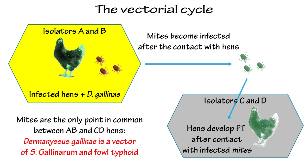
Each hen, identified by a unique marked ring on the left leg, was randomly selected and assigned to one of four groups, each consisting of 8 birds. The first two groups, A and B, were housed in two isolators Bioflex® B40 Rigid Body Poultry Isolator (Bell Isolation Systems, Livingston, UK) and the other two, C and D, in two isolators HM 1500 (Montair Process Technology, Kronenberg, The Netherlands). Isolators were located in two different rooms under controlled environmental conditions (temperature 25 ± 3 °C, relative humidity 65-75%). Light cycle consisted of 12 hours of light per day, from 8:00 a.m. to 8:00 p.m. The birds were fed ad libitum with unmedicated commercial complete feed; drinking water was provided in commercial drinkers.
The S. Gallinarum strain used for the experimental infection was a field strain of S. Gallinarum, isolated in 2009 during an outbreak of FT in a poultry farm. It was stored in 15% glycerol tryptic soy broth at -80°C and, before use, it was revitalized by streaking a loopful on tryptic soy agar (TSA, Oxoid, Milan, Italy), and incubated at 42°C overnight. A single, well-isolated colony was selected and passaged on TSA. After overnight incubation at 42°C, a single, well-isolated colony was selected and inoculated in 25 mL tryptic soy broth. The suspension was washed three times in 0.9% NaCl solution, titred by the plate count method, and administered after adjustment of the concentration to 6x10⁷ CFU/mL with 0.9% NaCl solution. One mL of suspension was administered by gavage to the hens of groups A and B (hereafter infected groups) on day D1, after they were acclimated for three days. Gavage was carried out using a soft plastic, sterile 1 mL Pasteur pipette for about two seconds. The study schedule is shown in Table S1.
After 7 days (D8), approximately 25,000 D. gallinae mites (mainly nymphs and adults) were introduced into each isolator, A and B, following a starvation period of three days to induce mites to more aggressively attack hosts to obtain a blood meal [21]. Contemporaneously, three mite traps were placed in each incubator to retrieve mites for transferring to isolators C and D, and to test for the presence of S. Gallinarum. Traps consisted of a pile of 5 wooden slats (200 mm long, 100 mm wide and 10 mm high) separated by 1 mm high wooden spacers and joint together by elastic bands. The mites came from a permanent colony reared at the facilities of MSD Animal Health MSD Innovation GmbH (Zur Propstei, Schwabenheim, Germany). The colony founders were collected from an industrial laying hen farm in Germany in 2001.
On day D10, all surviving hens of the infected group were humanely euthanized in accordance to the directive 2010/63/EU of the European Parliament and of the Council, and traps were removed from isolators A and B. Mites were recovered from the traps, starved for 24 h, and then, about 8,000 mites were introduced into each of isolators C and D, which held the other two groups of hens (hereafter, infested groups). The number of mites was estimated by weighing the mass of the mites, after establishing a standard by measuring a mass sample of five 5 aliquots consisting of 100 randomly selected mites.
Although a scheduled euthanasia program, the in-study deaths of 11 hens in the infested group meant that only five hens were euthanized at the end of the experimental program (the original schedule and its amendments are reported in Additional Table 1). Twenty-four days after the infestation of groups C and D, the experimental procedure was concluded by euthanizing the surviving hens. All remaining mites in all four isolators were collected by removing traps and manually recovering any mites present on the walls and floors of the isolators.
Health status of hens and clinical score
The health status of the hens was monitored twice a day for the duration of the experimental procedure and a score was assigned according to the observed clinical signs, as detailed in Additional Table 2. The sum of the two daily observations was considered as the daily score. Hens with daily scores greater than 4 were considered sick.
Post-mortem examination
All hens that died (spontaneously or euthanized) were necropsied, and the gross lesions recorded. Liver, spleen, ovary, and cecum were excised from all birds by using sterile scissors and surgical blades.
Mite samples
On D10, when traps were collected from isolators A and B, two 100-mite aliquots were prepared for analysis, and the other mites were starved for the infestation of groups C and D, as above described.
In addition, on D9 and D11, mites within the isolators A and B (infected groups) but outside the traps were collected, obtaining another 100-mite aliquot form each of the two isolators. No more aliquots were obtained from isolators A and B in order to avoid loss of mites for the infestations of isolators C and D.
Further mite samples were collected on D36, at the end of the procedures. Specifically, two 100-mite aliquots were retrieved from residual mites in isolators A and B, while ten 100-mite aliquots were prepared from isolator C, and seven 100-mite aliquots from isolator D. Six out of the aliquots from isolator C and four from isolator D were washed by formaldehyde 4% and rinsed with sterile distilled water three times. All aliquots underwent molecular detection and quantification of S. Gallinarum immediately after preparation.
Molecular detection and quantification of Salmonella entericasubsp.entericaser. Gallinarum
In order to carry out the total genomic DNA extraction, 30 mg of each collected tissue and the 100-mite pools were carefully ground with sterile mortar and pestle, and then processed by the means of the PureLink Genomic DNA kit (Thermo Scientific, Milan, Italy), according to the manufacturer's instructions. The quantification of DNA solutions was achieved by measuring optical density at 260 nm with NanoDrop 1000 spectrophotometer (Thermo Scientific).
The detection of S. Gallinarum was performed by seminested PCR (snPCR) as previously described [22]. The quantification of S. Gallinarum was carried out by real-time PCR (qPCR) from the snPCR-positive mite samples, according to the previously described protocol [20]. Briefly, amplification was carried out in 20 µL of mixture containing 1X SsoFast™ Probes Supermix with ROX (Bio-Rad, Milan Italy), 100 nm of each primer, 400 nm of FAM-labelled probe and 1 µL of template. Non-template controls were included in each run by adding sterile distilled water instead of DNA. The conditions were: one cycle at 95 °C for 5min for Taq polymerase activation, followed by 45 cycles of 95 °C for 30 s, 55 °C for 30 s and 65 °C for 30 s. Fluorescence was acquired during each extension step. Data were acquired and treated by the mean of the Sequence Detection Software Version 1.2.3 (Applied Biosystems, Milan, Italy). Cycle threshold and baseline were calculated automatically by the software and checked by the operators to ascertain possible inconsistencies. For each plate, the standard curve was set up by including 7 serial dilutions (specifically, 1:50, 1:100, 1:500, 1:1000, 1:5000, 1:10 000, 1:50 000) of purified DNA from a pure culture of S. Gallinarum. The initial concentration of the DNA solution was determined using the NanoDrop 1000 (Thermo Scientific). All unknown samples, non-template controls, and standards were analyzed in triplicate. The R2 value of the standard curve in all experiments was higher than 0.98.
Considering that 1 µL of DNA solution was included in each reaction, that DNA was extracted from 100 mites, and that it was eluted in a final volume of 200 µL, the S. Gallinarum genome size and the unicity of the target locus in the pathogen genome, the results have been normalized and expressed as S. Gallinarum cells/mite [20]. Considering the sample size, values below 0.01 S. Gallinarum cells/mite (corresponding to one cell per 100 mites) were treated as 0.
Serological investigation
One mL of venous blood was collected from hens on D-1 for confirming that there had been no prior contact with S. Gallinarum, by the mean of an indirect enzyme-linked immunosorbent assay (ELISA). Serum was separated soon after collection and anti-group-D Salmonella antibodies were measured using the Chicken Salmonella Antibody Test Kit (BioChek, Reeuwijk, Netherlands), according to the manufacturer’s instructions. Data were analyzed by BioChek II Version 2013.0.07. A sample to positive (S/P) ratio higher than 0.5 was considered positive.
Statistical analysis
The normal distribution of quantitative datasets was verified by the Shapiro–Wilk test, with a threshold of P = 0.05. Considering that no dataset contained normally distributed data, non- parametric analysis was performed. Therefore, the Hodges-Lehmann location estimator [23, 24] has been used to calculate the central values and the 95% confidence interval (CI). Datasets were compared by the Mann-Whitney U test. Mortality and morbidity of infested and infected groups, as well as the number of positive organs collected from the two groups, were compared by the Fisher’s exact test. In both cases, P = 0.05 was assumed as the significance threshold.
All statistical analyses were performed by the mean of R software v. 3.6.1 [25] and DescTools package v. 0.99.28.
Determination of the infection rate, of the entomological infection rate and of the vectorial capacity
The maximum likelihood estimation (MLE) of the infection rate (IR) [26] of D. gallinae was calculated by the PooledInfRate v. 4.0, an algorithm that takes into account the number of S. Gallinarum positive mite aliquots and the size of aliquots [27].
The IR was calculated using a combination of pathogen location within and on mites. To avoid potential bias by inclusion of washed samples (eliminating pathogens on the mite surface), only unwashed aliquots were considered in the IR calculation.
The entomological inoculum rate (EIR) was obtained by the equation:

where CpH is the number of mites that came in contact with each host and IR the infection rate (modified from [28]). The number of contacts was calculated by considering that, from previous observations, about 90% of starved mites were found to feed when they can, and that about 1,000 mites per hens were introduced in isolators C and D.
The vectorial capacity (VC) was calculated as previously described [29], with the equation:

where m is the number of total mites per host, a is the daily blood feeding rate, b is the transmission rate among exposed mites, p is the probability of daily survival and n the extrinsic incubation period in days. For this study, it was assumed that m = 1,000 (the number of mites per hen in incubators C and D); a = 0.9 (as above described); b = IR; n = 3 (as mites were introduced into the isolators C and D 3 days after their first contact with infected hens in isolators A and B). It should be underlined that m is a value that describes the mite population as a mix of stages and sexes, all but larvae capable of feeding on hosts. The assumed survival rate of mites following a blood meal was 0.836, derived from the mean of three earlier values: 89.1% of protonypmhs, and 66% of deutonymphs [30] and our own unpublished observations of adult mite survival rate of 95.83%.
Since mites were found to be positive as soon as one day after exposure to infected hens, n could also be assumed equal to 1, but the most stringent parameters were used. Vectorial capacity quantifies the probability that an arthropod may transmit a pathogen following exposure to an infected host [31, 32].