Reagents and antibodies
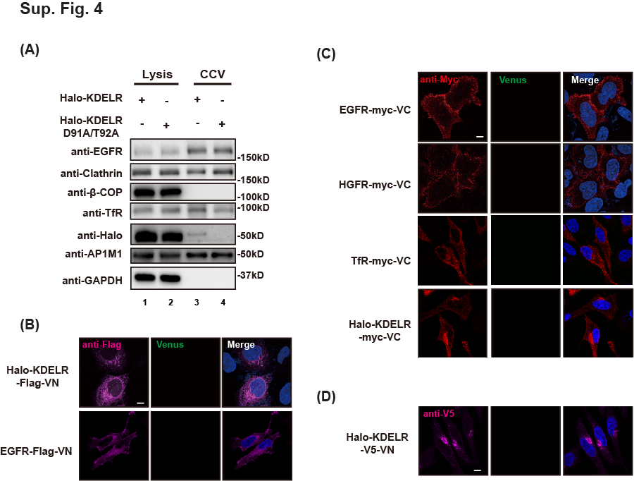
The following antibodies were used: anti-GAPDH (HRP-60004, Proteintech), anti-EGFR (4267S, Cell Signaling Technology), anti-Myc (2278S, Cell Signaling Technology), anti-Golgin97 (13192, Cell Signaling Technology), anti-mCherry (ab167453, Abcam), anti-Clathrin heavy chain (ab21679, Abcam), anti-Transferrin Receptor (ab214039, Abcam), anti-β-COP (ab2899, Abcam), anti-Halo (G9211, Promega), anti-AP1M1 (12112-1-AP, Proteintech), anti-E-Cadherin (20874-1-AP, Proteintech), anti-ARFGAP1(ab204405, Abcam), anti-Flag (F1804-1MG, Sigma), anti-TGN46 (AHP500GT, Bio-Rad), anti-p-EGFR (Tyr1068) (3777S, Cell Signaling Technology), anti-p-EGFR (Tyr1086)(ab32086, Abcam), anti-p-EGFR (Tyr45) (sc-57542, Santa Cruz Biotechnology), anti-STAT1 (9172, Cell Signaling Technology), anti-p-STAT1 (Tyr701) (9167, Cell Signaling Technology), anti-STAT3 (ab68153, Abcam), anti-p -STAT3 (Tyr705) (ab76315, Abcam), anti-p-STAT3 (Ser727) (ab76315, Abcam), anti-ERK (4695, Cell Signaling Technology), anti-p-ERK (MA515174, Invitrogen), anti-JAK1 (66466-1, Proteintech), anti-p-JAK1 (Y103/1035) (66245S, Cell Signaling Technology), anti-Src (2109S, Cell Signaling Technology), anti-p-Src Family (Tyr416) (6943S, Cell Signaling Technology), HaloTag Alexa Fluor 488 Ligand (G1002, Promega), HaloTag TMR Direct™ Ligand (G2991, Promega). Anti-Rabbit Alexa Fluor 488 (A21441), Alexa Fluor 568 (A10042), Alexa Fluor 647 (A21245), anti-Mouse Alexa Fluor 488 (A21200), Alexa Fluor 568 (A10037), and Alexa Fluor 647 (A21236) for immunofluorescence (IF) were obtained from ThermoFisher. Lentivirus concentration solution (AC04L442) was purchase from Life iLab Bio. TAEKDEL and TAEAAAA peptides were synthesized by GenScript.
Cell culture, transfection and lentivirus infection
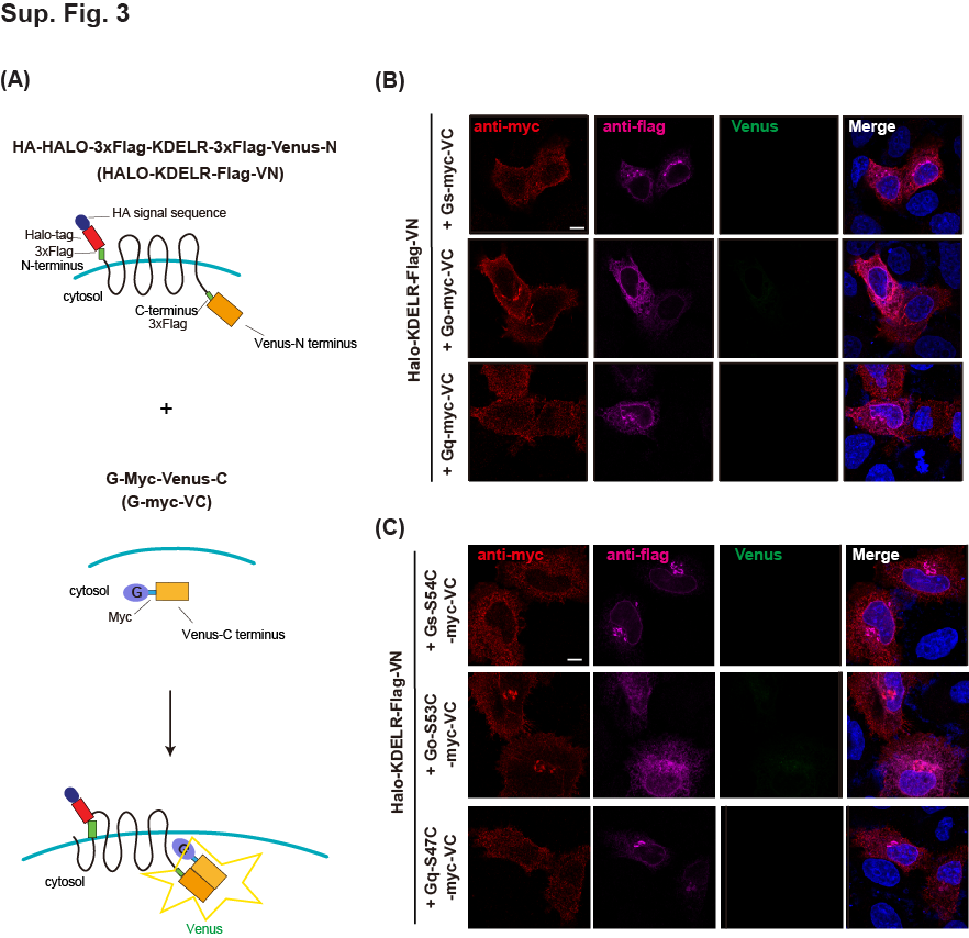
HeLa cells (ATCC, CCL-2) were grown in Delbecco’s modified Eagle medium (DMEM, Meilunbio) supplemented with 10% fetal bovine serum (FBS, ExCell Bio). HT-1080 cells (Stem Cell Bank, Chinese Academy of Sciences) were grown in Minimum Essential Medium (MEM, Meilunbio) with 10% fetal bovine serum (FBS, ExCell Bio). Transfection of plasmids was performed using Lipofectamine 2000 (11668019, Thermo-Fisher), according to the manufacturer’s instructions. For gene overexpression experiments, cells were transfected for 18 hours, prior to co-IP and IF experiments.
To stably knock down KDELR in HT1080 cell line, the lentivirus expressing KDELR-shRNA (GGTTGCCAAACACTAAATCTG, targeting 3′-UTR) was packaged and commercially provided by Shanghai GenePharma, China. Cells were infected with the lentivirus using polybrene overnight. Three days after infection, the cells were cultured in puromycin (0.6 μg/ml) for five days.
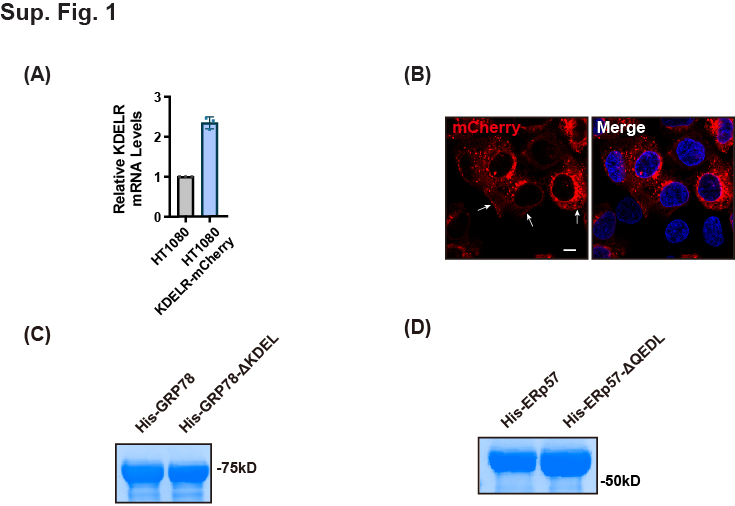
HT1080 cells stably overexpressing KDELR-mCherry or Halo-KDELR were prepared using lentivirus expressing KDELR-mCherry or Halo-KDELR respectively. To produce and pack lentivirus expressing KDELR-mCherry, a lentiviral vector containing KDELR-mCherry along with packing (psPAX2) and envelop (pMD2.G) vectors were transfected into 293FT cells using Lipofectamine 2000. The supernatants were collected after transfection for 48 hours and 72 hours, filtrated with 0.45 nm filters, and concentrated using a Lenti-concentration kit (AC04L441, life-ilab). The lentivirus expressing Halo-KDELR was packaged and commercially provided by OBiO Technology.
Scratch wound healing assay
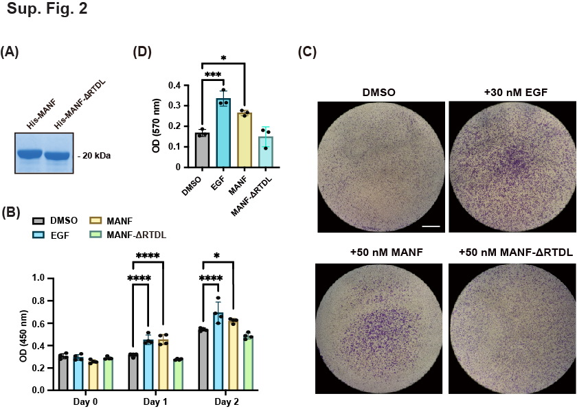
Cells were seeded into a 96-well (Corning) and gown to full confluency in MEM supplemented with 10% FBS. Then cells were scratched using a wound maker (IncuCyte) to create homogeneous, 700-800 μm wide wounds in cell monolayers. After wounding, cells were washed twice with sterile PBS and incubated in MEM supplemented with 0.3% FBS and DMSO, EGF, or recombinant ER proteins for 24 hours. During this time period, real-time gap distances were imaged and determined using a high-throughput screening system (IncuCyte ZOOM).
Reverse transcription and quantitative real time (qPCR) Assay
Total mRNA extraction was performed using using RNA Isolation kit (R0026, Beyotime). The reverse transcription was carried out by HiScript II Q RT SuperMix (R223-01, Vazyme) according to manufacturer’s protocol. Real time-PCR was performed by a QuantStudio 7 real-time PCR system (ThermoFisher) using ChamQTM Universal SYBR qPCR Master Mix (Q711-02, Vazyme). GAPDH mRNA was used for normalization. Primers are listed as follows: KDELR forward: AGCCACTACTTGTTTGCGCTA, KDELR reverse: CCTGCCACAATGGCGATGA; Cyclin-D1 forward: GCTGCGAAGTGGAAACCATC, Cyclin-D1 reverse: CCTCCTTCTGCACACATTTGAA; Bcl-2 forward: GGTGGGGTCATGTGTGTGG, Bcl-2 reverse: CGGTTCAGGTACTCAGTCATCC; Vimentin forward: GACGCCATCAACACCGAGTT, Vimentin reverse: CTTTGTCGTTGGTTAGCTGGT; ICAM-1 forward: ATGCCCAGACATCTGTGTCC, ICAM-1 reverse: GGGGTCTCTATGCCCAACAA; GAPDH forward: ACCACAGTCCATGCCATCAC, GAPDH reverse: TCCACCACCCTGTTGCTGTA
CCK-8 assay
Cell viability was analyzed by Cell Counting Kit-8 (Meilunbio) according to the manufacturer’s protocols. Cells were seeded at a density of 1×10⁴/well into a 96-well plate and cultured in MEM supplemented with 10% FBS. At 0, 24, and 48-hour time points, 10% volume of CCK-8 reagent was added to the medium and incubated for 1 hour. The absorbance was recorded at 450 nm using a microplate reader (ThermoFisher Scientific). The culture medium was replaced with fresh MEM medium supplemented with 0.3% FBS and DMSO, EGF, or recombinant ER proteins at each time point.
Mass spec-based proteomics for cell surface proteins
HeLa cells (three 10 cm dish of cells for each experimental condition) were transfected with Halo-KDELR for 18 hours and incubated with DMEM medium supplemented with 10% FBS and non-membrane permeable HaloTag PEG-biotin ligand (Promega) or DMSO for 30 minutes at 4oC. After incubation, the cells were washed three times with cold PBS, and harvested in 1 mL lysis buffer (20 mM HEPES, pH 7.4, 100 mM NaCl, 2 mM DDM (N-Dodecyl-β-D-Maltoside, Anatrace), 0.02% CHS (Cholesteryl hemisuccinate, Sigma), protease inhibitors (Bimake)). Then the cells were lysed by passing through a 1mL syringe needle for 15 times and incubated a 4oC for 1 hour. The lysates were cleared by centrifugation at 15,000 g for 20 minutes. The supernatants were incubated with Streptavidin beads for 1 hour at 4 oC. The beads were washed three times with cold PBS, and the proteins were eluted by boiling in Laemmli SDS sample buffer and subjected to SDS-PAGE for mass spectrometry. Samples were prepared by in-gel digestion, separated and analyzed on an Easy-nLC 1000 liquid chromatograph coupled to a Q Exactive HF mass spectrometer (ThermoFisher Scientific).
Co-IP and immunoblotting
For co-IP experiments, total lysates were prepared using lysis buffer (20 mM HEPES, pH 7.4, 100 mM NaCl, 2 mM DDM (n-dodecyl-β-D-maltoside, Anatrace), 0.02% CHS (cholesterol hemisuccinate, Sigma), protease inhibitors (Roche)). Then, the lysates were passed through a syringe needle for 15 times and incubated with agitation for 1 hour at 4oC. The supernatants were prepared by centrifugation at 15,000 g for 10 min and incubated with anti-RFP agarose beads (MBL life science) or anti-Flag agarose beads (GenScript) for 4 hours at 4 oC. The beads were washed three times with cold PBS. The protein were boiled in 2× SDS running buffer and subjected to SDS-PAGE for western blotting.
For immunoblotting, proteins were separated by SDS-PAGE (Gensript) and transferred to the nitrocellulose membranes (Amersham). Then, the membranes was blocked for 1 hour at room temperature with bovine serum albumin (BSA) in the blocking buffer (ABCONE), probed with specific primary antibodies overnight at 4 oC, and incubated with peroxidase-conjugated secondary antibodies (Jackson Immuno Research). The bands were visualized with chemiluminescence (Clarity Western ECL Substrate, Bio-Rad) and imaged by a ChemiDoc Touch imaging system (Bio-Rad).
IF staining and confocal microscopy
Cells grown on a 24-well glass bottom plate (Cellvis) were fixed with 4% paraformaldehyde (PFA) for 10 min, permeabilizated in permeabilization buffer (0.05% Triton-X100 in PBS) for 3 min, and blocked in blocking buffer (3% BSA, 0.05% Triton-X100 in PBS) for 60 min. Then the cells were incubated with primary and secondary antibodies in blocking buffer for 1 hour. The nucleus was statined with Hoechst-33342 Santa cruz Biotechnology). Cells were washed three times with wash buffer (0.2% BSA, 0.05% Triton-X100 in PBS) and twice with PBS. Images were acquired using a Zeiss LSM880 confocal microscope with a 63×oil immersion objective.
Split-ubiquitin membrane yeast two hybrid assay
Human KDELR cDNA was subcloned in frame and upstream of the C-terminal half of ubiquitin (Cub) and the artificial transcription factor LexA-VP16 in the pBT3-SUC bait vector. ACBD3, EGFR, GLUT4, and ITGA5 cDNA were individually fused to the mutated N-terminal half of ubiquitin (NubG) in the pPR3-N prey vector. The NMY51 yeast strain co-transformed with bait and prey pair was spread onto selective medium lacking leucine and tryptophan (SD/-Leu/-Trp, DDO, Clontech). The physical binding of bait and prey was identified by colony selection in selective medium lacking adenine, leucine, tryptophan, and histidine (SD/-Ade/-Leu/-Trp/-His, Clontech) supplemented with X-α-Gal. Co-transformation of KDELR-Cub and pOST1-NubI was performed as a positive control.
GST-pulldown assay
KDELR C-terminal (CT, residues 205-212) was inserted in pGEX-6p-1 plasmid for the expression of GST fusion proteins. The constructed plasmids were transformed into BL21 (DE3) (Vazyme) competent cells. Single colony of cells was grown in LB broth at 37°C until OD600 reading was between 0.6 and 0.8. Then proteins expression was induced by addition of 0.3 mM IPTG (Isopropyl β-d-thiogalactopyranoside) at 37°C for 3 hours. Subsequently, bacteria were collected in lysis buffer (50 mM Tris-HCl, pH 7.4, 100 mM NaCl, 1 mM dithiothreitol 0.5% Triton X-100) and lysed by sonication. The supernatants were collected by centrifugation at 15,000 × g for 10 minutes, and incubated with Glutathione HiCap Matrix (Qiagen) at 4°C for 1hour. The beads were washed by lysis buffer and the GST fusion proteins were eluted by 10 mM glutathione in lysis buffer.
For the pulldown assay, 100 ug of each GST-fusion protein was immobilized on glutathione Sepharose 4B (GE Healthcare). The resins were washed 3 times with lysis buffer and incubated with HeLa or HT1080 cell lysates at 4°C for 1 hour. The resins were washed three times, resuspended in 2×SDS loading buffer, and subjected to SDS-PAGE analysis.
Image processing and statistical analysis
Pearson coefficient was analyzed by Fiji software. Statistical analyses were performed with GraphPad Prism 9.0 software using student’s t-test or ANOVA and represented as mean ± SD. n is noted in the figure legends.