Bc Patient Samples
30 BC patients’ cancer tissues and their paired normal adjacent tissues were obtained from the patients who have not received any anti-cancer therapy before surgery. The samples were stored in liquid nitrogen immediately after resected from patients. All patients participating in the study gave them consent forms and the procedures were approved by Institution Ethics Committees of the hospital.
Cell Culture
Human normal breast MCF-10A cells (Zhongqiaoxinzhou Biotech, Shanghai China) were cultured in Mammary Epithelial Cell Medium (MEpiCm, ScienCell, Research Laboratoried, Carlsbad, CA, USA). BC cell lines, MDA-MB-231, MDA-MB-468, HCC-1937, MCF-7 (Chinese Academy of Science, Shanghai, China) and SKBR-3 (iCell Bioscience Inc, Shanghai, China) were cultured in Dulbecco’s modified Eagle’s medium (DMEM) (Gibco, Grand Island, NY, USA) with 10% Fetal Bovine Serum (FBS) (Gibco), penicillin (100 U/ml) and streptomycin (100 µg/ml) (PS, Enpromise, Hangzhou, China). Cells were incubated at 37 ̊C supplemented with 5% CO2, and subcultured when the cell density was around 80%.
Transfection Assay
siRNA circ-LATS2 and circ-NC, miR-4686, miR-NC, pcDNA3.1 (+) circRNA Vector-circ-LATS2 (OE-circ) and Vector-circ-NC (OE-NC) were synthesized by Intergrated Biotech Solutions (Shanghai, China) (Supplementary table 1). Cells were added into six-well-plate and cultured with DMEM medium with FBS and PS. When the cell confluency reached 30–50%, 0.8 µmol siRNA circ-LATS2, siRNA circ-NC, miR-4686, miR-NC, OE-circ-LATS2 or OE-circ-NC and 4 µl Lipofectamine 2000 (Invitrogen, Carlsbad, CA, USA) were added into the cells with DMEM medium. After 4 hrs of incubation, the medium was replaced by DMEM with FBS and PS. After 48 hrs, the cells were harvested for further experiments.
Fluorescence In Situ Hybridization (fish)
Cells were cultured on coverslips, then hybridization buffer was added and incubated overnight with circ-LATS2 probe. The circ-LATS2 probe was chemically synthetized by Guangzhou RiboBio Co. Ltd (Guangzhou, China). Cell nuclei were counterstained with 4,6-diamidino-2-phenylindole (DAPI). The images were obtained by Thermo Fisher microscope (Thermo Fisher Scientific, Massachusetts, USA).
Qrt-pcr
Total RNA was extracted using TRIzol reagent (Invitrogen, USA). Nuclear and cytoplasm RNA was extracted from MCF-10A and MDA-MB-231 cells using Paris™ kit (Thermo Fisher Scientific, Massachusetts, USA). cDNA was generated by reverse transcription using the PrimeScript RT-PCR kit in accordance with the manufacturer's instructions (Takara, Tokyo, Japan). Real-time PCR was performed on a 7900HT Fast RT-PCR instrument (Applied Biosystems, Singapore). The primer of circ-LATS2 and miR-4686 were synthetized by Intergrated Biotech Solutions (Shanghai, China). The qRT-PCR results were analysed with the 2−△△CT method.
Colony Formation Assay
The treated cells were then harvested and plated into a six-well-plate at 500 cells/well. The plates were incubated for 7 to 10 days. When the colonies were visible, the medium was removed, and the plates were washed with phosphate buffered saline and allowed to dry. After that, the dried colonies were fixed in 95% ethanol, then dried and stained with 0.1% crystal violet solution. Finally, the colonies were washed, dried and immediately photographed.
Mtt Assay
The treated cells were seeded into 96-well-plate at 500 cells/well. Cell viability was estimated using an MTT assay kit (Sangon, Shanghai, China). After 4 hrs incubation in MTT assay reagent, the medium was replaced with 150 µl dimethylsulfoxide (DMSO, Sangon, Shanghai, China). The absorbance at 490 nm was measured using a microplate spectrophotometer (BioTek, Vermont USA).
Wound-healing Assay
Cells were transfected in 6-well-plate with a range of constructs as indicated. When cells reached about 90% confluent, a scratch was produced in the cell monolayer by drawing a 200-µl-pipette tip over the surface of each well, holding the tip perpendicular to the plate. The monolayers were washed and cultured with DMEM medium without FBS and PS. Wound-healing was observed under a light microscope and pictures were taken at 0, 24 and 48 hrs at the same position to observe cell movement.
Western Blotting
Whole cell protein extracts were prepared by lysis of cells using cold RIPA buffer (100 µl/well, Beyotime, Shanghai, China). The concentration was measured using a bicinchoninic acid (BCA) Protein Assay Kit (Beyotime, Shanghai, China). Equal protein amounts of each of the samples were denatured with 6 × sodium dodecyl sulfate (SDS) loading buffer (Beyotime, Shanghai, China) at 100 ̊C for 10 min. Protein samples were separated by electrophoresis on a 10% polyacrylamide SDS gel (Beyotime, Shanghai, China) and transferred onto 0.45-µm nitrocellulose membranes (Beyotime, Shanghai, China). Following 60 min of blocking with 5% fat-free milk in double distillation H2O, membranes were incubated with the primary antibodies in antibody diluent (Beyotime, Shanghai, China) overnight at 4 °C. Blots were washed with PBST (PBS with Tween), and incubated for 1 hr with the anti-rabbit or anti-mouse secondary antibody, as appropriate (1:2000, Santa Cruz Biotechnology, USA). Immunoreactive protein bands were detected with an Odyssey Scanning system (Li-Cor, USA). The density of the bands was measured by ImageStudio.
The following primary antibodies and dilutions were used:
β-actin (1:2000, Santa Cruz Biotechnology, USA), LATS2 (1:1000, Bioworld, Shanghai, China), PCNA (1:2000, Danvers, MA, USA) and WNT5A (1:1000, Proteintech, Chicago, USA).
Dual-luciferase Reporter Assay
PmirGLO-hsa_circ_0029693 mutant and wild type reporter plasmids, pmirgGLO-WNT5A 3'-UTR mutant and wild type reporter plasmids were purchased from Integrated Biotech Solutions (Shanghai, China). 293T cells were seeded in 48-well plates, and co-transfected with pmirGLO-hsa_circ_0029693 mutant or wild type reporter plasmids, and miR-4686 or NC and vector using Lipofectamine 2000 reagent. These reagents were added into Opti-MEM according to the manufacturer’s instruction. 293T cells were co-transfected with pmirgGLO-WNT5A 3'-UTR wild type or mutant reporter plasmids, and miR-4686 or NC using Lipofectamine 2000 reagent. After 24 hours, firefly and Renilla luciferase activities were measured using a Dual Luciferase Reporter Assay (Promega, Madison, WI, USA). Firefly luciferase activity was normalized to the Renilla control, and the ratio of firefly/Renilla was recorded.
Animal Experiment
The five-week-old female athymic nude mice were used to do tumorigenicity in vivo. MDA-MB-231 cells were infected by lentiviruses. Mice were divided into two groups randomly. Over expressing circ-LATS2 or circ-NC MDA-MB-231 cells were implanted into the right side of second mammary fat pad of the mice (3 × 10⁶ cells/mouse). After 5 weeks of observation, mice were sacrificed. The mice tumors were harvested, weighed and photographed.
Immunohistochemical Staining (ihc)
The mice tumors were formalin-fixed and paraffin-embedded (FFPE). 5 µm FFPE sections were cut. Antibody LATS2 was used to do IHC following the protocol. The images were captured using Thermo Fisher microscope (Thermo Fisher Scientific, Massachusetts, USA). The ratio of brown areas was analyzed using Image-Pro Plus 6.0 software (Media Cybernetics, Silver Spring, MD, USA).
Databases Analysis
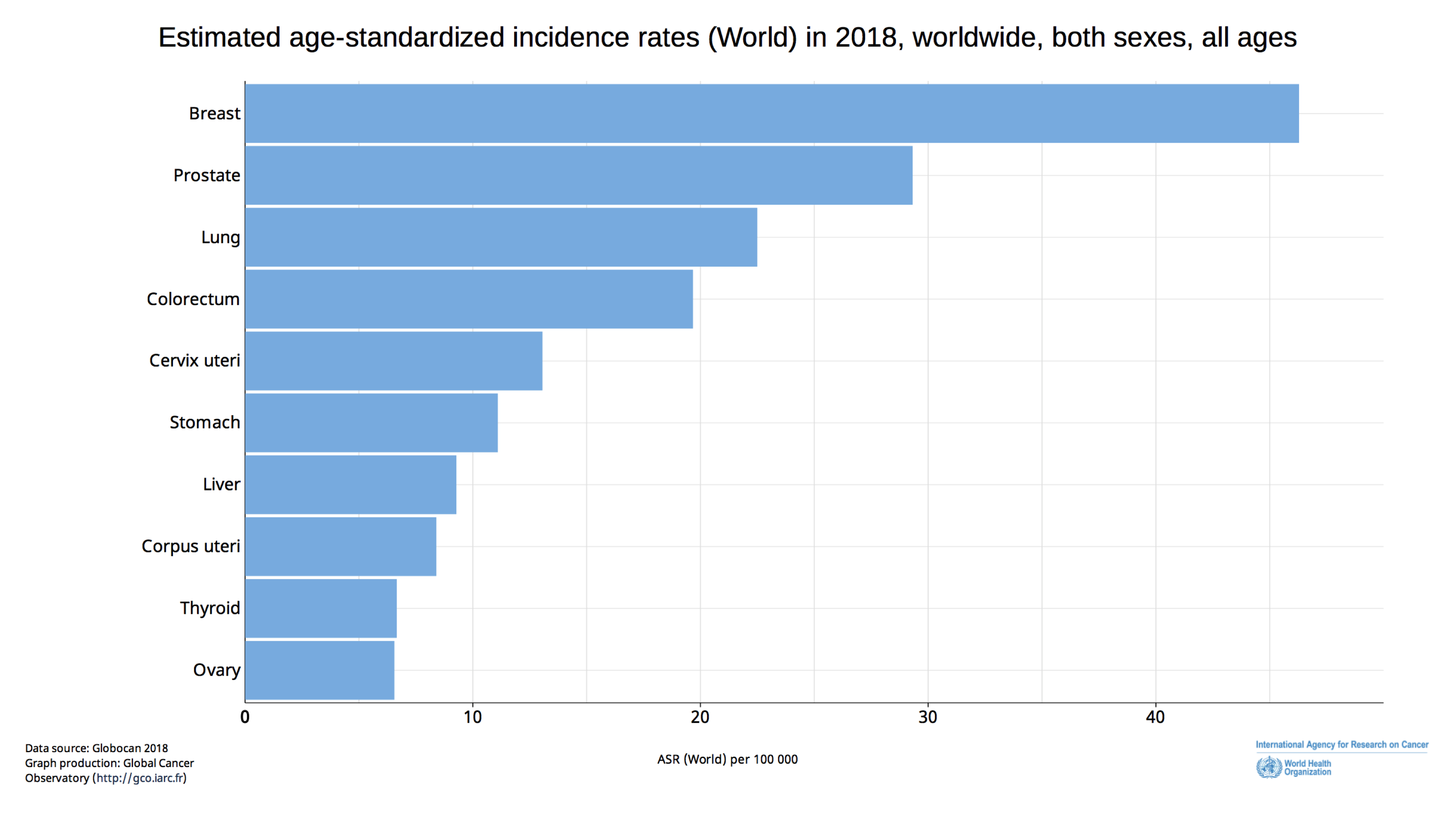
RegRNA 2.0 (http://regrna2.mbc.nctu.edu.tw) was used to identify regulatory circRNA motif and its binding site for miRNA 15. CircBase (http://www.circbase.org) was used to search for the sequence of circRNA 16. USCS Human Genome Browser (https://genome.ucsc.edu/index.html) was used to identify the position and the component of circRNA 17. Data from GEO, EGA, TCGA and Pubmed were combined in KMplotter (https://kmplot.com/analysis/), and Kaplan-Meier survival analysis was used to determine the association between expression levels of potential prognostic biomarkers and clinical outcome in a range of cancers including BC 18. KMplotter was used to evaluate the association between LATS2 expression and BC survival. TargetScan 7.2 (http://www.targetscan.org/vert_72/) was used to predict potential target genes of miRNAs 19. The Global Cancer Observatory (GCO) is a web-based program presenting global cancer statistics 20. GCO was used to estimate age-standardized cancer incidence rates in 2018 worldwide.
Statistical analysis
Data were obtained from three independent experiments which are presented as the means ± standard deviation. Differences were considered significant for P-values less than 0.05. GraphPad Prism version 8.0 was used for statistical analyses.