2.1. Main reagents
Reagents were obtained as follows: Cobalt nanomaterials (particle size 100nm) (Shanghai Yunfu Technology Co., Ltd), anhydrous ethanol (C2H5OH) (Tianjin Baishi Chemical Co., Ltd), silver nitrate (AgNO3) (Sinopharm Chemical Reagent Co., Ltd), Mueller-Hinton (M-H) AGAR culture medium (Wenzhou Kangtai Biotechnology Co., Ltd), LB liquid culture medium (dry powder (Beijing Solarbio Science and Technology Co., Ltd). Yellow River water was obtained from the Lanzhou section of the Yellow River.
2.2. Standard strains
Pseudomonas aeruginosa (ATCC27853, Gram-negative bacteria), Escherichia coli (ATCC25922, Gram-negative bacteria), Staphylococcus aureus (ATCC25923, Gram-positive bacteria) and Candida albicans (ATTC90029, yeast) were stored in the Microbiology Laboratory of Xi 'an Ninth Hospital.
2.3. Treatment of the Yellow River water
Firstly, large debris were removed from the Yellow River water through 200-mesh sieve. Then, the Yellow River water stood for precipitation to obtain the supernatant under sterile conditions. A volume of 100μl Yellow River water supernatant was absorbed and evenly applied on LB solid medium, which was removed to be cultured at 37℃ for 12h. The turbidity of Yellow River water as 6 ~ 7×10³CFU/MLand the bacterial assemblage of the Lanzhou section was mainly composed of fecal coliform[15-16].
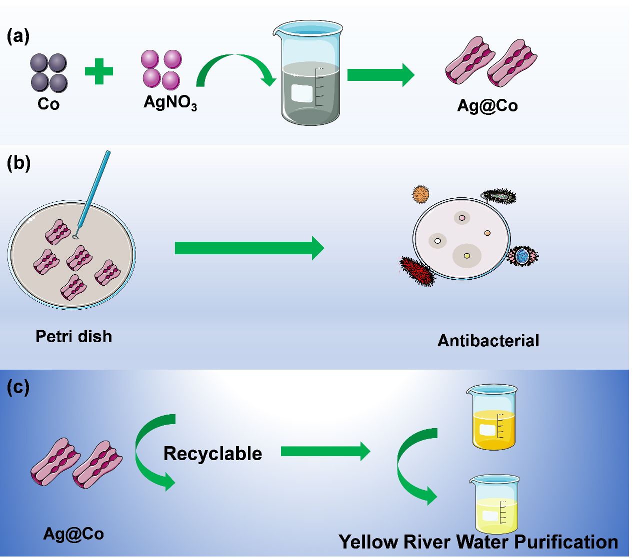
2.4. Preparation of Ag@Co nanomaterials
70.8mg cobalt nanomaterials and 272mg AgNO3 powder were accurately weighed and each dissolved in 50mL deionized water. The two solutions were added to a three-pot flask, and the volume of reaction system was increased to 200mL.The mixture was stirred (800rmp) for 4h at room temperature. Ag@Co nanomaterials can be obtained after centrifugation at 5000rmp. The samples were successively washed with deionized water and anhydrous ethanol by ultrasonic and centrifugation for 3 times, and then freeze-dried for further use.
2.5. Characterization of Ag@Co nanomaterials
The morphologies of the synthesized Ag@Co nanoparticle samples were characterized by transmission electron microscope (TEM, Kevex JSM-6701F, Japan). The type of elements, valence and mass ratio of Ag@Co nanomaterials were determined by X-ray photoelectron spectroscopy (XPS, ESCALAB 250, ThermoFisher Scientific Technology, USA). The phase composition of the Ag@Co nanomaterials was determined by X-ray diffraction (XRD, Bruker D8 Advance). The magnetic properties of Ag@Co nanomaterials were detected by an oscillating sample magnetometer at room temperature (VSM, Lakeshore Cryotronics Inc., Ohio, USA).
2.6. Measurements of antibacterial properties of Ag@Co nanomaterials in vitro
The standard strains of Pseudomonas aeruginosa, Escherichia coli, Staphylococcus aureus and Candida albicans were selected as representative bacteria for investigating the antibacterial properties of Ag@Co nanomaterials. The minimum inhibitory concentration (MIC) values of Ag@Co nanomaterials were determined by the disc agar diffusion method and broth dilution method. 10mg/ml of Ag@Co nanoparticle suspension were mixed into blank drug-sensitive paper, which was shook overnight to prepare drug-sensitive paper containing Ag@Co nanomaterials. The prepared drug-sensitive papers were pasted on the M-H agar medium inoculated with the above four kinds of standard bacteria overnight to measure the antibacterial effect.
Under sterile conditions, a single colony of Pseudomonas aeruginosa, Escherichia coli, Staphylococcus aureus and Candida albicans were selected and diluted with LB liquid medium to reach a level of 1.0×10⁵ CFU/ML. 2mL diluted bacterial solution was added to each well of the 24-well plate sequentially. Then, a certain amount of bacteriostatic solution containing Ag@Co nanomaterials was added in order and the concentrations of the bacteriostatic solution was as follows: 0 (positive control, bacteria solution added without Ag@Co nanomaterials), 100, 200, 300, 400, 500, 600, 700, 900, 1000, 2000g/ml, with the last well as a negative control (2ml Ag@Co nanomaterials bacteriostatic solution added); all the groups were with the duplicate holes set. The plates were incubated at 37℃ on a rotary shaker (180 rpm) for 12h. The antibacterial effect of Ag@Co nanomaterials was measured on the next day, and the minimum concentration without bacterial growth was the MIC.
The minimum inhibitory concentration (MIC) of Ag@Co nanomaterials for the Yellow River water: under sterile conditions, the Yellow River water was diluted into the LB liquid medium at 1:100. After that, 2ml diluted Yellow River water LB liquid medium was added to the 24-well plate, and then a certain amount of bacteriostatic solution containing Ag@Co nanomaterials was added sequentially. The bacteriostatic concentration of each well was as follows: 0 (positive control, only bacteria solution added without Ag@Co nanomaterials), 100, 200, 300, 400, 500, 600, 700, 900, 1000, 2000g/ml, with the last well as negative control (2ml Ag@Co nanomaterials bacteriostatic solution added); all the groups were with the duplicate holes set. The plates were incubated at 37℃ on a rotary shaker (180rpm) for 12 h. The antibacterial effect of Ag@Co nanomaterials was measured on the next day, and the minimum concentration without bacterial growth was the MIC.
2.7. Measurement of antibacterial properties of Ag@Co nanomaterials reclaimed
Measurement of minimum inhibitory concentration (MIC) of Ag@Co nanomaterials reclaimed for Pseudomonas aeruginosa, Escherichia coli, Staphylococcus aureus and Candida albicans: After the MIC of Ag@Co nanomaterials were measured, the magnets were put under the 24-well plates for 30 min to make sure that Ag@Co nanomaterials were attracted at the bottom of the plate. After slowly discarding bacteria liquid in the 24-well plates, 2ml of diluted bacteria liquid was added. The plates were incubated at 37℃ on a rotary shaker (180rpm) for 12h. The bacteriostatic effect of Ag@Co nanomaterials was observed by naked eyes on the third day, and the minimum concentration without bacteria growth was MIC.
Measurement of minimum inhibitory concentration (MIC) of Ag@Co nanomaterials reclaimed for Yellow River water: After the MIC of Ag@Co nanomaterials for Yellow River water were measured, the magnets were put under the 24-well plates for 30 min to ensure that Ag@Co nanomaterials were attracted to the bottom of the plate. After slowly discarding the bacteria liquid in the 24-well plates, 2 ml diluted bacteria liquid were added. The plates were incubated at 37℃ on a rotary shaker (180 rpm) for 12h. The bacteriostatic effect of Ag@Co nanomaterials was observed by naked eyes on the third day, and the minimum concentration without bacteria growth was MIC.
2.8. Measurements of the reclaim rate of reclaimed Ag@Co nanomaterials
5mg of Ag@Co nanomaterials were precisely weighed and dissolved into a centrifuge tube containing 25mL Escherichia coli dilution solution with a bacterial concentration of 1.0×10⁵CFU/ML. The bacteria were incubated at 37℃ on a rotary shaker (180rpm) for 12h. The next day, the magnet was placed under the centrifuge tube for about 30min to make sure that Ag@Co nanomaterials were attracted to the bottom of the tube. Then slowly discarding the bacteria liquid in the centrifuge tube and dried the left Ag@Co nanomaterials at 60℃ until the centrifuge tube was at constant weight. The reclaimed Ag@Co nanomaterials were weighed, and the reclaim rate was calculated according to the following formula: recovery (%) = recovery amount/input amount*100%.