Ethic statement
All experiments were authorized by the Ethical Committee of Guangzhou First People's Hospital, School of Medicine, South China University of Technology.
The animal experiments were conducted following the guidelines for the care and use of laboratory animals.
All procedures were strictly implemented by the code of ethics.
Establishment and grouping of permanent middle cerebral artery occlusion (pMCAO) mice
A total of 96 adult male C57BL/6 mice (6–8 weeks, 20–28 g) were purchased from Vital River Laboratory Animal Technology [SYXK (Beijing) 2017-0033, Beijing, China]. The mice were placed in separate cages at 22°C ± 2°C and 65 ± 5% relative humidity on a 12 h light/12 h dark cycle with free access to food and water.
The pMCAO mouse model was established 18. Mice were anesthetized with 2% pentobarbital sodium (50 mg/kg) (Sigma Chemical Co., St. Louis, MO, USA) by intraperitoneal injection. The right common carotid artery (CCA) was exposed and ligated using (5/0) surgical suture through a ventral midline neck incision. Mice were placed in left lateral decubitus position. An incision was made from the lateral orbit to the external auditory meatus, the base the parotid gland and the upper part of the temporal muscle were isolated to expose the cortical branches of middle cerebral artery (MCA) under the right side of skull. A craniotomy was performed directly above the distal part of the MCA using a 0.8 mm high-speed micro drill. Coagulator was used to cauterize the exposed cortical branches of MCA on the right side of the brain. The operation should be performed with caution to avoid damage to the surrounding brain tissue. The incision was sutured using conventional method. Mice should be kept warm during operation. The sham-operated mice only received the separation of cervical vessels and exposure of skull and drilling holes without ligation of right CCA and cauterization of corresponding arteries. The neurological function score and cerebral infarct area were analyzed 24 h after model establishment.
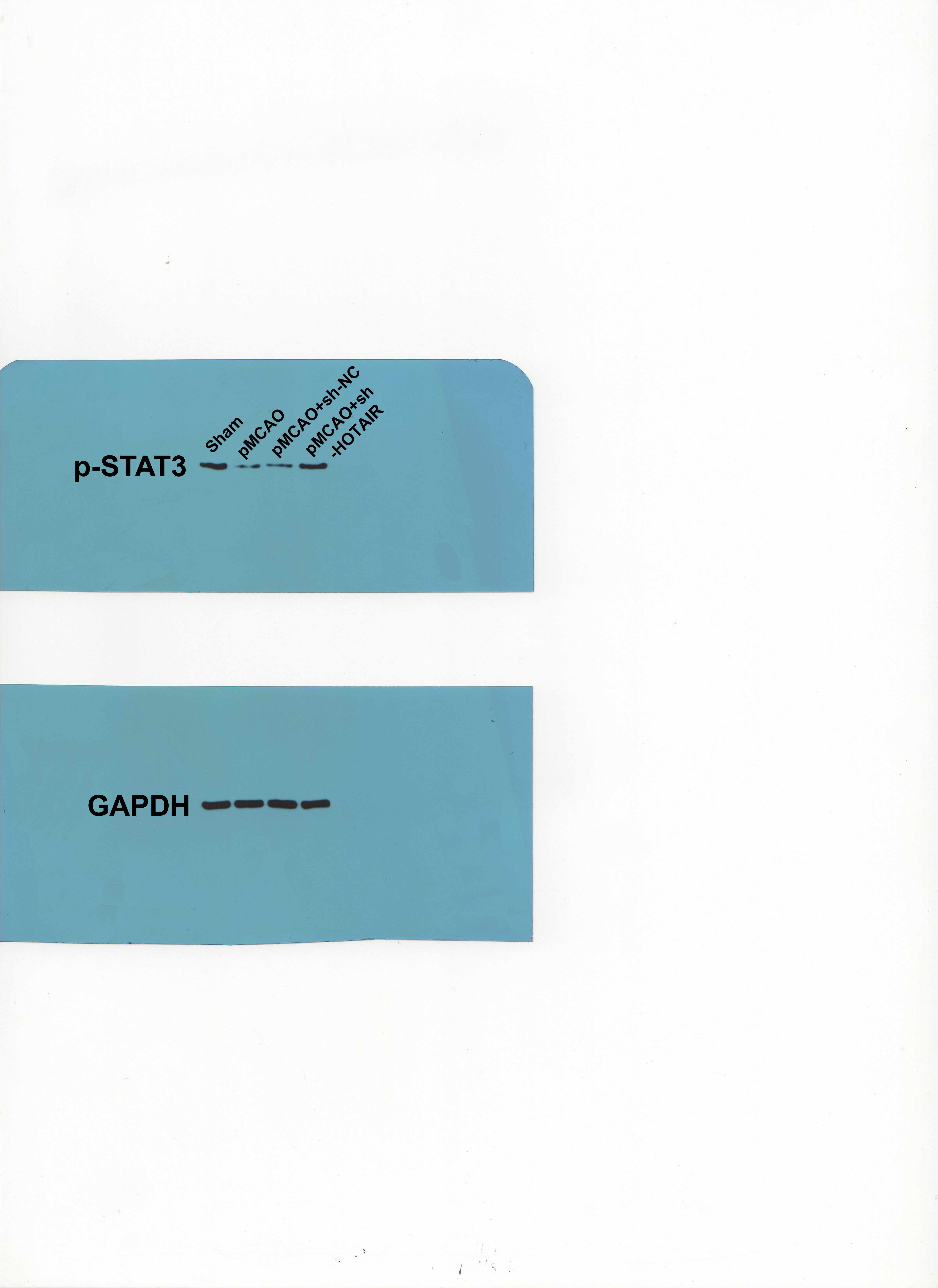
Mice were randomly assigned into 4 groups (N = 24 in each group): sham group, pMCAO group, pMCAO + sh-HOTAIR group, and pMCAO + sh-NC group. The low expression lentivirus shRNA HOTAIR (sh-HOTAIR) and its control (sh-NC) were constructed and packaged by GenePharma (Shanghai, China). Lentivirus processing was applied 2 weeks before pMCAO operation. Mice were anesthetized and fixed in a stereotactic apparatus (RWD Life Science Co., Ltd, Shenzhen, China). The lentivirus was injected via the right lateral ventricle at the following coordinates: 0.3 mm posterior to bregma, 1 mm right lateral to sagittal suture, 2.2 mm deep to the dura. The 5 µL lentivirus was slowly injected at a rate of 1 µL/min using a Hamilton microinjector, and the needle was retained in place for 5 min following the injection. The titer for sh-HOTAIR and its control were 3 × 10⁸ CFU/mL 19.
Neurobehavioral evaluation
Mice were evaluated for neurological function after 24 h of pMCAO modeling based on the modified neurological severity score (mNSS) 20 in a double blind manner from perspectives of movement, sensation and reflex. Evaluation standards were: motor tests (1–9 points) including raising mice by the tail, placing mice on the floor and abnormal movement; sensory tests (1–2 points) including placing test (visual and tactile test) and proprioceptive test; reflex tests (1–3 points) including pupil reflex, corneal reflex and startle reflex. The mNSS score was divided into: minor injury (1–4 points), moderate injury (5–9 points) and severe injury (10–14 points). Higher score meant severer deficit.
Beam balance test
Beam balance test was applied to observe the ability of coordinating muscles and keeping balance of mice 20. The wooden beam (length 1 m, width 5 mm, diameter 15 mm, horizontally positioned 1 m above the underlying surface) was supported by platforms at both sides. The mouse was placed at the start of the beam and scored based on its performance on the beam. The three-day trial was performed twice a day with a 5 min interval. The average value was taken as the experiment result. Performance on each trial was scored as follows: reach the platform or balance in steady posture, 0 point; grasp one side of the beam, 1 point; hang the beam and one limb falls off the beam, 2 points; hang the beam and two limbs fall off the beam or rotate on the beam for over 60 s, 3 points; try to keep balance on the beam for over 40 s but fall, 4 points; try to keep balance on the beam for over 20 s but fall, 5 points; fall with no intention to keep balance, 6 points. Higher score meant worse balance ability.
Morris water maze test
Water maze test was applied to evaluate the learning-memory, spatial location and orientation capability of mice 21. Titanium dioxide (Shanghai Jianghu Titanium Dioxide Chemical Industry Co., Ltd, Shanghai, China) was added into the water at concentration of 125 g/L. Morris water maze system (Mobile Datum, Shanghai, China) was applied for animal behavior analysis and water temperature was kept at 25°C. The tests included two parts: orientation navigation experiment and space exploration experiment. In the orientation navigation experiment, mice were placed in the water and the time (s) to find the platform was recorded (escape latent period). If the mice were unable to find the platform within 60 s, the experimenter guided the mice to the platform and time was recorded as 60 s. Each mouse was trained for 4 times per day with 15–20 min interval for 4 days. In the space exploration experiment, on day 5, the platform was removed. Mice were placed in the water randomly and swam for 60 s. The time of the mice crossing the original platform was recorded.
2,3,5-triphenyltetrazolium chloride (TTC) staining
Mice were euthanized by injection of 800 mg/kg pentobarbital sodium after beam balance test. Brain was taken out and made into continuous slices at 2 mm. The slices were incubated in 2% TTC solution (2530-85-0, Guidechem, Shanghai, China) at 37°C in the dark for 30 min. Normal brain tissues showed a pink or red color, and infarcted tissues were in light grey. TTC-stained slices were photographed using a digital camera. The cerebral infarcted area was calculated using Image J (National Institutes of Health, Bethesda, MD, USA) software.
TdT-mediated dUTP Nick-End Labeling (TUNEL)
The brain tissues were cut into sections at 5 µm and dewaxed by xylene and dehydrated with ethanol, and treated according to the instructions of TUNEL kit (C1089, Beyotime, Shanghai, China). Briefly, 20 µg/mL protease K working solution (Solarbio, Beijing, China) was added at room temperature for 15 min. The sections were washed using phosphate buffered saline (PBS) for 3 times, and then added with 50 µL TUNEL detection solution and incubated in the dark for 60 min. After being washed by PBS for 3 times, the samples were added with primary antibody NeuN (ab177487, Abcam Inc., Cambridge, MA, USA) and the Alexa Fluor® 488-labeled secondary antibody (ab150077, Abcam). Then sections were counterstained using 4,6-diamidino-2-phenylin-dole (DAPI). The images were obtained by a fluorescence microscope (Leica, Wetzlar, Germany). The apoptosis rate = apoptotic cells (red)/total cells (blue)∗100%.
Oxygen–glucose deprivation (OGD) model establishment
N2a cells (ATCC, Manassas, VA, USA) were cultured in Dulbecco's modified Eagle's medium (DMEM) (A4192101, Gibco, Grand Island, NY, USA) + 10% fetal bovine serum (FBS) (Thermo Fisher Scientific, Shanghai, China) in 5% CO2 incubator at 37°C.
The in vitro OGD model was established as previously described 22. Cells in good growth conditions were cultured in glucose, serum and oxygen free DMEM with 95% N2 and 5% CO2 at 37°C for 3 h. The cells were washed with PBS and then added with 10% FBS high glucose medium and cultured with 5% CO2 at 37°C for the following experiments.
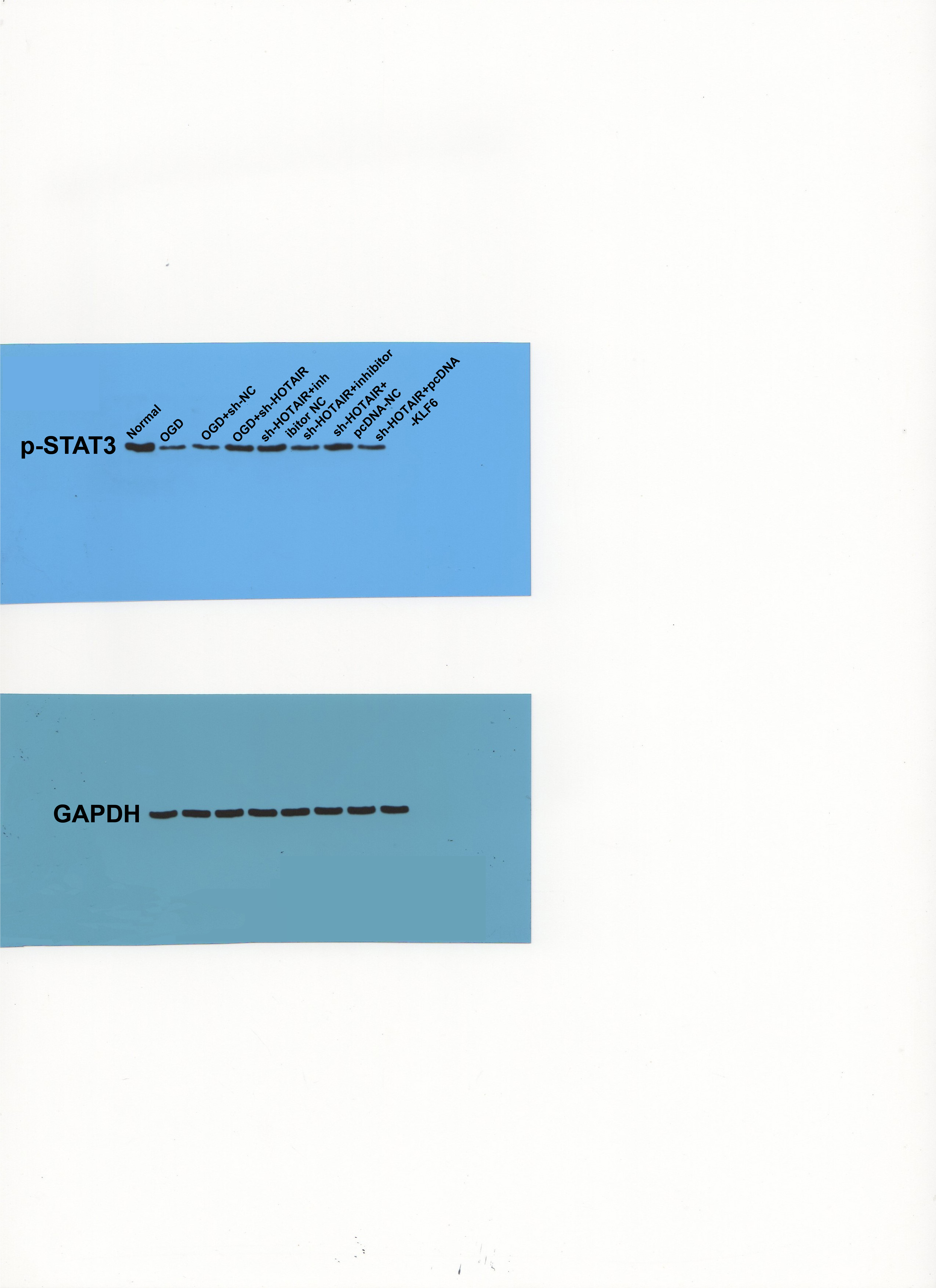
The design and synthesis of miR-148a-3p inhibitor, inhibitor NC and pcDNA3.1-KLF6, and pcDNA3.1-NC were processed by RiboBio Company (Guangzhou, China). The cells were transferred in accordance with the instructions of Lipofectamine 3000 (Thermo Fisher Scientific). For HOTAIR lentivirus treatment, cells in plates at a density of 5×10⁶ cells/well were cultured for 24 h and then cultured in 2 mL fresh complete medium containing 10ug/mL polybrene for 1h at 37°C. Then 15 mL lentivirus with a multiplicity of infection (MOI) of 30 was added to the cell suspension and incubated for 6 h. The medium was then replaced with equal volume of fresh medium again and the cells were incubated for further experiments 23. Cells were assigned into normal, OGD, OGD + sh-NC, OGD + sh-HOTAIR, OGD + sh-HOTAIR + inhibitor NC (sh-HOTAIR + inhibitor NC), OGD + sh-HOTAIR + miR-148a-3p inhibitor (sh-HOTAIR + inhibitor), OGD + sh-HOTAIR + pcDNA3.1 NC (sh-HOTAIR + pcDNA-NC), OGD + sh-HOTAIR + pcDNA3.1-KLF6 (sh-HOTAIR + pcDNA-KLF6).
Cell counting kit-8 (CCK-8) assay
Cells from each group were extracted and made into single cell suspension and seeded in 96-well plates (1×10⁴ cells/well) with 100 µL per well for 24 h. CCK-8 solution (Dojindo Molecular Technologies, Kumamoto, Japan) was added (10 µL/well) into each well, and the cells were incubated for 2 h at 37°C in a 5% CO2 incubator. The absorbance was detected using an EnVision multimode plate reader (Plate Reader, PerkinElmer, Waltham, MA, USA) at 450 nm and relative cell viability was calculated.
Flow cytometry
The cells were detached by trypsin. A total of 1 × 10⁵ cells/mL suspended cells were harvested and detected using Annexin V-FITC cell death detection kit (BD Bioscience, Franklin Lakes, NJ, USA). First, 100 µL 1× Annexin buffer was used to suspend cells. Then 5 µL Annexin V-FITC and 1 µL propidium iodide were added to stain the cells for 15 min at 4°C without light exposure. Apoptosis was then detected on a flow cytometer (FACScan, BD Biosciences).
Fractionation of nuclear and cytoplasmic RNA
The PARIS™ kit (#AM1921, Thermo Fisher Scientific) was applied for fractionation of nuclear and cytoplasmic RNA to detect expression of lncRNA HOTAIR in nucleus and cytoplasm of mouse N2a cells. The expression of HOTAIR was analyzed by RT-qPCR.
Florescence in situ hybridization (FISH)
Firstly, online software LncATLAS (http://lncatlas.crg.eu/) was applied to predict the distribution of HOTAIR. Then, FITC-labeled HOTAIR was designed and synthesized by GenePharma. Nuclei were stained with DAPI. All processes were conducted using the FISH kit (GenePharma) following the manufacturer’s instructions. Images were obtained by a FV3000 microscope (Olympus, Tokyo, Japan).
Bioinformatics analyses
HOTAIR related miRs were retrieved using starBase database (starbase.sysu.edu.cn/) and RAID database (http://www.rna-society.org/raid2/index.html). IS expression chip GSE35338 of mouse samples was obtained through GEO database (https://www. ncbi.nlm.nih.gov/geo/), including 10 normal samples and 11 disease samples. The “limma” package in R language was used for differential analysis. False discovery rate (FDR) was used to rectify P value, and differentially expressed genes from IS mouse model were screened out with |logFC|>1 and adj.p.val < 0.05 as criteria. The miR-148q-3p target gene in human and mice was predicted using TargetScan database (http://www.targetscan.org/vert_71/) and binding sites were obtained. Interaction analysis of 11 candidate genes was performed using GeneMANIA database (http://genemania.org/), and then interaction scores were obtained.
Dual-luciferase reporter assay
The potential binding sites of lncRNA HOTAIR and miR-148a-3p were predicted using RNA22 v2 (https://cm.jefferson.edu/rna22/Interactive/) 24. The potential binding sites of miR-148a-3p and kruppel-like factor 6 (KLF6) were predicted using starBase (http://starbase.sysu.edu.cn/index.php) 25. The wild type (wt) and mutant type (mut) sequences of HOTAIR and KLF6 were cloned to pmiR-GLO luciferase vector (Promega, Madison, WI, USA) to construct HOTAIR-wt, KLF6-wt and HOTAIR-mut and KLF6-mut. HEK293T cells (Shanghai Institute of Biochemistry and Cell Biology, Chinese Academy of Sciences, Shanghai, China) were transferred with the constructed plasmids and mimic NC and miR-148a-3p mimic (GenePharma), respectively using lipofectamine 3000 (Thermo Fisher Scientific). Luciferase activity was detected after 48 h by the Dual-Luciferase Reporter Assay kit (Vazyme, Nanjing, China) following the manufacturer’s instructions.
RNA pull-down
The biotinylated mut or wt miR-148a-3p (GenePharma) or its negative control (NC-bio) was transfected into the cells. The cells were lysed using lysis buffer at 48 h post-transfection. Then, streptavidin agarose beads (Invitrogen, Carlsbad, CA, USA) were incubated with the cell lysates, and the RNA complex that bound to the beads was eluted and purified using TRIzol reagent (Takara, Shiga, Japan). Gene level in the RNA complex was analyzed by RT-qPCR 26.
Enzyme-linked immunosorbent assay (ELISA)
The homogenate or cell suspension from each group was centrifuged at 4°C for 10 min at 1,000 g and the supernatants were collected. Protein contents of tumor necrosis factor-α (TNF-α) (MTA00B), interleukin (IL)-6 (M6000B) and IL-10 (M1000B) were measured using ELISA kits (R&D Systems, Minneapolis, MN, USA).
RT-qPCR
Total RNA and miRs were extracted separately from mice brain tissues and cells with TRIzol reagent and miRNeasy Mini Kit (Invitrogen). cDNA was synthetized using M-MLV reverse transcriptase (Invitrogen) or TaqMan MicroRNA Reverse Transcription Kit (Thermo Fisher Scientific). RT-qPCR was performed using SYBR Green Real-Time PCR Master Mix (Thermo Fisher Scientific) or TaqMan miRNA assay kit on Applied Biosystems7900 Real-Time PCR System. GAPDH and U6 were used as internal controls. Relative expression levels were calculated using the 2−△△ct method. Amplified primer sequences are illustrated in Table 1.
Table 1
|
Gene
|
Forward 5’-3’
|
Reverse 5’-3’
|
|
LncRNA HOTAIR
|
CAGGACGCTGTTCTTGTTGA
|
CGGAACAGCTACTCCTCCTG
|
|
miR-148a-3p
|
TCAGTGCACTACAGAA
|
CAGTGCGTGTCGTGGAGT
|
|
KLF6
|
ATGAAACTTTCACCTGCGCT
|
TCAGAGGTGCCTCTTCATGT
|
|
U6
|
CGCTTCGGCAGCACATATAC
|
AAATATGGAACGCTTCACGA
|
|
GAPDH
|
ATGGCGTAGCAATCTCCT
|
TTACTCCTTGGAGGCCAT
|
| Note: LncRNA HOTAIR, long-non-coding RNA HOX transcript antisense intergenic RNA; miR, microRNA; KLF6, kruppel-like factor 6; GAPDH, glyceraldehyde-3-phosphate dehydrogenase. |
Western blot
Total proteins from homogenate and cells were extracted with radio immunoprecipitation assay (RIPA) lysates (Beyotime). Proteins were quantitatively detected by a bicinchoninic acid kit (Thermo Fisher Scientific). Extracted protein was denatured at 100°C for 5 min. A total of 50–100 µg protein was separated by sodium dodecyl sulfate-polyacrylamide gel (SDS-PAGE), and then proteins were transferred onto polyvinylidene fluoride (PVDF) membranes and incubated with 5% skim milk for 2 h, followed by Tris-buffered saline Tween (TBST) washing for 3 times. The primary antibody p-STAT3 (1:20000, ab76315, Abcam) or GAPDH (1:1000, ab9485, Abcam) was added and the membranes were incubated overnight at 4°C. After washing in TBST, the secondary antibody Goat Anti-Rabbit IgG H&L (HRP) (1:5000, ab97051, Abcam) was added and the membranes were incubated for 2 h at 37°C and detected using chemiluminescent method. Grey value analysis was performed using Image J (National Institutes of Health).
Statistical analysis
Data analysis and drafting were performed using SPSS 21.0 software (IBM Corp. Armonk, NY, USA) and GraphPad Prism8.0.1 (GraphPad Software Inc., San Diego, CA, USA). Data were in normal distribution and presented as mean ± standard deviation (SD). Differences between groups were analyzed using one-way analysis of variance (ANOVA) or two-way ANOVA, followed by Tukey's multiple comparisons test or Sidak's multiple comparisons test. P value was obtained using a two-sided test. P < 0.05 was considered a statistically significant difference.