Materials
MgO NPs (average particle size 20 nm and 99% purity) was purchased from US Research Nanomaterials, Inc. Film grade LDPE resin pellets (LF0200, MFI 2 g/10 min, density 0.92 g/ml and softening point 94 ºC) were obtained from Bandar-Imam Petrochemical Co., Iran. Commercial ε-poly-L-lysine powder (Epolyly TM, Belgium, 50% w/w) was purchased from Hendry Co, Belgium and prepared at concentrations of (500, 1000, and 2000 µg/ml) by dissolving it in distilled water. The solutions were sterilized through a 0.22 µm filter and stored at 4°C.
Bacterial Culture
E. coli O157:H7 (ATCC 135218) and L. monocytogenes (ATCC 191181) were obtained from the Department of Food Hygiene and Public Health, School of Veterinary Medicine, Shiraz University, Iran. Stock cultures of E. coli O157:H7 and L. monocytogenes were prepared by inoculating in tryptic soy broth (TSB, Merck, Germany) and incubating at 37°C for 24 h. Then, they sub-cultured individually on MacConkey agar (Merck, Germany) and Palcam agar (Merck, Germany), respectively. A single colony from plates of bacteria transferred into TSB and incubated at 37 ºC for 24 h. The optical density (OD) of the bacterial suspensions was adjusted to the standard of McFarland No. 0.5 (1.5×10⁸ CFU/ml) by adding sterilized normal saline.
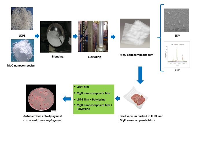
Preparation of MgO NC film
The combination of MgO NPs powder and LDPE resin pellets (10: 90) were directly blended for 1 h. The mixture was introduced to a twin screw extruder (Brabender, Germany) to be cut into masterbatch nano-granules. The heating profile was set to six zones of the extruder including 160, 160, 175, 150, 150, and 140 ºC. For production of MgO NC film proper amounts of masterbatch were added to pure LDPE resin pellets into a single-screw blowing machine (Brabender, Germany) at 190 ºC in the two barrel zones. MgO NC films were prepared in concentrations of 5 and 10% W/W MgO NPs with a thickness of 60 µm [6, 31–33]. Pure LDPE resin pellets without MgO NPs were analogously prepared and used as a control film.
Characterization Of MgO NC Film
The X-ray diffraction (XRD) pattern of the MgO NC film was obtained with X-ray diffractometer (D8 Advance, Bruker AXS, US) in the range of 2ɵ values from 2 º-40 º and λ = 1.54 ºA. The morphology of MgO NC films was characterized by a scanning electronic microscope (SEM, Cambridge Stereo Scan, Leica, Germany). For this work, films were protected with a thin gold layer using a sputter coater. SEM images were taken at an acceleration voltage of 15 kV. [32].
Enumeration of E. coli O157:H7 and L. monocytogenes in the culture media (In vitro)
Effect of MgO NC film (10 % w MgO NPs) and LDPE film (control) with and without polylysine on survival of E. coli O157:H7 and L. monocytogenes were evaluated [34–37]. MgO NC and LDPE films were cut 2×2 mm2 cubes and sterilized with 70% ethanol and dried up in air. 200 mg of sterilized film cubes were immersed in 4.5 ml filter sterilized TSB and/or TSB containing 2.5, 5.0 and 10.0 mg polylysine for alone and combined treatments, respectively. Then, 0.5 ml of E. coli O157:H7 or L. monocytogenes suspensions (105 CFU/ml) was inoculated to each flask. Final concentration of polylysine was 500, 1000 and 2000 µg/ml.
The flasks were incubated at 37°C in shaking incubator at 100 rpm for 5 days. Bacteria were enumerated on days 0, 1, 2, 3, 4, and 5 of incubation. Ten fold serial dilutions of samples were made and the surface plated on MacConkey agar and/ or Palcam agar, respectively. The plates were incubated at 37°C for 24–48 h. The bacterial colonies were enumerated and expressed as log CFU/ ml, and their mean value was recorded. All treatments were done in three independent replicate.
Enumeration of E. coli O157:H7 and L. monocytogenes in fresh beef (Invivo)
Freshly cattle muscles (Longissimus dorsi) were prepared from a slaughterhouse and stored in 4°C for overnight. To sterilize surface of the cylindrical beef, it was immersed in boiling water
for 10 seconds, and after removing the cooked sections with a sterile knife under aseptic conditions, they were cut in a transverse direction in size 2×5×5 cm. Two slices were evaluated for the likelihood of E. coli O157:H7 and L. monocytogenes [ 38]. The other slices were placed on a sterile tray in a biological safety cabinet, and the upper surface of each beef slice was inoculated with 100 µl of selected bacterial suspensions (106 CFU/ml) to obtain an initial bacterial count about 1× 10⁵ CFU/sample, and spread evenly over the surface of beef using sterile spreaders. Samples were held for 10 min to allow sorption of the tested bacteria. For polylysine treatments, 1 ml of polylysine solutions (500, 1000 and/ or 2000 µg/ml) was poured to the surface of the treated beef and spreaded. Distilled water was used for control samples instead of polylysine. Afterward, 10×10 cm of ethanol sterilized MgO NC film and/ or LDPE film (as control) was put on the top of each inoculated beef slice. Then they were inserted into polyethylene bag (thickness: 85 micron, density: 1.1 g/ ml) and vacuum packed using a table vacuum machines (Webomatic D-44866, Germany) at 99 % vacuum. The vacuum packed samples were stored at 4 ºC for 20 days and were evaluated on days 0, 5, 10, 15, and 20 of storage.
At each selected time, three samples of each treatment were weighted and homogenized by a stomacher with 9 times normal saline for 2 min. Serial decimal dilution was prepared and appropriate dilutions were plated on the selective media. The plates were incubated at 37 ºC for 24–48 h. The bacterial colonies were enumerated and expressed as log CFU/sample [34, 39, 40].
Measurement of MgO migration from MgO NC film to beef
For migration study, the 10×10 cm slice of beef samples were weighted and vacuum packaged with the MgO NC film and/ or LDPE film in size 10×10 cm and stored for 20 days at 4°C. For digestion, 0.4 g of homogenized beef samples with 3 ml of HNO3 (65%) and 1 ml of H2O2 (30%) poured into the to vessels of microwave oven (Panasonic NN-ST757W, China). After cooling down the vessels, the digested solution was diluted with deionized water and analyzed by inductively coupled plasma mass spectrometry (ICP-MS) (Vista PRO, US). The control was prepared under the same conditions, containing all reagents except the beef. The calibration of ICP-MS was performed using an external Mg standard solution[37, 41].
The amount of migration (µg/ cm2 of MgO NC film) was calculated from the difference in MgO content of the beef samples covered by MgO NC film from the beef samples covered by LDPE film.
Statestical Analysis
The results were analyzed by variance analysis (ANOVA) using the SPSS software (version 16). The differences between means were compared through the Duncan test at a level of 5% of significance.