Chemicals
PCR reagents, T4 DNA ligase, and restriction endonucleases were obtained from Promega (Madison, WI, USA). Isopropyl-β-D-thiogalactopyranoside (IPTG), lysozyme, DOPA, mushroom tyrosinase (m-tyrosinase), BCN, DBCO, Doxorubicin hydrochloride (Doxo), Dulbecco's Modified Eagle Medium (DMEM) high glucose, Fetal Bovine Serum (FBS), Antibiotic Antimycotic solution (100X), Trypsin-EDTA solution and most of the chemicals required for these experiments were procured from Sigma (St. Louis, USA). The host bacterium Escherichia coli (E. coli) strain XL1-blue (Strata gene, USA) was used for plasmid DNA preparation. The nickel nitrilotriacetic acid (Ni-NTA) affinity column was purchased from clone tech (Takara Bio, USA). E. coli strain JW2581 Tyr auxotroph was obtained from E. coli Genetic Stock Centre (Yale University, USA) and plasmid pQE-80L was obtained from Qiagen (Valencia, USA).
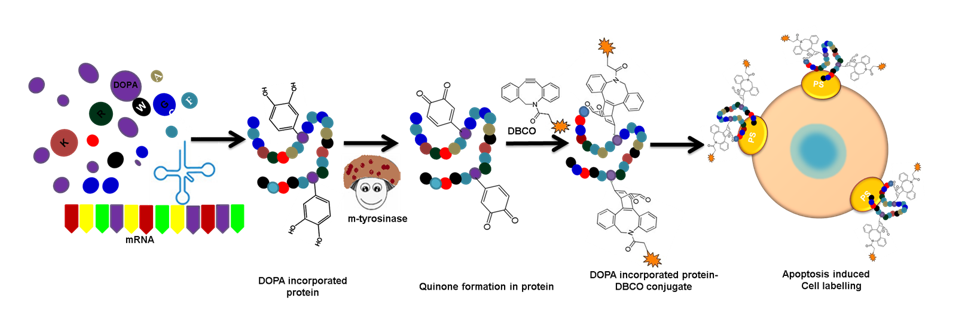
Residue-specific incorporation of DOPA
Residue-specific incorporation of DOPA was carried out in E. coli JW2581 containing pQE80L-anX and GFP vector as described by George et al [27]. The cells harboring pQE80L-anX and GFP plasmids for residue-specific incorporation were grown in LB broth containing ampicillin and incubated overnight at 37 ºC. The cultures were collected after centrifugation at 5000 rpm, 4 ºC, 15 mins and 1 % inoculum were added into 20% glucose minimal media (MM) supplemented with 20 amino acids (40 mg/L), 1 mM CaCl2, 1 mM MgSO4, thiamine HCl (1 mg/mL) and ampicillin (100 µg/mL). Subsequently, cells were centrifuged at 5000 rpm for 15 min and washed thoroughly in 1X phosphate buffer saline (1X PBS, pH = 7.4), and further cells were inoculated into the same 20% glucose MM supplemented with 19 amino acids (40 mg/L) except Tyr, 0.03 mM Tyr, and ampicillin (100 µg/mL). The cells were grown to mid-log phase (OD600 = 0.6-0.8) at 37 ºC and 180 rpm. At this point, 1 mM of DOPA was added to the culture flask before IPTG induction. The optimum expression of DOPA incorporated anX and GFP was achieved upon 1 mM IPTG induction for 6 h at 37 ºC. As a result, dihydroxyphenylalanine incorporated Annexin A5 (anX-DOPA) and dihydroxyphenylalanine incorporated Green Fluorescent protein (GFP-DOPA) were obtained from residue-specific incorporation methods. Then, the cells were harvested and frozen at -80 ºC for further experiments.
Cycloaddition of BCN with DOPA
50 µL of DOPA stock solution (10 mM) was treated with 60 µL of tyrosinase (60 U) in Tris buffer, pH 6.8, and mixtures were incubated at room temperature (RT) in dark condition for 5 min. Subsequently, 100 µL of 2.5 molar excess of BCN stock solution (10 mM) in a 1:1 ratio of methanol and MilliQ (MQ) water was added to the tyrosinase treated DOPA and the reaction was carried out in dark condition at RT for 2 h. Then the reaction mixture was filtered through a 0.22 µm Millipore filter device (Millipore, USA). The tyrosinase-treated DOPA with BCN and tyrosinase-treated DOPA without BCN was subjected to Ultraviolet-Visible (UV/Vis) spectroscopy.
Mass Spectrometric Analysis (HR-MS)
Tyrosinase catalyzed dihydroxyphenylalanine conjugated with bicycle [6.1.0] non-4-yne (DOPA-BCN) reaction mixture was lyophilized and dissolved in 1 mL of HPLC grade methanol and was thoroughly filtered using Millex-syringe driven filter unit. Then, 10 µL of samples (DOPA and DOPA-BCN) were injected into the High-resolution mass spectrometry (HR-MS) (Finnigan LCQ advantage max, Thermo Fisher Scientific) equipped with a syringe pump.
Preparation of protein conjugates
The conjugation of fluorescein isothiocyanate with recombinant Annexin A5 (anX-FITC) was prepared by incubation of protein with Fluorescein isothiocyanate (FITC) at 37 °C in the dark for 1 h as described earlier [27]. Initially, 100 µg of FITC was dissolved in Dimethyl sulfoxide (DMSO), and it was sequentially added to 1 mg of protein in 1X PBS at pH 7.4. Eventually, an tyrosinase catalyzed dihydroxyphenylalanine incorporated Annexin A5 conjugated with Dibenzocyclooctyne-PEG4-Fluor545 (anX-DOPA-DBCO) conjugate was prepared by the treating 100 µl of anX-DOPA (0.5 mg) with 120 U of tyrosinase in Tris buffer pH 6.8 and incubated at RT for 5 min. At the same time, similar reactions were performed by mixing the tyrosinase treated anX-DOPA (quinone formation in protein) with DBCO (200 µM) in 0.1% DMSO for 2 h at RT. After labeling, anX-FITC and anX-DOPA-DBCO conjugate were separated from free FITC and DBCO by a 10 kDa cut-off filter (Millipore, USA). Both protein conjugates were excited using fluorescence spectrometric analysis.
Nitroblue Tetrazolium (NBT) Staining
The formation of dopaquinone and BCN conjugation with the tyrosinase-treated DOPA incorporated proteins (GFP-DOPA and anX-DOPA) was confirmed by NBT staining. The reaction mixtures were loaded into the Sodium dodecyl sulfate-polyacrylamide gel electrophoresis (SDS-PAGE) and the proteins were transferred onto the methanol-activated polyvinylidene difluoride (PVDF) membrane. Protein bands on the membrane were incubated in NBT staining solution (2 M sodium glycinate, 0.24 mM NBT, pH 10) in a dark room for 3 h. Subsequently, the membrane was washed with a sodium borate solution overnight for protein band visualization.
Matrix-Assisted Laser Desorption Ionization Time of Flight (MALDI-TOF) Analysis
The protein samples were subjected to MALDI-TOF analysis as described earlier [28] and were desalted by a C18 zip-tip (Millipore, USA). After desalting, protein samples were examined by MALDI-TOF with a sinapinic acid matrix on a Micro flex LT instrument (Bruker Daltonik, Germany). The MALDI-TOF was operated in a linear model to provide optimal resolution with a 25 kV extraction voltage and 13 kV reflectron potential.
Cell line and culture conditions
The Endothelial cell type (EA.hy926) cells were obtained from ATCC, Washington DC, USA. The cells were grown as a monolayer in Dulbecco's Modified Eagle Medium (DMEM) high glucose supplemented with 10% Fetal Bovine Serum (FBS) and 1% antibiotic antimycotic solution in humidified air with 5% CO2 and 37 °C.
Cell viability assay
The EA.hy926 cells were grown till 80-90% confluence and trypsinized using Trypsin-EDTA solution for 2 min [29]. After trypsinization, 2×10⁴ cells from the parent flask were cultured in a 96-well plate for 24 h and different concentrations of Doxo were diluted using serum-free DMEM high glucose-containing 1X antibiotic antimycotic solution and added with the cells for 24 h. After the indicated treatment period, the cells were incubated for 3 h at 37 °C in a culture medium containing 1 mg/ml of 3-(4,5-dimethylthiazol-2-yl)-2,5-diphenyltetrazolium bromide (MTT) in 1X PBS at pH 7.4. The blue MTT formazan precipitate was then dissolved in 100 μL of DMSO, and the absorbance was measured at 570 nm with a multi-well plate reader. The cell viability was expressed as the percentage of the absorption values in the treated cells relative to the non-treated (control) cells.
Fluorescence microscopy analysis
As described, the above cells (2×10⁴) were grown and Doxo (50 µg/mL) was used to induce apoptosis in a 100 µL medium. After the treatment, the cells were washed with 1X PBS, fixed with 4% paraformaldehyde for 10 to 15 min at RT, and washed with 1X anX binding buffer (0.2 µm sterile-filtered 0.1 M HEPES (pH 7.4), 1.4 M NaCl, and 25 mM CaCl2 solution). Then, the cells were incubated with 0.5 µg/mL concentrations of anX-FITC and anX-DOPA-DBCO with the addition of 1 µg/mL of 4′,6-diamidino-2-phenylindole (DAPI) and were diluted by 1X anX binding buffer at pH 7.4 and incubated for 15 min at RT (in dark). Thereafter, cells were washed with 1X anX binding buffer and sealed with a coverslip using glycerol mounting medium. The cells were then viewed by a 10 X objective lens under an inverted fluorescence microscope (Leica DMi8, Germany) using a three-color filter and the image was analyzed by Leica Application suite 4.7 (Leica, Munich, Germany).
Flow Cytometric Analysis
EA.hy926 cells (1×10⁵) were grown as described above and were collected by centrifugation at approximately 500 x g for 5-10 mins at RT (18-24°C) and washed by resuspending in 500 μL ice-cold 1X PBS and pelleted by centrifugation. Again, the cells were washed once with 1X anX binding buffer and were stained with anX-FITC, anX-DOPA-DBCO, anX-FITC+Propidium iodide (PI), and anX-FITC+anX-DOPA-DBCO. The staining solution was prepared as follows; working concentration of anX-FITC and anX-DOPA-DBCO (0.5 µg/mL), PI (5 µg/mL), and were diluted in 1X anX binding buffer at pH 7.4. Cells were resuspended in 100 µL of the staining solution and stained for 15 min at RT in the dark. Then, the cells were diluted in 1X binding buffer to a final volume of 400 µL and analysed by flow cytometric analysis (FCM) in BD FACS CantoII (BD Biosciences, CA) having three color filters. The labeled cells were excited according to the reported excitation wavelength and suitable emission filters were used to detect fluorescence signals. Relative change in the fluorescence was determined by FACS-DiVa software.