Chemicals and materials:
Doxorubicin and ε-Caprolactone were purchased from Aladdin co. (Shanghai, China). IR780 was purchased from Sigma-Aldrich co. (St. louis, USA), PEG was obtained from Peng Sheng Biological Co., Ltd (Shanghai, China). Poly(lactic-co-glycolic acid) (PLGA) and human serum albumin was obtained from Aladdin co. (Shanghai, China). MTT Cell Proliferation and Cytotoxicity Assay Kit was obtained from Beyotime (Shanghai, China).1×Phosphate buffer solution (PBS) and deionized water were used in the experiments. All C57BL/6 female mice (18-20 g) were obtained from Yangzhou University Medical Center. All other reagents were purchased from Nanjing Wanqing Chemical Glassware Instrument Company and used as received.
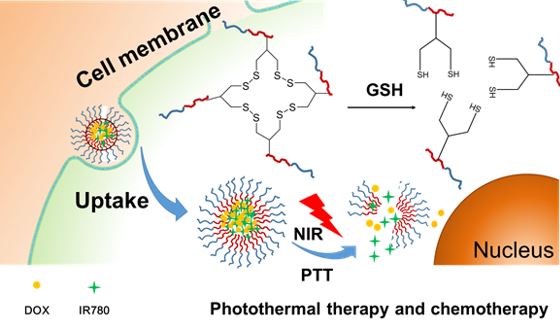
Synthesis of PCL-PEG-SS
We firstly synthesized HO-PCL-b-PEG114 with the terminal group of hydroxyl group. The specific synthesis steps are shown in Figure 1. The synthesis of HO-PCL-b-PEG114 is carried out by using PEG114-OH as an initiator and stannous octoate (Sn(Oct)2) as a catalyst to initiate ring-opening polymerization of monomer ε-caprolactone (ε-CL). Specifically, 2 g of dry treated CH3O-PEG114-OH (0.4 mmol) and 4 g of de-vaporized ε-CL (35.1 mmol) were weighed and dissolved in 10 mL of vacuum-distilled anhydrous toluene, followed by one drop of Sn(Oct)2. The liquid nitrogen was frozen, evacuated, purged with nitrogen, thawed, and cycled three times. After reacting for 24 h in a 110 °C oil bath, it was added dropwise to 500 mL of ice diethyl ether under stirring to precipitate. It was suction filtered, washed with diethyl ether three times, and dried in vacuo to get white solid HO-PCL-b-PEG114. The degree of polymerization of the PCL segment was calculated by nuclear magnetic resonance to be Dp=87. The block copolymer was HO-PCL87-b-PEG114. To synthesis PCL-PEG-SS, 0.5g HO-PCL87-b-PEG114 were dissolved in 20mL DMSO and thenadded 4-(dimethylamino) pyridine (DMPA, 0.2 g) solution in DMSO (3 mL) and lipoic acid anhydride (0.6 g) in DMSO (3 mL), respectively. The reaction was stirred for 48 h under nitrogen at 30 °C. The product was isolated by precipitation in cold ethanol, washed several times with ethanol, and dried in vacuo.
Preparation of DOX&IR780@PEG-PCL-SS NPs
After hydrophobization of doxorubicin, certain amount of DOX (1.0 mg), IR780 (1.0mg) and polymer PEG-PCL-SS (20mg) were dissolved in DMSO, and then added to PBS (pH7.4) under ultrasonic conditions. The solution was concentrated by ultrafiltration to remove free DOX and IR780. The drug loading was calculated by the drug amount in NPs, according to the standard curve of DOX and IR780 measured by UV absorption spectroscopy. Nanoparticles loaded with near-infrared photosensitizers were prepared by hydrophobic interaction of PCL and hydrophobic small molecules. The DTT was accurately weighed and the nanoparticles were reacted using DTT (10μL, 10mg/mL). Because DTT can partially hydrolyze the disulfide bond of the nanoparticles within a controllable range, the spatial structure of the nanoparticles after reflection is more compact. The nanoparticles loaded with photosensitizer and chemotherapeutic drugs were obtained by dialysis and concentration. As a control, non-crosslinked PEG-PCL NPs were prepared the same as PEG-PCL-SS NPs. To evaluate the delivery advantages of PEG-PCL-SS NPs, two other NPs (PLGA and Albumin) were prepared as described before (19-21). Briefly, The PLGA NPs were prepared in a solvent displacement process. DOX (1mg), IR780 (1mg) and 20mg PLGA first dissolved in DMSO (1mL). 1 mL of the solution was added dropwise to 10 mL of water. The mixture was then stirred in open air for 2 h. Then the solution was concentrated by ultrafiltration to remove free DOX and IR780. DOX and IR780-loaded Albumin nanoparticles were prepared via a molecular switch method as described previously (21, 22). The drug loading was calculated by the drug amount in NPs, according to the UV standard curve of DOX and IR780.
Characterization of DOX&IR780@PEG-PCL-SS NPs
Particle size and surface potential were measured by a Brookhaven BI-90Plus laser particle size analyzer. Transmission electron microscopy was used to observe the morphology of the nanoparticles. Samples were prepared by dropping a suitable concentration of the nanoparticle solution on a 200 mesh copper mesh and dried overnight.
Cell culture
Murine bladder carcinoma MB49 cells were obtained from Shanghai Institute of Biochemistry and Cell Biology, Chinese Academy of Sciences. The cell line was maintained in RPMI 1640 cell culture media supplemented with 10% fetal calf serum (Hyclone, Logan, UT) and antimicrobial-antimycotic (Gibco/Invitrogen, Carlsbad, CA) in a humidified incubator at 37 oC in an atmosphere composed of 5% CO2 . The cell line was transduced with the firefly luciferase gene by a lentivirus vector.
In vitro temperature curve
We prepared internal cross-linked polymeric nanoparticles containing IR780, internal cross-linked polymeric nanoparticles containing doxorubicin and IR780, and a PBS solution (pH7.4). The 808 nm near-infrared laser irradiation system was applied on the samples for 1-3 minutes, and the temperature probe measurement system was conducted. The photothermal effect of the IR780-containing nanoparticles was evaluated (50μg/mL IR780).
In vitro drug release
DOX and IR780 were encapsulated into the inner crosslinked polymeric nanoparticles. After dialysis, a small amount of organic solvent was removed. The dialysis bag filled with nanoparticles was placed in a PBS solution (pH7.4) with or without GSH (5mM or 10mM). The internal chemical structure of the internally crosslinked polymeric nanoparticles is changed when interacted with GSH, and the drugs arae continuously released from the drug-loaded nanoparticles. We then used near-infrared laser irradiation to irradiate the cross-linked polymeric nanoparticles. The near-infrared laser can promote the photothermal reaction of IR780 in the drug-loaded nanoparticles, and further promote the release of the drug from the nanoparticles. The drug content of nanoparticles in PBS (pH7.4) was measured at different time points (0 to 72h) under different experimental conditions, and the drug release curve was obtained.
Cellular uptake
MB49 cells were cultured for 24 h and then treated free doxorubicin solution (1.0μg/mL), free IR780 solution (1.0μg/mL), and drug-loaded nanoparticles containing doxorubicin and IR780for 6 h. The cells were washed with PBS twice and imaged by a fluorescence microscopy to determine the cellular uptake of the nanoparticles.
In vitro cytotoxicity
Different concentrations of doxorubicin-containing nanoparticles (DOX, from 0.11μg/mL to 1.775μg/mL), IR780-containing nanoparticles (IR780, from 0.078 to 1.25μg/mL), drug-loaded nanoparticles containing doxorubicin and IR780, and nanoparticles without any drugs were added to the cells. . After 24 hours, the cell viability and optimum concentration of the formulations was evaluated by MTT assays by measuring the absorbance of the solution at 570 nm.
In vitro therapeutic efficacy of DOX&IR780@PEG-PCL-SS NPs
Different concentrations of free doxorubicin solution, free IR780 solution, doxorubicin-containing nanoparticles, IR780-containing nanoparticles, doxorubicin and IR780 nanoparticles were prepared and added to the cells. The tumor cells were fully ingested with drugs or drug-loaded nanoparticles for 12hours, IR780 containing groups were irradiated with 808 nm near-infrared laser for 3 minutes, and then cultured for 24 hours. MTT assays were conducted with the same procedure described previously.
Establishment of an orthotopic bladder cancer model in C57BL/6 mice
All mice received care following the guidelines of the Care and Use of Laboratory Animals and their use followed the terms of the Institutional Animal Care regulations and Use Committee of Nanjing University. All animal experiments were approved by the Administration Committee of Experimental Animals in Jiangsu Province and the Ethic Committee of Nanjing University.
After anesthetizing C57BL/6 mice with 2% pentobarbital, the midline incision was taken to expose the bladder position of the mouse. After using the syringe to absorb the urine in the bladder, the MB49 bladder cancer cell suspension was injected into the bladder muscle layer. Small animal CT examination was performed at day 7 after successful surgery to observe whether there was a tumor in the bladder area.
Pharmacokinetics study
Pharmacokinetics study following single-dose intravenous injection was conducted in tumor-free male mice. The mice were randomly divided into PEG-PCL-SS NPs group, PEG-PCL NPs group, PLGA NPs group and Albumin NPs group (three mice per group). The formulations were administered via intravenous injection with the IR780 dose of 10 mg/kg. Blood samples were collected by retro-orbital bleeding at different time points (1 to 72 h) after administration. The content of IR780 in the serum samples was measured using a Varioskan Flash Spectral Scanning multimode plate reader (Thermo Fisher Scientific, Waltham, MA, USA). PK Solver Version 2.0, was used to calculate pharmacokinetic parameters from the plasma concentration versus time data (23).
In vivo biodistribution
The free IR780 solution and the nanoparticles loaded with doxorubicin and IR780 (0.3 mg IR780/kg body weight) were injected into the tumor-bearing mice by tail vein administration. Images were taken at 6, 12, 24 and 48 h after injection using the in vivo imaging system (IVIS Lumina XR III, USA). As a control, normal mice (without tumor) were injected with DOX&IR780@PEG-PCL-SS NPs and imaged at 6, 12, 24 and 48 h after injection using the in vivo imaging system. The mice were sacrificed at 48 h after injection, and the major organs including the heart, liver, spleen, lung, kidney and bladder tumor were collected for ex vivo imaging. The excitation wavelength of IR-780 is 745 nm and its emission spectrum is 780–900 nm.
Therapeutic efficacy of DOX&IR780@PEG-PCL-SS NPs using orthotopic bladder cancer model
The luciferase-expressing MB49 cell line was constructed and used for in vivo imaging of the tumor. Saline, doxorubicin-containing nanoparticles (4 mg/kg IR780), IR780-containing nanoparticles (2.5 mg/kg DOX), "drug-loaded nanoparticles containing doxorubicin and IR780" (2.5 mg/kg DOX, 4 mg/kg IR780) were injected into randomized mouse groups by tail vein administration. The experimental groups were irradiated with 808 nm near-infrared laser after 24 h, and repeated administration after one week. The mice were sacrificed after 3 weeks, and the heart, liver, spleen, lung, kidney and bladder of the mice were harvested for further study.
In vivo toxicity study
Polymeric nanoparticles loaded with different concentrations of doxorubicin (0mg/kg, 1.25 mg/kg, 2.5mg/kg and 5.0 mg/kg) and IR780 (0mg/kg, 1.25 mg/kg, 2.5mg/kg and 5.0 mg/kg) were synthesized and injected into the mice by tail vein administration. After 24 hours, the mice were sacrificed and blood samples were harvested by retro-orbital bleeding. ALT, AST, BUN, and Cr in the blood samples were immediately detected.
Histology
After the mice were sacrificed, the heart, liver, spleen, lung, kidney and bladder of the mice were harvested for hematoxylin and eosin (H&E) staining, the TUNEL assay and Ki67 immunohistochemistry. After dehydration and fixation, the sections were stained with H&E, and the bladder and tumor tissues were stained with TUNEL to observe the apoptosis and necrosis in the bladder cancer tissues.