Fermentation, extraction and purification of active compound from B. radicata
20% NaCl elution fraction from fermentation broth of B. radicata was named as SPAF by DEAE-cellulose column. The strongest antimicrobial activity fraction from SPAF was Griseococcin(1) by Sephadex LH-20 column. The UVmax of all the fraction was 215nm, the HPLC chromatograms of SPAF and Griseococcin(1) were shown in figure 1 (A~B). The chromatogram of B showed a single and symmetrical peak for Griseococcin(1) (fig1.B)
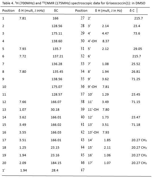
1D and 2D NMR of Griseococcin(1)
Griseococcin(1) was isolated as a white amorphous solid powder with the molecular formula of C37H43NO10 derived from the high-resolution electrospray ionization mass spectrum (HR-ESI-MS). The complete assignments for all protons and carbons were shown in Table 1. The 13C NMR spectra of Griseococcin(1) displayed signals of thirty seven carbons, including five carbonyl carbons (δC215.7–175.1), five aromatic/olefinic methine carbons (δC 128.86, δC215.7–175.1), seven non-protonated aromatic/olefinic carbons (δC 161.06-109.99), four methyl carbons (δ C20.27)), and four olefin carbons (δC 166.01). The 1H NMR spectrum of 1in D2O exhibited signals of four methyls at δ H 2.14 (3H, s, H-14’), δ H 2.12 (3H, s, H-15’), δ H 1.06 (3H, s, H-16’)and 1.07 (3H, s, H-17’), five aromatic protons δH 7.80 (1H, s, H-1), δH 7.93(1H, s, H-5), δH 7.72 (1H, s, H-6), δH 7.81 (1H, s, H-8) and 7.66 (1H, s, H-12)], four hydroxyl groups at δ H 8.37 (1H, br s, 4’-OH), δ H 7.81 (1H, br s, 9’OH) and δ H 7.80 (1H, br s, 11’-OH) and 9.63 (1H, br s, 13’-OH).
The structure of Griseococcin(1) was deduced by comprehensive analysis of 1H-1HCOSY, HMBC, and HSQC spectra (Fig.2A). In Griseococcin(1), the naphthoquinone substructure could be identified by the observation of HMBC correlations from H-8 (δH 7.80) to C-6 (δ C 137.21), C-4 (δC 138.60) and C-13 (δC 30.18), from H-1 (δH 7.81) to C-3 (δC 175.11), C-12 (δC 166.07) and C-1’ (δC 28.40), from H-5 (δH 7.93) to C-3 (δC 175.11) and C-9 (δC 138.56), from H2-13 (δH 1.07) to C-8 (δC 135.45) and C-6 (δC 137.21), from H3-14’ (δH 1.85) to C-2’ (δC 215.7) and C-4’ (OH) (δC 73.60), from H3-15’ (δH 2.11) to C-6’ (δC 215.70) and C-4’ (OH) (δC 73.60), from H2-7’ (δH 1.08) to C-9’ (δC 71.25) and C-13’ (δC 71.18). The 1H, 1H three-bond couplings observed in the COSY spectrum from H-8’ (δH 1.94) to H-9’ (δH 3.62) , from H-10’ (δH 1.29) to H-11’ (δH 3.49) , from H-12’ (δH 1.73) to H-13’ (δH 3.51), together with the chemical shifts of the 13C resonances (C-8’-13’) observed at alternating higher and lower fields, revealed the presence of cyclohexane with alternating hydroxyl and methyl groups. 1H-1H COSY correlations from H2-13 (δH1.07, m) to H2-14 (δH3.62, m), from H2-14 (δH3.62, m) to H2 -15 (δH 3.49, m) and from H2-16 (δH 3.55, m) to H2 -17 (δH 3.51, m) and HMBC correlations from H2-13 (δH 1,07, m) to C-15 (δC 166.02), from H2-14 (δH3.62, m) to C-16 (δC 166), from H2-15 (δH3.49, m) to C-17 (δC 166.01) and from H2-16 (δH 3.55, m) to C-18 (δC 23.15) identified coupled olefins. The key HMBC correlations from H2-1’ (δH1.94, m) to C-3’ (δC 23.4), from H -3’ (δH2.14, m) to C-5’ (δC 29.05), from H3-14’ (δH1.85, m) to C-2’ (δC 215.7)and C-4’-OH (δC73.6), from H3-15’ (δH2.11, m) to C-6’ (δC 215.7) and C-4’-OH identified two meta position carbonyl group and one ortho position hydroxyl group (Fig
2B).
This connectivity was also secured by the observation of the HSQC correlations from H3-14’ to C-3’ and from H3-15’ to C-6’. Therefore, the complete structure of naphthoquinone was determined as shown in Fig 2C.
Physico-chemical characterization of Griseococcin(1)
Griseococcin(1) was white powder and its solubility was 0.063 g/ml in water. It could be slight soluble in methanol and DMSO, but insoluble in n-hexane, dichloromethane, chloroform, ethyl acetate and acetone.
The FT-IR spectrum of Griseococcin(1) showed (Fig. 3) an intense and broad characteristic absorption peaks at 3417.2 cm−1 represented the stretching vibration of O–H. The weak absorption peaks at 2356 and 2925.5 cm−1 were resulted from the stretching vibration of C–H. The absorption bands at 1637.4 and 1618.1 cm−1 are due to the vibration of C=C and C=O in the ester group. The absorptions peaks at 1456.1, 1414 and 624 cm−1 were attributed to the presence of an internal C–H deformation. The strong absorption peak at 866 cm−1 was resulted from aromatics. In conclusion, the typical absorption peak indicated that Griseococcin(1) was naphthoquinone with group O–H,C-H,C=C,C=O and so on [23].
In Vitro antagonistic assay
Griseococcin(1) was assessed for antimicrobial and microbicidel activity against selected Trichophyton rubrum(ATCC 28188), Trichophyton mentagrophytes(ATCC 9533), Epidermophyton floccosum(ATCC 52066), Candida albicans (ATCC 10231), Staphylococcus aureus (ATCC 6538), Bacillus subtilis (ATCC 6051), Escherichia coli (ATCC 8739) and Pseudomonas aeruginosa (ATCC 27582) The results were shown in table 1, it displayed strong antifungal activity against T. rubrum, T. mentagrophytes with ZOI values of 18.06±0.85, 15.01±1.02 mm, as compared to the positive control with ZOI= 20.67 ± 1.58, 28.33 ± 2.15 mm, respectively. While antibacterial activity was weak.
J.Meletiadis et al reported that compounds were considered bactericidal or fungicidal when the MBC/MIC or MFC/MIC ratio is ≤4 [24]. In this study, it was inportant to discern whether the Griseococcin(1) possesses bactericidal and fungicidal activities. The fungicidal activities of Griseococcin(1) were assessed as MIC, MFC and MFC/MIC. The results were shown in Table 2. Griseococcin(1) showed the high fungicidal activities by means of lowest values of MIC and MFC against the four fungi, especially for main pathogenic fungi (T. rubrum ), the MIC, MFC and MFC/MIC values were 31.2±2.7, 31.2±3.1 μg/ml and 1, while MIC, MFC and MFC/MIC values of Terbinafine were 15.6 ± 1.2, 31.2 ± 1.6 μg/ml and 6. Fungicidal activities of Griseococcin(1) are revealed more effective than that of commercial reagents (Terbinafine).
Griseococcin(1) also showed high bactericidal activities with MIC, MBC and MBC/MIC values ranged between 62.5~125 μg/ml, 125-500 μg/ml and 2-4 against examined bacteria(S. aureus, E. coli and P. aeruginosa). The results were shown in table 3. Griseococcin(1) showed the highest bactericidal activity for S. aureus and E. coli,. MBC/MIC value of Griseococcin(1) was 2.0, while the MBC/MIC ratio was 3.0 and 4.0 for positive control (Gentamicin sulfate).
Due to side effects and the continuous drug resistance, commercial reagents might be unsafe for patients [11], Therefore, the development of fungicidal therapies is crucial, above results (MIC,MFC or MBC and MFC/MIC or MBC/MIC) add more value to Griseococcin(1)