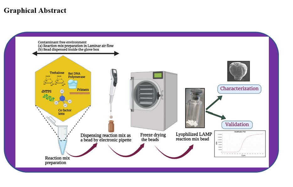
In this study, the process of making bioactive freeze-dried LAMP beads was developed to establish knowledge about long-term storage with the desired result. The lyophilized beads will be used as reagents inside a Lab-on-a-chip platform in an automated analysis system. The purpose is to carry out on-site environmental monitoring of rivers and lakes using LAMP, and to detect the eDNA from Esox lucius (regionally alien). The specificity and sensitivity of the designed Esox lucius LAMP primers, which were used in the lyophilized beads, have been previously determined [24]. Here, some of the characteristic properties of the freeze-dried beads were examined. Three trehalose concentrations: 10,15 and 20 % (w/v), were used in the LAMP mixture beads (Fig. 1). Several batches of 5 µL lyophilized beads were stored over time, in separate sealed glass ampoules both at room temperature 20 °C and at 4 °C in a refrigerator.
Surface Dimensions and Morphology
A scanning electron microscope was used to observe the objects in the micron to nano level range. The prepared beads were subjected to SEM observation and their dimension was marked and surface morphology observed (Fig. 2 (i) and (ii)). The physical shape of the 5 µL beads was found to be round to oval shape, depends on the needle tip and dispensing time. The dimension of the lyophilized beads was found to be in the range of 1.1mm to 1.8 mm among the three trehalose concentration beads (Fig. 2 (i)). The surface nature of the lyophilized LAMP mixture beads was examined at higher magnification, revealing the dense and porous nature, various nanofiber formations connecting the voids of the beads, as clearly seen (Fig. 2 (ii)). It was noticed that a trehalose concentration of 10 % in the LAMP mixture beads readily absorb moisture and shrinks immediately, while the increasing concentration of 15 % and 20 % were more stable when exposed to atmospheric air. In this context, the shrinkage pattern of the surface is seen for the 10 % trehalose- LAMP mixture bead, a dense structural surface with a porous nature is visible for both the 15 % trehalose-LAMP mixture bead and 20 % trehalose-LAMP mixture bead. The freeze-dried LAMP beads, regardless of the trehalose concentration, appeared in similar, yet the surface morphology varied between the different beads, and this was the case both with the same and with different trehalose concentrations. It was of interest to investigate the possible difference that could be linked to the morphology of the freeze-dried beads and to the formation of varieties of networks through glycosidic linkage, based on the difference in concentrations of the trehalose sugar. It was found that the different structure of the freeze-dried LAMP beads did not have a significant contribution to the reaction time of the LAMP mixture.
Molecular Vibrations by RAMAN Spectroscopy
Trehalose-water-protein interactions have been previously and extensively studied using spectroscopic methods. In particular, Raman spectroscopy has been used because of the advantage of label free detection in biological samples [25–27]. RAMAN spectra analysis was therefore performed on different freeze-dried beads; completely pure trehalose beads, and some of the trehalose-LAMP mixture beads, and the spectra are given (Fig. 3). In the completely pure trehalose bead, the vibrations in the C-O-C skeletal structure produced strong C-C bond stretching peaks in the range 400 cm-1 to 1800 cm-1, which is considered to be a fingerprint region of trehalose. Clearly prominent peaks at 529 cm-1, 1105 cm-1, 1363 cm-1, (Fig. 3 (a)), are also characteristic of trehalose. Addition of enzyme and organic matter resulted in a change of the intensity of trehalose in the fingerprint region (Fig. 3 (b) and (c)). The intensity of the shoulder peak in the region around 2900 cm-1, which corresponds to C-H stretching, is different for completely pure trehalose beads and for trehalose-LAMP mixture beads, and this also reinforces the presence of biomolecules. The region of ~3400 cm-1 indicates O-H symmetric and anti-symmetric stretching because free water may be present during measurement [28–30]. From these RAMAN spectra, the difference in the molecular vibration of both completely pure trehalose beads and trehalose LAMP mixture beads can be seen. In addition, the literature on Raman spectroscopy describes that trehalose has an ability to make the protein inflexible, which is an essential factor for protein stabilization [31]. This has also been observed in our results of trehalose both with and without biomolecules.
Stability Study of Lyophilized Bead
The trehalose-LAMP mixture beads were stored at room temperature to evaluate the activity of lyophilized reagents at different time intervals and the average Ct values (Cycle threshold) of the amplification (Fig. 4). Our results reveal that the duration of the stability of the lyophilized reagents without losing any sensitivity, was up to 30 days at room temperature storage. The bioactivity was completely lost after 30 days and showed zero Ct for two months of storage. The average Ct value for 10 % trehalose-LAMP mixture bead is 50.3, 42.4, 50.5, 45.5, and 55.4 for storage after the first day, third day, fifth day, seventh day and one month, respectively. After storage of one month, the average Ct for 15 % trehalose- LAMP mixture bead is slightly less than 10 % trehalose-LAMP mixture beads and much less than 20 % trehalose-LAMP mixture beads. The 95% confidence interval for population mean of the15 % trehalose-LAMP mixture beads was considerably less in one month storage, compared to the other two composition of beads as shown in Tables 2 (a), (b), and (c). Only the 15 % trehalose LAMP mixture beads had a significantly lower Ct, when stored at room temperature for one month. Although the 10 % trehalose-LAMP mixture beads showed effective bioactivity over the entire storage period, these beads are not physically stable at room temperature because they readily absorb moisture and become hygroscopic. Besides, 15% and 20% trehalose-LAMP mixture beads maintain their physical bead shape to a greater extent, even when exposed to atmospheric moisture.
The trehalose-LAMP mixture beads were also refrigerated (4 °C) over time to evaluate the bioactivity of lyophilized reagents at different time intervals, and the average Ct values of the amplification was calculated (Fig. 5). We observed that the activity of lyophilized beads was not lost after one year storage at 4 °C and the statistical significance are shown in Table 3(a), (b) and (c) for 10 %, 15 %, and 20 % trehalose-LAMP mixture bead, respectively. Although the 20 % trehalose LAMP mixture bead has a lower Ct value, our results showed that these values are not consistent and had a standard error of ± 42. The bioactivity of the 15 % trehalose LAMP mixture beads was significant after one year of storage with a lower Ct value.
Quantification and Gel Electrophoresis
The template Northern Pike (Esox lucius) DNA was quantified both before and after the amplification reaction and amplicons were seen in 1 % agarose gel (Fig. 6 (a) and (b)). Before amplification, the Northern Pike DNA concentration was 0.0002 µg/µL and this concentration increases drastically after amplification with lyophilized LAMP reagent beads. The results indicate that the number of DNA copies would be slightly reduced on day seven of storage of LAMP beads at room temperature. The LAMP amplicons were also observed as smear band in agarose gel electrophoresis together with the 1Kb ladder and negative control.