Plant materialandCallus induction
In our study, two alfalfa lines (Erzurum, and Muş) were used as the material for the response to CaO, CuO and ZnO NPs nanoparticulate. The mature seeds were sterilized with 1% NaOCl for 5 min, washed several times with sterile distilled water and rinsed with several changes of sterile distilled water overnight at 40C. The mature seeds were cultivated in Petri dishes containing full MS medium (Murashige and Skoog 1962) for 30 days at 25±1 and in 16 hours light / 8 hours dark photoperiod at 1500 lux illumination intensity. Leaves were removed aseptically using forceps and placed on MS medium (Murashige and Skoog 1962) with 2 mg L-1 glycine, 4 mg L-1 2,4-D, 100 mg L-1 myo-inositol, 0.5 mg L-1 nicotinic acid, 0,5 mg L-1 pyridoxine HCl, 0.1 mg L-1 of thiamine HCl vitamins, 1.95 g of MES, 50 mg L-1 of ascorbic acid, 20 g of sucrose, solidified with 7 g of agar and the pH adjusted to 5.8 before autoclaving. In sterilization of the vitamins and hormones, 0,22 µm of porous cellulose nitrate filters were used and added 0.8 ppm CaO, CuO and ZnO NPs. The leaves were incubated in total darkness at 25±1°C temperature for one month.
Green synthesis and structural characterization of CaO, CuO and ZnO NPs
Preparation of Plant Extract
The walnut shells to be used in the green synthesizing of CaO, CuO and ZnO NPs NPs were collected from the walnut gardens in Erzurum in August-September 2020 and kept in the refrigerator at +4 oC until they were studied.
25 grams of walnut shells were first washed with distilled water, then the walnut shells were broken. The solid particles were then separated from the solution by filtration using filter paper (Watman 1). CaO, CuO and ZnO NPs were synthesized using obtained walnut shell extract 0,1 M Ca(NO3)2, Zn(NO3)2 and Cu(NO3)2 solutions.
Green Synthesis of CaO, CuO and ZnO NPs Nanoparticles
The synthesis of CuO, CaO and ZnO NPs was synthesized by using the walnut shell extract as reducing agent, using the green synthesis method previously used by Nadaroglu et al 2017.
Characterization of CaO, CuO and ZnO NPs
The CaO, CuO and ZnO NPs characterization obtained was carried out within the Eastern Anatolia High Technology Application and Research Center (DAYTAM) affiliated to Atatürk University. Scanning Electron Microscopy (SEM) and XRD analysis were used for the characterization of CaO, CuO and ZnO NPs NPs. Information about the size and morphological properties of nanoparticles synthesized in this way was obtained.
Salt stress treatment
Erzurum and Muş leaf explants were used for callus formation in MS (Murashige and Skoog 1962) medium containing 4 mg L-1 2,4-D (2,4-dichlorophenoxyacetic acid) and 0.125 mg kinetin including 0.8 ppm CaO, CuO and ZnO NPs nanoparticulate. The total culture duration was one month. Then, callus was obtained from 50 mM salt stress, such as one week and two week medium in terms of salt stress treating callus exposure times. Callus was transferred to hormone MS medium (Murashige and Skoog 1962) 1.0 mg/l 2,4-D 2,4-dichlorophenoxyacetic acid) and 1 mg/l kinetin in the presence of 50 mM NaCl including 0.8 ppm CaO, CuO and ZnO NPs.
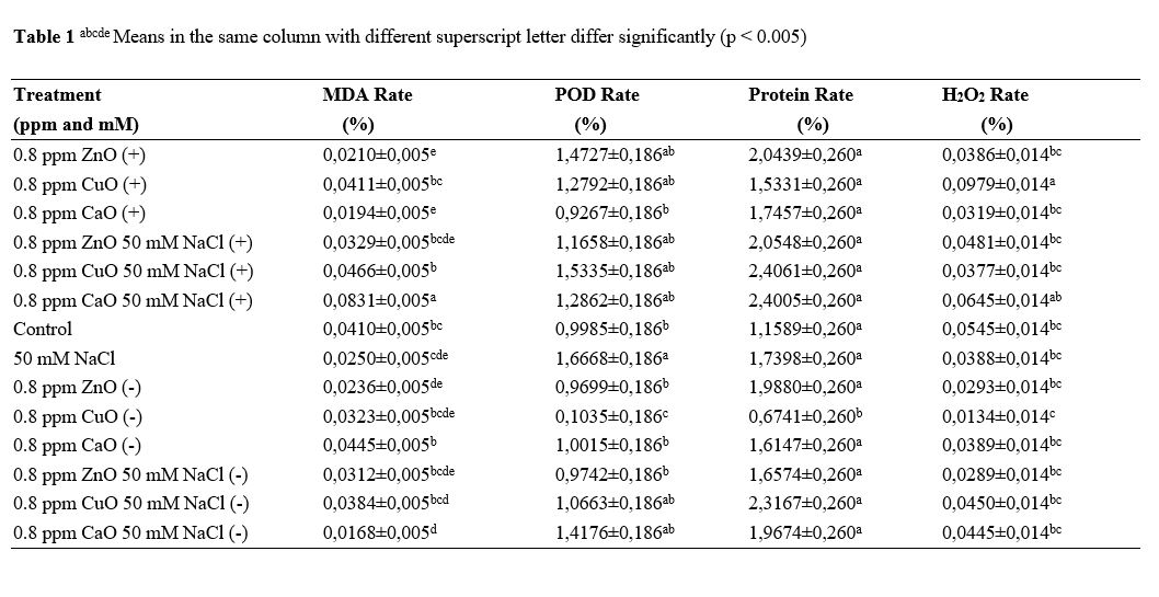
MDA (Malondialdehyde)
Malondialdehyde was measured using the method of (Heath and Packer 1968) using liquid nitrogen. 0.4 grams of ground callus material was dispersed in 0.5% (w/v) thiobarbituric acid solution containing 20% (w/v) tricholoroacetic acid. The sample was boiled at 98 ◦C for 30 min. and then quickly taken into an ice bath. The sample content was centrifuged at 3000 ×g for 10 min. and the value of the supernatant was monitored at 532 and 600 nm (Heath and Packer 1968; Jaleel et al. 2007; Erdal 2012).
H2O2 (Hydrogen peroxide)
H2O2 (Hydrogen peroxide) content was measured using the method of Sergiev et al. (1997). 0.4 g of callus material was homogenized in 4 ml of trichloroacetic acid and centrifuged at 4 °C for 15 min. at 13000 rpm. 2 ml of extract was mixed with 0.8 ml of KH2PO4 and 1.6 ml of KI in test tubes. The absorbance of the callus sample product was measured at 390 nm using a standard curve with H2O2 solutions (Velikova et al. 2000).
POD (Peroxidase)
The activity of POD (Peroxidase) was measured following the procedure established by Chance and Maehly (1955) by adding 100 μL of the callus extract to 3 mL of assay solution, which contained 13 mM guaiacol, 5mM H2O2 and 50Mm Na-P buffer (pH 6.5). The POD (Peroxidase) activity was determined in absorbance at 470 nm of protein. The total soluble protein contents were determined by BCA (Bicinchoninic Acid) protocol (Yee et al. 2003; Erdal 2012).
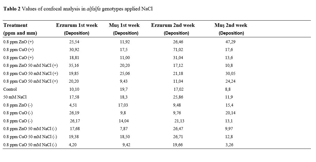
Sectioning with microtonal
Having been kept in 10% formaldehyde for 3 days, callus structures were taken to the cassettes and left for an overnight wash. Then, it was kept in 70, 80, 96% ethyl alcohol, one hour apart, respectively. Absol-I, Absol-II, Xylol-I, and Xylol-II were kept for 1 hour, respectively. The calluses were embedded in parafilm, and a section of 6µm was taken (Rolls et al. 2012).
Laser scanning confocal microscope (CLSM)
Callus, sectioned with microton, are kept for about 30 minutes with 1% rhodamine. Then it is passed through distilled water 3 times. Fluorescence images were obtained with a Nikon Eclipse TE2000 Confocal Laser Scanning Microscope C1si. Samples were excited with the 488 nm line of an argon laser and dye emission was collected at 520 and 610 nm. The DCF fluorescence was visualized in a single optical section of the callus. All images were obtained at the same depth (Minta et al. 1989).
Scanning Electron Microscopy
Alfalfa callus tissues were prefixed in 5% buffered glutaraldehyde (0.1 M phosphate buffer, pH 7.2) for 2 h at room temperature. After dehydration through a graded ethanol series, samples were dried with a CPD (CO2 critical-point drying) system, sputter-coated with gold (Jeol JFC-1100 E ion-sputtering system) and observed with a scanning electron microscope (HITACHI S-4700).
Statistical Analysis
Each experiment was repeated three times. Analysis of variance was conducted using a one-way ANOVA test using SPSS 13.0 and means were compared by Duncan test at the 0.05 confidence level.