Mice
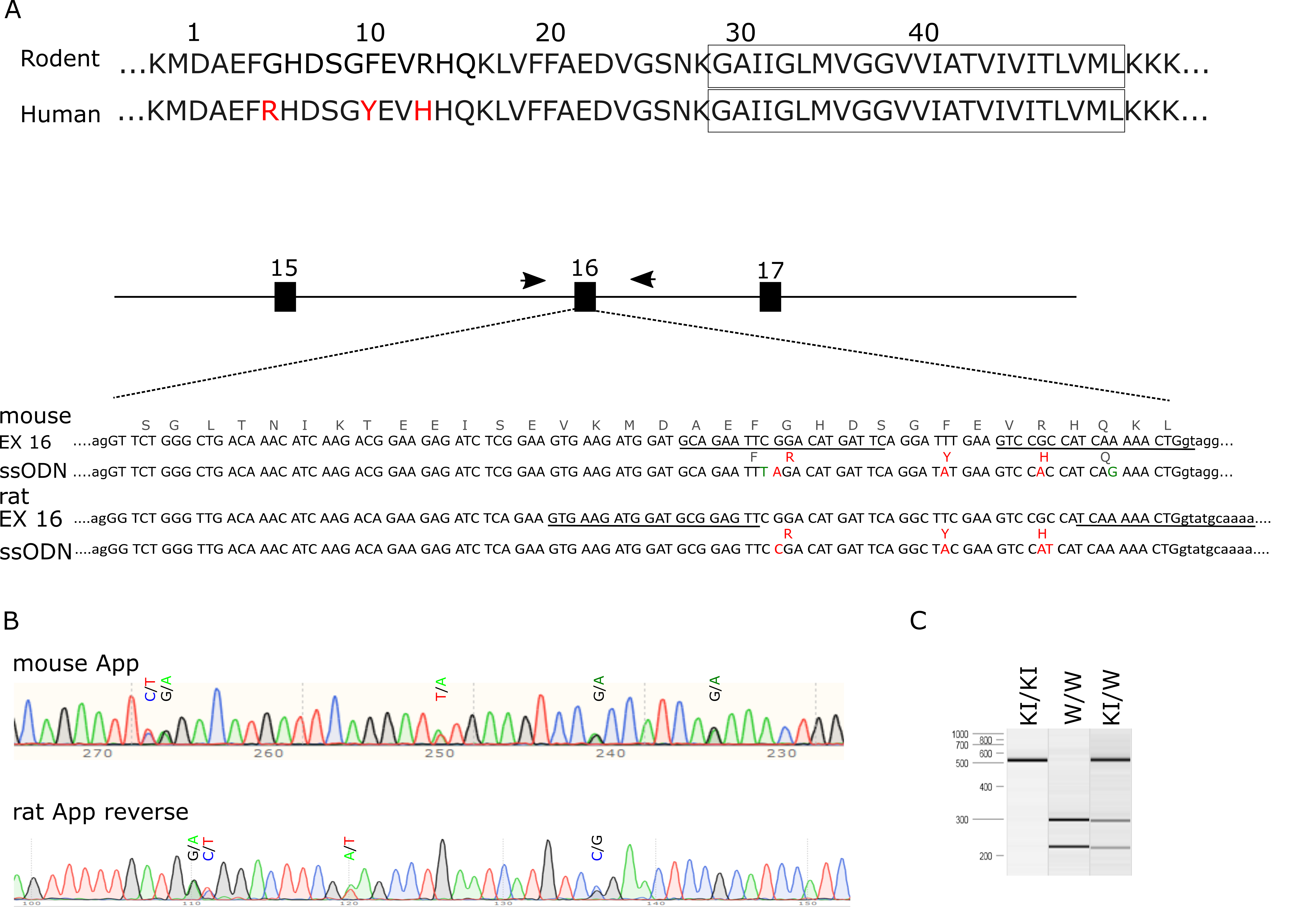
Mice Appem1Bdes with a humanized Aβ sequence (G676R, F681Y, R684H) were generated using CRISPR/Cas9 technology to target exon 16 of the mouse App gene. RNA guides were selected using the CRISPOR web tool. Guide 5’-GCAGAAUUCGGACAUGAUUC-3’ and 5’-GUCCGCCAUCAAAAACUGGU-3’ were selected and tested in mouse embryonic fibroblast (MEF) cells for cleaving efficiency. To promote homologous recombination directed repair(34) we made use of a ssODN repair template to mutate the target amino acids and to introduce two silent nucleotide substitutions. The first silent substitution destroys an EcoRI restriction cleaving site, facilitating genotyping. The second silent substitution prevents Cas9 cleaving the modified locus. Ribonucleoproteins (RNPs) containing 0.3 μM purified Cas9HiFi protein (Integrated DNA Technologies, IDT), 0.6 μM CRISPR RNAcrRNA, 0.6 µM trans activating crRNA (IDT) and 10 ng/µl ssODN (5’-tactttgtgtttgacgcagGTTCTGGGCTGACAAACATCAAGACGGAAGAGATCTCGGAAGTGAAGATGGATGCAGAATTtaGACATGATTCAGGATaTGAAGTCCaCCATCAgAAACTGgtaggcaaaaataaactgcctctccccgagattgcgtctggccagatgaaatacgtggcacctcgtggcttgtcctgtgt-3’) were injected into the pronucleus of 72 C57Bl6J embryos by microinjection in the Mouse Expertise Unit of KU Leuven. One positive pup was identified by PCR and restriction analysis. Sanger sequencing of App exon 16 region, as well as the 5 most likely off target sites predicted by the CRISPOR web tool, confirmed correct targeting (Additional file1) and absence of spurious events at other sites. The founder mouse was backcrossed over two generations using C57BL6J mice before a homozygous colony was established, which was designated Apphu/hu. The strain is maintained on the original C57Bl6J background by backcrossing every 5th generation. Standard genotyping is performed by PCR with primers 5’-taggtggtggttaatggtt-3’ and 5’-cgtagctgcaacgttggact-3’ followed by digestion of the PCR product with EcoRI.
Apptm3.1Tcs (6) also known as App NL-G-F and Tg(Thy1-MAPT)22Schd (35) also known as Thy-Tau22 mice were used as positive controls during histological examination. Mice are kept on a C57Bl6J background and both females and males were included in the study. Mice are housed in cages enriched with wood wool and shavings as bedding, and given access to water and food ad libitum. All experiments were approved by the Ethical Committee for Animal Experimentation at the University of Leuven (KU Leuven).
Rats
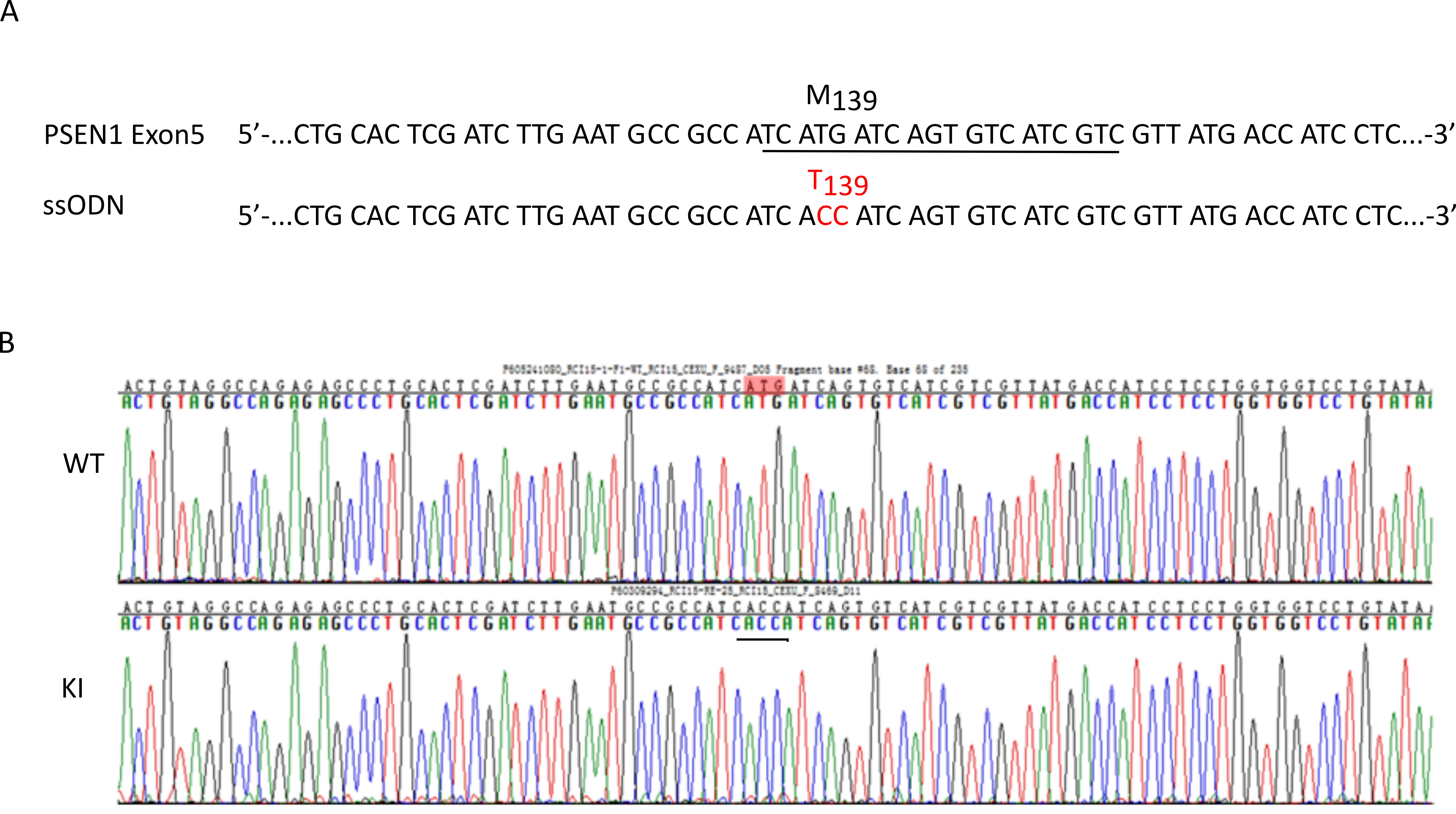
As the rat is one of the most studied model organisms(36), and until recently no knock-in rat models of AD were available(27), we set out to humanize the Aβ sequence in rats using a similar strategy as we used in the mouse. Two gRNAs (Additional file1) , 5’-GUGAAGAUGGAUGCGGAGUU-3’ and 5’-UUUUGCAUACCAGUUUUUGA-3’, Cas9 mRNA and an oligo donor with targeting sequence (flanked by 120 bp homologous sequences on both sides) were co-injected into zygotes of Long Evans rats We also introduced the early-onset familial Alzheimer’s disease (FAD) mutation M139T(23) into the endogenous Rat Psen1 gene. To target Psen1. Cas9 mRNA, sgRNA 5’-GAUGACACUGAUCAUGAUGG-3’ and an oligo donor containing the ATG/ACC substitution with 120 bp homologous sequences were co-injected into zygotes (Additional file2). F0 rats were genotyped after weaning using PCR and Sanger sequencing. Founder rats carrying the humanized Aβ sequence and M139T mutant allele were crossed twice with WT Long Evans rats (Charles River). Rats homozygous for the humanized APP KI were obtained after crossing heterozygous offspring. A breeding colony homozygous for the humanized Aβ sequence and heterozygous for the Psen1M139T allele was established and designated Apphu/hu;Psen1M139T. Standard genotyping for the APP KI mutation is done with the forward primer 5’-caTGATTCAGGCTaCGAAGTCCat-3’ and a common reverse primer 5’-CTCAGTGGTAATACGCCTGCCTAGC-3’. 5’-TGATTCAGGCTtCGAAGTCCgc-3’ is the forward primer for amplification of the wildtype App allele. For Psen1, genotyping is performed by PCR with a WT specific forward primer 5’-cgatcttgaatgccgccatcatg -3’, or a M139T specific forward primer 5’-cgatcttgaatgccgccatcacc-3’, together with a common reverse primer 5’-ctgcacatgtacactctggcaag-3’. Rats are kept on a Long Evans background and backcrossed every fifth generation to WT rats. Both females and males were included in the study. Rats are housed in cages enriched with wood wool and shavings as bedding, and given access to water and food ad libitum. All experiments were approved by the Ethical Committee for Animal Experimentation at the University of Leuven (KU Leuven).
Human tissue samples
Human brain samples were resected from the lateral temporal neocortex and were obtained from patients who underwent amygdalohippocampectomy for medial temporal lobe seizures. Samples were collected at the time of surgery and immediately transferred to the laboratory for processing. All procedures were conducted according to protocols approved by the local Ethical Committee of KU Leuven (protocol number S61186).
Sample collection and protein analysis
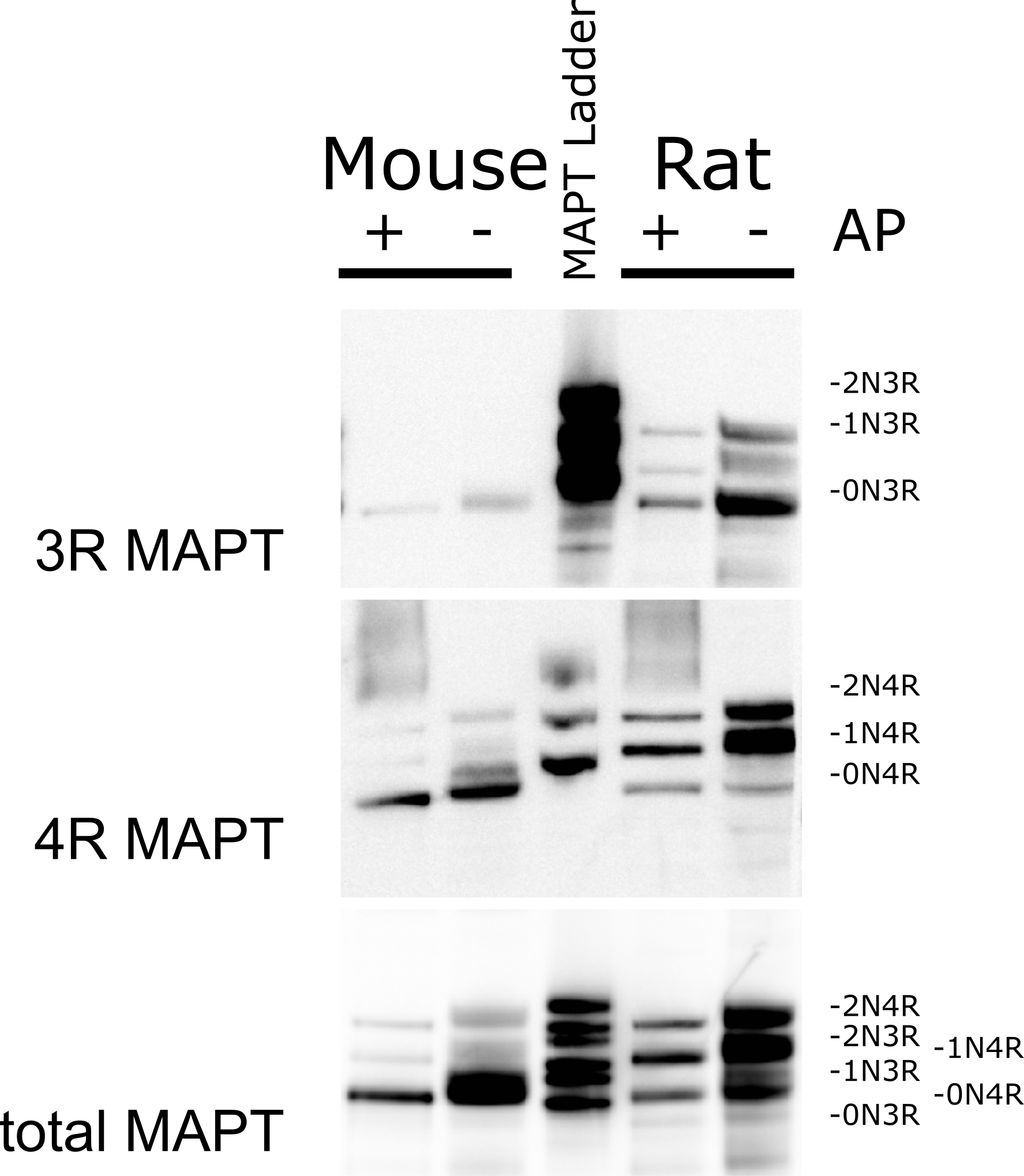
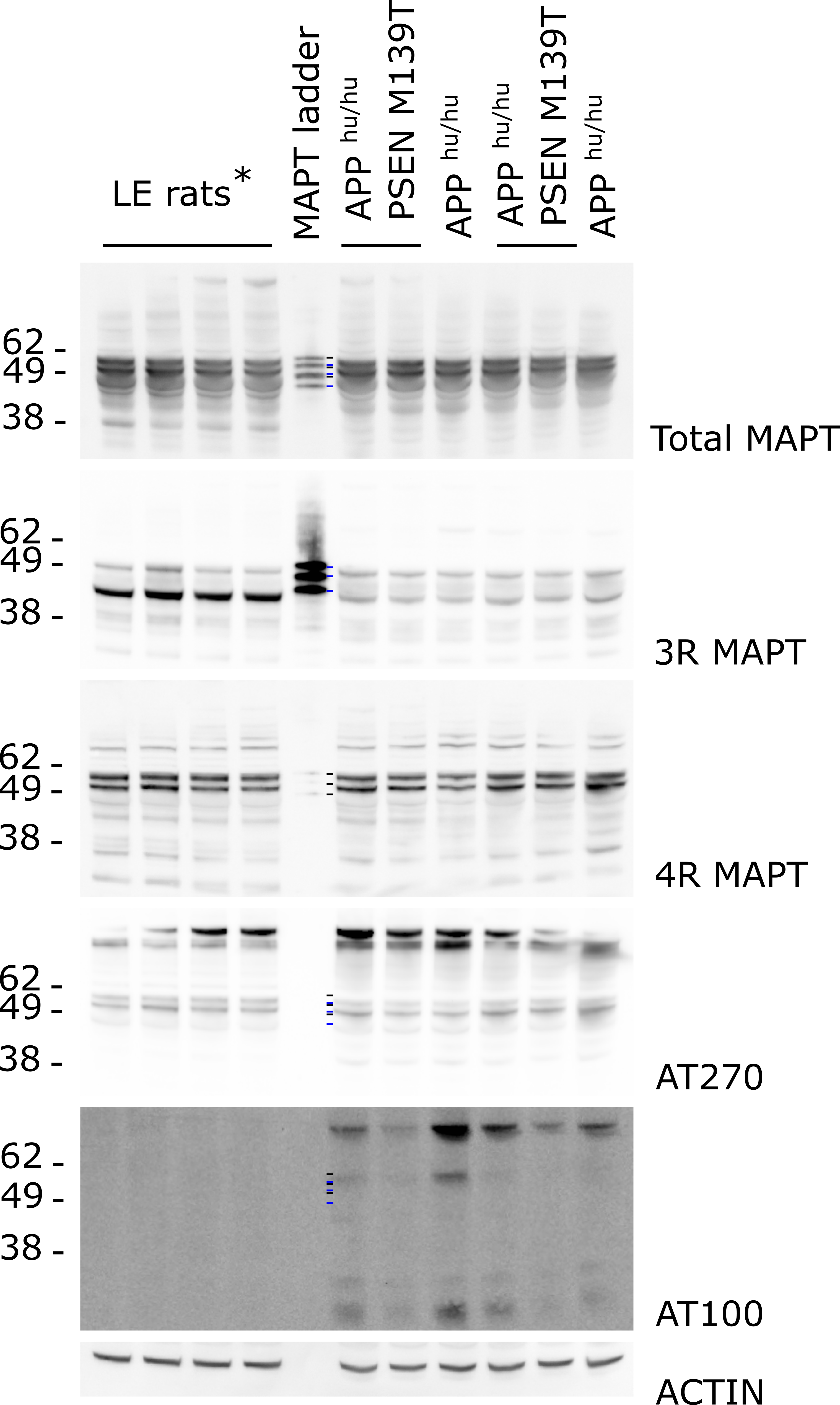
Three female and 3 male mice and rats of the indicated genotypes were aged to 14 weeks and then euthanized by carbon dioxide overdose, followed by intracardial perfusion of ice-cold phosphate buffered saline. Brains were removed from the skull, and the cerebrum was snap frozen using liquid nitrogen and stored at -70°C until further processing. Half a brain hemisphere was weighed and homogenized in a bead mill using 5 volumes of buffer containing 20 mM Tris , 250 mM sucrose, 0.5 mM EDTA,0.5 mM EGTA (pH 7.4 HCl) supplemented with cOmplete™ protease inhibitor cocktail (Roche) and PhosSTOP™ (Sigma). This homogenate was divided in three fractions of 250 µl. One fraction was used to extract soluble Aβ using 0.4% Diethylamine treatment for 30 min at 4°C. Following high speed clearing at 100,000g for 1h (at 4°C), the sample was neutralized by adding 1/10 volume of 0.5 M Tris-HCl (pH 6.8) and analyzed by ELISA. To obtain the guanidine HCl (GuHCl)-soluble fraction, the 100,000g pellet was washed with 0.4% Diethylamine before solubilization in 6M GuHCl, 50 mM Tris-HCl (pH 7.6), supplemented with cOmplete™ protease inhibitor cocktail (Roche) and PhosSTOP™ (Sigma), and sonication using a micro-tip for 30 s at 10% amplitude (Branson). After incubation for 1h at 25°C with agitation (600 rpm), the sample was cleared by spinning at 100,000g, diluted to 0.1M GuHCl and analyzed by ELISA. Aβ38, Aβ40 and Aβ42 levels were quantified on Meso Scale Discovery (MSD) 96-well plates using ELISA and antibodies provided by Dr. Marc Mercken (Janssen Pharmaceutica). Monoclonal antibodies JRFcAβ38/5, JRFcAβ40/28 and JRFcAβ42/26, which recognize the C terminus of Aβ species terminating at amino acid 38, 40 or 42, respectively, were used as capture antibodies. JRF/rAβ/2 (rodent specific antibody) or JRFAβN/25 (human specific antibody) labeled with sulfo-TAG were used as the detection antibodies. Human Aβ43 was measured using the amyloid-beta (1-43) high sensitivity ELISA kit from IBL. To measure APP protein in the brain samples, 250 µl of homogenate were supplemented with 1% Triton X100, incubated for 30 minutes on ice and cleared for 30 minutes at 14,000g (at 4°C). For the extraction of soluble MAPT protein, 250 µl of a buffer containing 300 mM NaCl, 50 mM Tris-HCl, 150 mM NaCl, 1 mM EDTA, 2% NP-40, 0.5 % sodium deoxycholate (pH 7.5), supplemented with cOmplete™ protease inhibitor cocktail (Roche) and PhosStop™ (Sigma), was added to 250 µl of brain homogenate. The sample was sonicated and incubated for 30 min on ice and cleared by centrifugation at 14,000g (at 4°C) for 30 min. When indicated, cell lysates were dephosphorylated after dialysis against 50 mM Tris-HCl (pH7,6) using Calf Intestine Phosphatase (Bioke). Total protein content of the cell lysates was measured using a Biorad protein assay kit. Fifty micrograms of protein were loaded in reducing and denaturing conditions on NuPAGE™ (Thermo) gels and subjected to electrophoresis. Following separation, proteins were transferred to nitrocellulose membrane for western blotting. Membranes were blocked with 5% non-fat milk Tris buffered saline, containing 0.1% Tween 20, and incubated with the indicated primary antibodies, washed, and incubated with horseradish peroxidase conjugated secondary antibodies (Biorad). Blots were developed using the ECL Renaissance kit (Perkin Elmer). Primary antibodies used in this study were B63 (against the C-terminal amino acids of APP(previous used in (37), 1/1000)), 82E1 (IBL, 1/500), JRF/rAβ/2 (Janssen, 1/1000), anti-human TAU (Dako, 1/1000), anti-3RTAU (Millipore, 1/1000), anti-4RTAU (CosmoBio, 1/1000) and Anti-Actb clone AC-15 (Sigma, 1/20000). Intensities of the bands were quantified with Aida/2D densitometry software.
All data are presented as mean±SD, and were analyzed by GraphPad Prism 8. Unpaired two way Student’s t test and one-way ANOVA were used for group comparisons. P < 0.05 was considered statistically significant.
Histology
Rats and control mice were euthanized with an overdose of carbon dioxide and transcardially perfused with PBS. Brain tissue was subsequently harvested and post-fixed overnight in 4% PFA in PBS. Brains were cut in serial sections of 40 μm thickness with a vibrating microtome (Leica). For each sample, six series of sections were sequentially collected in free-floating conditions and permeabilized for 30 minutes with PBST (0.2% Triton X-100 in PBS) and blocked for 2 hours with 5% normal donkey serum in PBST. Antigen retrieval was performed by boiling the sections for 1 minute in 10 mM sodium citrate (pH6) in a microwave. After three rinses with 0.1% Tween 20 in PBS, sections were incubated for 20 minutes with 10 µM X34 (Sigma) in 0.1% NaOH made in 40% Ethanol washed and incubated overnight at 4°C with primary antibodies against Aβ: 82E1 (IBL, 1/150) or 6E10 (BioLegend, 1/200). The antibody stained sections were washed three times with 0.2% TritonX100-PBS and incubated with Alexa594 Donkey anti-mouse IgG (Thermo Fisher, 1/300) for 2 hours at RT. After final washes with 0.2% TritonX100-PBS, sections were counterstained with TO-PRO (Thermo Fisher, 1/1000) and after three final washes were mounted on super frost microscope slides. Sections were visualized on a Nikon A1R Eclipse confocal system.
Computational model of the humanized β-CTF bound to BACE1
We have modelled the interaction of humanized β-CTF with BACE1, using as a template the published crystal structure of BACE1 in combination with an active site peptide inhibitor (PDB ID 5MCQ)(16). The sequence of the humanized β-CTF was aligned to the peptide in 5MCQ, aligning residue E11 from the humanized β-CTF sequence to the STA query (Threonine) of the crystallized peptide (Fig1).