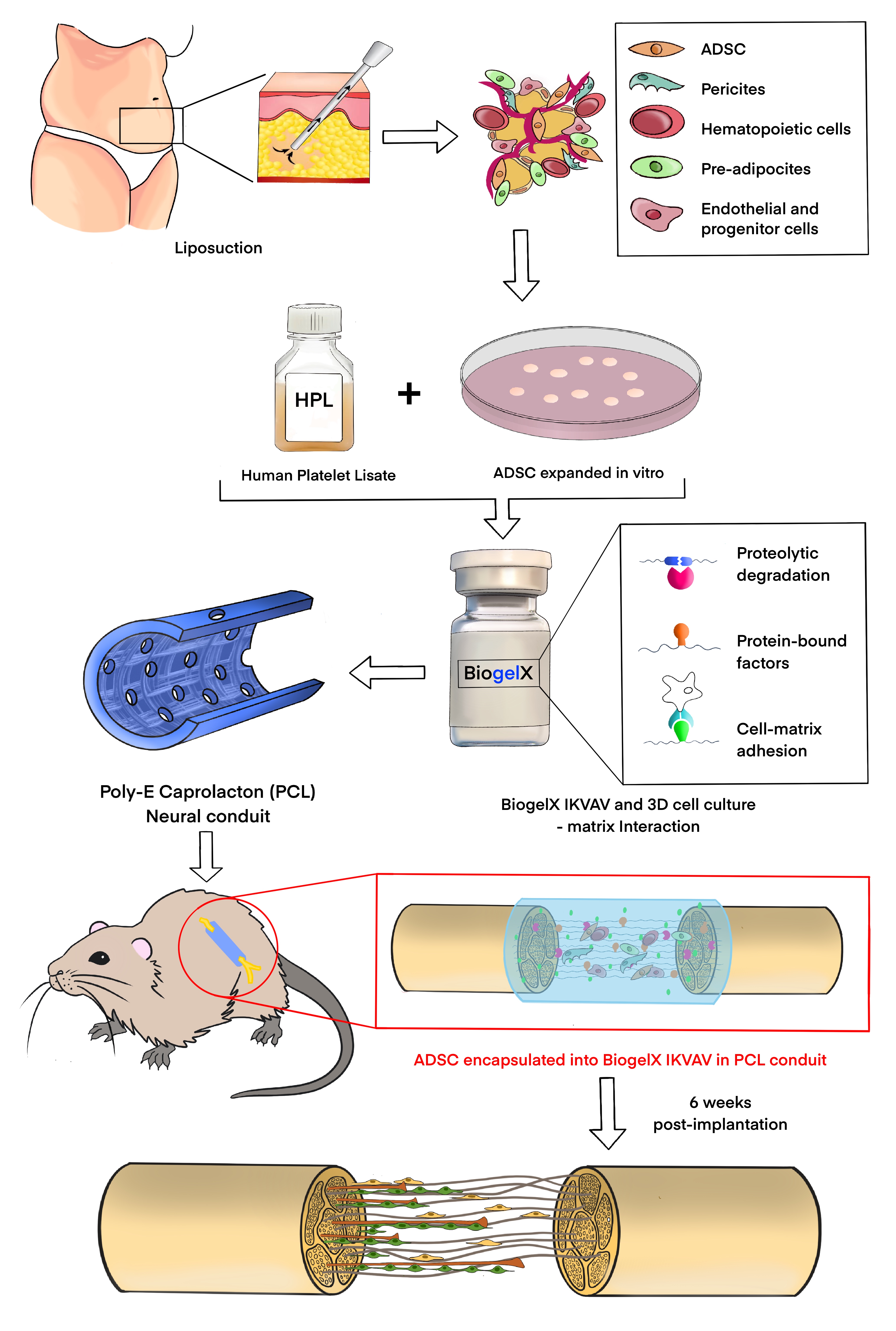
Cell therapy is a promising treatment to approach peripheral nerve injuries, as demonstrated by a vast number of pre-clinical in vitro and in vivo research studies. (28, 29, 30) For autologous cell therapy, ADSC have been identified as ideally suited, as they are abundant, easily expandable and have been shown to have positive effects on nerve regeneration. (31, 32, 33, 34)
Although cell-therapy is a valuable approach, significant concerns remain around cell survival following transplantation in vivo, the persistence and the duration of ADSC neurotropic properties/effects in the recipient neural microenvironment. (10, 35, 36) One of the open questions is the delivery method in an in vivo scenario, which permits cell survival; and supports and exploits the properties of the transplanted cells longer term. In fact, direct cell injection into the injury site has shown poor engraftment outcomes: ADSC injected at the nerve gap has only had a survival rate of about 0.5% and are undetectable 2 weeks after their application. (36, 37, 38) Multiple hypotheses have been advanced, such as the intense local inflammatory response after nerve transection, mechanical damage endured during injection, (39) or the high concentration of adenosine triphosphate that has been shown to induce cell death via P27X receptors in ADSC and Schwann cells after peripheral nerve injury. (37, 40)
For all these reasons, a wide range of biomaterial carriers have been tested, with the aim to maintain therapeutic cells in a suitable state for sufficient time to permit exploitation of their beneficial effects. Recently, research efforts are being focused on developing a bio-engineered scaffold modified with ECM proteins, or their bioactive moieties, with adjusted viscoelastic properties and tailored degradation profiles, to optimize cell retention and functional maintenance at the injury site. (3, 41, 42, 43, 44, 45, 46)
The introduction of ECM molecules provides an enriched environment for cell adhesion and migration, activating and/or inhibiting cell signalling pathways that in turn can act on regenerative and survival biochemical factors. (47, 48, 49) Specifically, LN was selected in developing an engineered nerve construct, due to its critical role in myelin formation and axonal sorting (50), with similar findings obtained using laminin-derived peptide sequences, particularly RGD and IKVAV. (51)
Short peptide molecules were shown to self-assemble into water-swollen networks forming highly biocompatible and biodegradable SAP hydrogels with ideal properties for biomedical applications. (52, 53) The SAP hydrogels used here, and synthesized by Biogelx, consist of interwoven nanofibers of ca. 10 nm in diameter with pore sizes ranging between ca. 10 and 200 nm which provide a suitable 3D environment for cell growth and allow the diffusion of small soluble factors. The Biogelx scaffold fibers and pores are smaller than the cells’ diameter and similar to the dimensions seen in the native ECM thus, cells can adhere to the microfibers. While they are somewhat embedded in the nanofibrous scaffold, they are free to move and interact with each other and their environment. (15, 52, 54, 55)
Previous studies demonstrated that functionalised SAP hydrogels promoted cell attachment, proliferation and migration of multiple cell types including osteoblasts (56), human umbilical vein endothelial cells, hADSC and SC. (57, 58)
Considering the development of ADSC-therapy strategy for peripheral nerve repair, we chose one of the LN-derived peptides, the IKVAV, which was affirmed earlier as a supportive factor of neuronal growth cone adhesion and elongation (50). As shown in our previous studies with LN, and widely confirmed by the literature, IKVAV also contributes to the support of ADSC viability, proliferation and neurotrophic cell commitment. (5, 7, 9) Our ADSC viability assessment confirmed the significant enhanced proliferation in Biogelx-IKVAV between the 3rd and 7th day compared to the S type gel, particularly in a 2D system.
Consistently, previous studies found that YIGSR (another LN-peptide) and RGD mediated SC adhesion and proliferation, while IKVAV drove neural differentiation and enhanced nerve regeneration by supporting the axonal elongation both in vitro and in vivo. (59) Santiago et al. found that the hADSC adhere preferentially to the IKVAV peptidescompared to two other LN-derived peptide counterparts RGD and YIGSR. (60)
Moreover, recent studies investigated the combination of LN or LN peptide (IKVAV) and growth factors (e.g., BDNF) for nerve repair: Park et al. fabricated a hyaluronic acid-based hydrogel with the IKVAV peptide and full length BDNF molecule incorporated into a 3D scaffold for the treatment of spinal cord injury. The introduction of this conduit resulted in faster differentiation of stem cells and facilitated nerve regeneration. (61) Then, Frick et al. developed an injectable IKVAV-functionalised SAP hydrogel, which incorporates BDNF, promoting attachment and favourable neurite outgrowth of spiral ganglion neurons in cochlear implants. (62) Again, LN-binding BDNF was widely applied in pre-clinical models, for the treatment of cerebral ischemia, facial nerve injury and recurrent laryngeal nerve injury. (63, 64, 65)
Therefore, using the ADSC expanded in hPL and encapsulated in Biogelx IKVAV, we combined the synergetic effects of ADSC-therapy, the ECM and the hPL growth factor enrichment (14) with a 3D SAP support. Biogelx-based laminin scaffold could be a simplified method that overcomes the drawbacks of current delivery systems, stimulating the transplanted cells through peptide functionalization, adaptable stiffness and guaranteeing biosafety during clinical translation.
In this work, such hybrid cell–gel systems, loaded into microfabricated PCL conduits to support regeneration, were tested initially in vitro in terms of cell viability and proliferation, then in vivo over a period of six weeks, in terms of peripheral nerve sprouting and gastrocnemius muscle histology.
The axonal regeneration through the 15 mm sciatic nerve defect was evaluated by the distances reached by regenerating axons and SC in longitudinal sections of the explanted conduits. Scaffolds filled with hADSC encapsulated into Biogelx-IKVAV (hADSC-IKVAV) achieved significantly higher values both in terms of longest axonal sprouting and furthest SC migration along the regenerative growth cone compared to the empty conduit (empty, p < 0.0001 ****), the ones filled with hydrogel only (S, IKVAV, p < 0.001 ***) or cells only (hADSC, p < 0.01 **). Moreover, cells alone or hydrogel only achieved significantly higher axonal length than the empty conduit (S p < 0.05 *, IKVAV p < 0.01 **), but only IKVAV gained a similar significant trend in SC migration distance (p < 0.05 *). However, no significant differences, both in terms of axonal elongation and SC migration distance were found between cells alone and gels only: these results underline how the effects of hADSC are dependent on an adequate delivery/support system and how the polyvalent value of IKVAV can impact both ADSC and neural cells.
Overall, these effects could be most likely ascribed to the complementary roles of hADSC-IKVAV to assure cell-to-cell interactions with regrowing axons, release and maintenance of the hADSC secretome (e.g., growth factors such as NGF, vascular endothelial growth factor, and BDNF) with its slow progressive release in the local milieu (31, 66). Each of these molecules is essential for the recruitment of endogenous SC at the site of a peripheral nerve injury, improving tissue regeneration and functional repair. Furthermore, the functionalization with LN-derived IKVAV-peptide can further enhance survival and neurotrophic properties of transplanted hADSC and directly stimulate axonal regrowth.(5, 7)
Regarding the histological muscle analysis, no quantitative significant differences were found among the experimental groups, as could probably be expected after a 15mm critical sciatic nerve gap and only 6 weeks experiment. Indeed, although axonal regrowth through the nerve gap was observed, the muscle changes at the level of muscle fiber composition take place over a longer time period than six weeks, as other studies have shown (67). The trend of a higher ratio nuclei/muscle fiber area could be explained by a denser infiltration of inflammatory cells or a more intense recruitment of satellite cells around the muscle fibers, particularly in the experimental conditions (IKVAV, hADSC) with an additive effect between the cells and hydrogel components (hADSC-IKVAV). Both inflammatory cell and satellite cell tissue invasion/recruitment are initial signs of muscle regeneration, but again, the earlier timeline considered here cannot be significantly diriment in supporting our hypothesis. (68, 69, 70)
A longer-term study (12 weeks) will also be critical to evaluate potential muscle recovery after denervation, together with behavioural, functional, and histological outcomes.
