-
Confirmation and differentiation of NSCs
To investigate the mechanism in vitro, primary cultured hippocampal NSCs were used. The NSCs were isolated from fetal hippocampi of Sprague-Dawley rats on embryonic (E) day 14 to E15. Observation under microscope showed that NSCs grew in a typical round shape (Fig. 1A), which was confirmed using immunofluorescence with the NSC marker nestin (Fig. 1B). For NSC differentiation, NSCs were induced to differentiate for 120 h which was confirmed using immunofluorescence with neuron marker β-tubulin III and astrocyte marker glial fibrillary acidic protein (GFAP; Fig. 1C).
Confirmation and differentiation of NSCs. (A) Primary cultured hippocampal NSCs observed under microscope. Scale bar = 100 µm, 50 µm. (B) Immunofluorescence staining of NSC marker nestin. Scale bar = 100 µm. (C) Results of β-tubulin III (red) and GFAP (green). Scale bar = 50 µm.
-
Repeated exposure to sevoflurane led to early differentiation of hippocampal NSCs
To determine whether sevoflurane can affect NSC differentiation, the expression of NSC marker nestin, neuron marker β-tubulin III, and astrocyte marker GFAP was examined in fetal brains and primary cultured hippocampal NSCs using western blotting and immunofluorescence at 24 h and 72 h after single or repeated exposure to sevoflurane (Fig. 2, 3). After repeated maternal exposure to 3% sevoflurane, the β-tubulin III (Fig. 2A, B, D) and GFAP levels (Fig. 2A, C, E) were increased and the nestin level (Fig. 2A–C, F) was decreased in fetal brain tissue. However, significant difference was not observed between control (CON) group and single 3% sevoflurane exposure (SEV × 1) group; Fig. 2A–F). Primary cultured NSCs exposed to 4.1% sevoflurane once or 3 times (SEV × 3) showed β-tubulin III (Fig. 3A, B, D), GFAP (Fig. 3A, C, E), and nestin (Fig. 3A, C, F) levels corresponded to in vitro results. The results showed that repeated exposure to sevoflurane led to early differentiation in hippocampal NSCs.
Repeated maternal exposure to 3% sevoflurane led to early NSC differentiation in fetal brains. (A) Western blotting images of β-tubulin III, GFAP, and nestin. (B) Immunofluorescence images of β-tubulin III (green) and nestin (red). Scale bar = 100 µm. (C) Immunofluorescence images of GFAP (green) and nestin (red). Scale bar = 100 µm. (D) Quantitative analysis of β-tubulin III. (E) Quantitative analysis of GFAP. (F) Quantitative analysis of nestin. Values are means ± SEM (n = 5/group). *p < 0.05, **p < 0.01, ***p < 0.001 compared with CON group; # p < 0.05, ## p < 0.01, ### p < 0.001 compared with SEV × 1 group. One-way ANOVA followed by Tukey’s post hoc multiple comparison test were used for data analysis.
Repeated exposure to 4.1% sevoflurane led to early differentiation in primary cultured hippocampal NSCs. (A) Western blotting images of β-tubulin III, GFAP, and nestin. (B) Immunofluorescence images of β-tubulin III (green) and nestin (red). Scale bar = 100 µm. (C) Immunofluorescence images of GFAP (green) and nestin (red). Scale bar = 100 µm. (D) Quantitative analysis of β-tubulin III. (E) Quantitative analysis of GFAP. (F) Quantitative analysis of nestin. Values are means ± SEM (n = 3/group). *p < 0.05, **p < 0.01 compared with CON group; #p < 0.05, ##p < 0.01 compared with SEV × 1 group. One-way ANOVA followed by Tukey’s post hoc multiple comparison test were used for data analysis.
-
Repeated exposure to sevoflurane led to long-term neuron reduction and astrocyte proliferation in hippocampus
Our previous study showed that a single maternal 3% sevoflurane exposure does not cause long-term neurocognitive impairment in fetal rats, while repeated exposure of 3% sevoflurane can cause learning and memory impairment in the offspring (Wu et al., 2018). The β-tubulin III and GFAP levels in rat brains were examined on postnatal day 28 (Fig. 4) as well as in the cultured NSCs (Fig. 5). After repeated exposure to sevoflurane, β-tubulin III protein was reduced in both postnatal fetal hippocampus (Fig. 4A, B) and cultured NSCs (Fig. 5A, B) compared with the CON and SEV × 1 groups. GFAP expression was upregulated in postnatal fetal hippocampus (Fig. 4A, C) and cultured NSCs (Fig. 5A, C). Immunofluorescence showed similar results regarding the quantity of neurons and astrocytes in CA1 hippocampal region (Fig. 4D, E). Significant difference was not observed between CON and SEV × 1 groups.
Effects of sevoflurane exposure on the expression of β-tubulin III and GFAP in fetal hippocampi on postnatal day 28. (A) Western blotting images of β-tubulin III and GFAP. (B) Quantitative analysis of β-tubulin III. (C) Quantitative analysis of GFAP. (D) Immunofluorescence images of β-tubulin III (green) in hippocampal CA1 region. Scale bar = 100 µm, 50 µm. (E) Immunofluorescence images of GFAP (green) in hippocampal CA1 region. Scale bar = 100 µm, 50 µm. Values are means ± SEM (n = 5). **p < 0.01, ***p < 0.001 compared with CON group; ##p < 0.01 compared with SEV × 1 group. One-way ANOVA followed by Tukey’s post hoc multiple comparison test were used for data analysis.
Effects of sevoflurane exposure on β-tubulin III and GFAP expression in cultured NSCs on postnatal day 28. (A) Western blotting images of β-tubulin III and GFAP. (B) Quantitative analysis of β-tubulin III. (C) Quantitative analysis of GFAP. Values are means ± SEM (n = 3). *p < 0.05 compared with CON group; #p < 0.05 compared with SEV × 1 group. One-way ANOVA followed by Tukey’s post hoc multiple comparison test were used for data analysis.
-
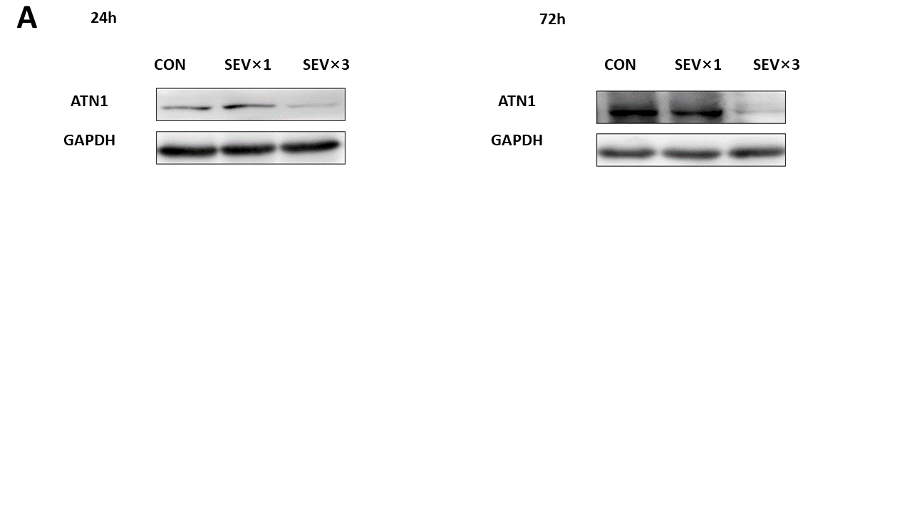
Exposure to sevoflurane affected NSC differentiation by downregulating ATN1 expression
The expression of ATN1 was downregulated in both fetal brain tissues (Supplement 1A, B) and primary cultured NSCs (Fig. 6A, E) after repeated exposure to sevoflurane. After transfecting ATN1 overexpression lentivirus into NSCs, in the 4.1% SEV × 3 + ATN1-overexpression lentivirus (LV-ATN1 + SEV × 3) group, the levels of β-tubulin III (Fig. 6A, C, F), GFAP (Fig. 6A, D, G), and nestin (Fig. 6A, H) were not significantly different than in the CON group at 24 h and 72 h. In the LV-ATN1 + SEV × 3 group, β-tubulin III (Fig. 6A, C, F) and GFAP (Fig. 6A, D, G) expressions were significantly reduced and nestin (Fig. 6A, H) expression was upregulated compared with the SEV × 3 group. ATN1 mRNA expression level was detected using reverse transcription quantitative polymerase chain reaction (RT-qPCR; Fig. 6B). β-tubulin III (Fig. 6A, I) and GFAP (Fig. 6A, J) protein levels were rescued in the LV-ATN1 + SEV × 3 group at day 28. Results confirmed that sevoflurane affected differentiation of hippocampal NSCs by downregulating ATN1 expression.
ATN1 overexpression alleviated sevoflurane-induced early NSC differentiation. (A) Western blotting images of β-tubulin III, GFAP, nestin, and ATN1. (B) RT-qPCR analysis of ATN1 mRNA expression. (C) Immunofluorescence images of β-tubulin III (green). Scale bar = 100 µm. (D) Immunofluorescence images of GFAP (green). Scale bar = 100 µm. (E) Quantitative analysis of ATN1. (F) Quantitative analysis of β-tubulin III. (G) Quantitative analysis of GFAP. (H) Quantitative analysis of nestin. (I) Quantitative analysis of β-tubulin III on day 28. (J) Quantitative analysis of GFAP on day 28. Values are means ± SEM (n = 3). *p < 0.05, **p < 0.01, ****p < 0.0001 compared with CON group; #p < 0.05, ##p < 0.01, ####p < 0.0001 compared with SEV × 3 group. One-way ANOVA followed by Tukey’s post hoc multiple comparison test were used for data analysis.
-
ATN1 is the direct target of miR-410-3p
Whether ATN1 is the direct target of miR-410-3p was investigated using TARGETSCAN database (Agarwal, Bell, Nam, & Bartel, 2015) (Friedman, Farh, Burge, & Bartel, 2009). The analysis showed ATN1 was the target site of miR-410-3p as described in Fig. 7A. The site was further verified using the dual-luciferase reporter assay (Fig. 7B).
ATN1 is a target gene of miR-410-3p. (A) Target prediction program predicted a specific binding region between the ATN1 gene and miR-410-3p sequence. (B) Relative luciferase activity assay analysis. The experiment was performed three times. Paired Student's t-test was used for data analysis.
-
Exposure to sevoflurane affects NSC differentiation by upregulating miR-410-3p
The miR-410-3p level was upregulated in NSCs repeatedly exposed to sevoflurane. The levels of ATN1 (Fig. 8A, F), β-tubulin III (Fig. 8A, D, G), GFAP (Fig. 8A, E, H ), and nestin (Fig. 8A, I) in NSCs treated with miR-410-3p suppression lentivirus and repeatedly exposed to sevoflurane were significantly different than levels in the SEV × 3 group at 24 h and 72 h. In the 4.1% SEV × 3 + miR-410-3p-suppression lentivirus (LV-410-SEV × 3) group, β-tubulin III expression (Fig. 8A, J) was upregulated and GFAP (Fig. 8A, K) expression was downregulated compared with the SEV × 3 group in cultured NSCs on day 28. RT-qPCR showed that miR-410-3p was suppressed by lentivirus (Fig. 8B). ATN1 mRNA level in LV-410 + SEV × 3 group was upregulated compared with the SEV × 3 group (Fig. 8C).
MiR-410-3p suppression alleviated sevoflurane-induced early NSC differentiation. (A) Western blotting images of β-tubulin III, GFAP, nestin, and ATN1. (B) RT-qPCR analysis of miR-410-3p expression. (C) RT-qPCR analysis of ATN1 expression. (D) Immunofluorescence images of β-tubulin III (green). Scale bar = 100 µm. (E) Immunofluorescence images of GFAP (green). Scale bar = 100 µm. (F) Quantitative analysis of ATN1. (G) Quantitative analysis of β-tubulin III. (H) Quantitative analysis of GFAP. (I) Quantitative analysis of nestin. (J) Quantitative analysis of β-tubulin III on day 28. (K) Quantitative analysis of GFAP on day 28. Values are means ± SEM (n = 3). *p < 0.05, **p < 0.01, ***p < 0.001, ****p < 0.0001 compared with CON group; #p < 0.05, ##p < 0.01, ###p < 0.001 compared with SEV × 3 group. One-way ANOVA followed by Tukey’s post hoc multiple comparison test were used for data analysis.