Cell culture
Mouse bEnd.3 cells was provided by Shanghai Biology Institute (Shanghai, P.R. China) and cultured in DMEM with the extra addition of 10% FBS, 2 mM l-glutamine, and 1% penicillin/streptomycin (Solarbio, China). A continuous culture condition of 5% CO2 atmosphere and 37°C was supplied for cell experiments.
In Vitro Model
TBI model cells were established by scratch bEnd.3 cells method. bEnd.3 cells were grown in a 24-well plate until the formation of the cell monolayer. Scratch injury of a 0.5mm wide gap was made with a sterile 26G syringe needle. The scraped cells were rinsed off with PBS. bEnd.3 cells in the control group were with no scratch injury.
ELISA
The medium or serum content of IL-18 and IL-1β was analyzed using the ELISA method with ELISA kits basing on the manufacturer’s instruction.
QRT-PCR
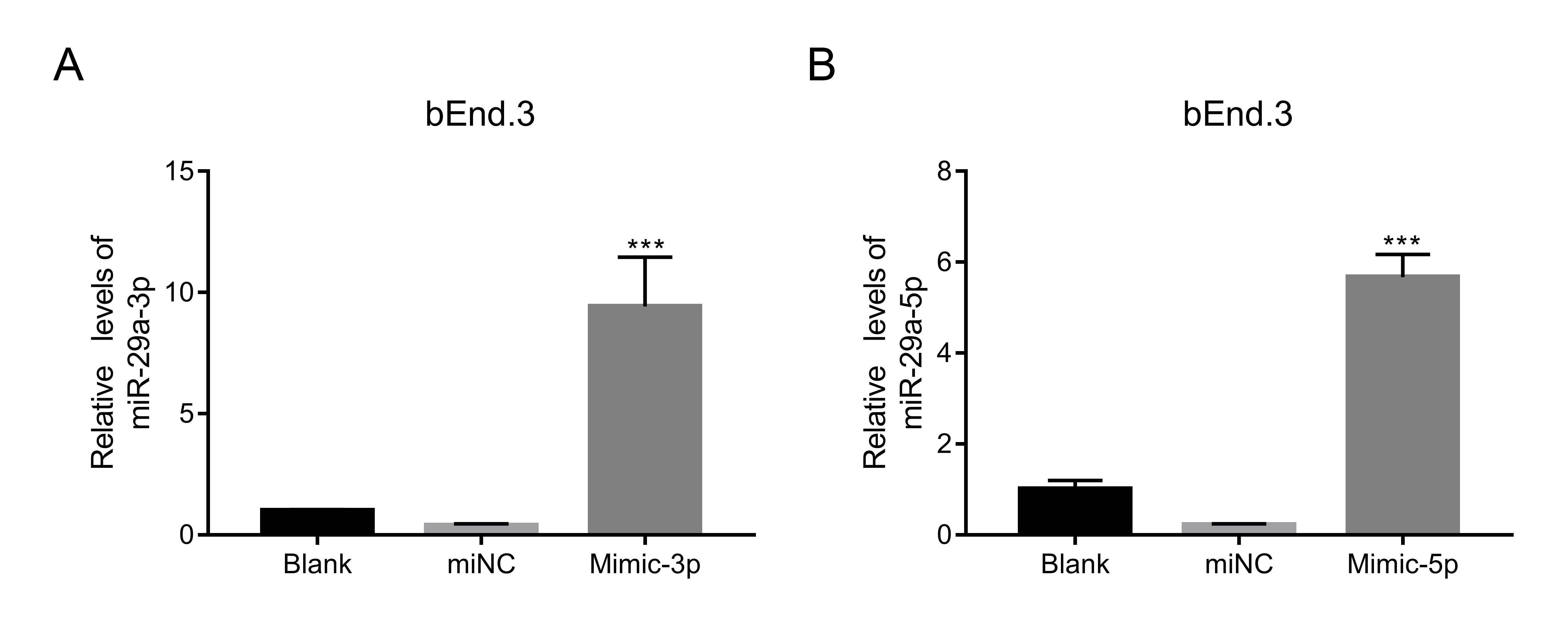
Total RNA were made by using TRIzol Reagent (Invitrogen, USA) supplementary with RNase inhibitor. For the first step of the qRT-PCR procedure, a total RNA sample was added to the reverse transcriptional reaction solution of the cDNA synthesis kit to produce cDNA. Then the cDNA product was added to a real-time PCR reaction solution (SYBR™ Green Master Mix Applied Biosystems™) and amplified based on the primers with the reaction conditions of 95°C for 10 minutes, 40 cycles of 95°C for 15 seconds, and 60°C for 45 seconds. Primer sequences were listed in Supplemental file1. Gene expressions were calculated by the 2−ΔΔCt method and employed U6 or GAPDH as the internal control.
Western blot
Total protein were made by using RIPA lysis buffer added with protease inhibitor cocktail (Roche, Germany). After a quantitative analysis using the BCA protein assay kit, test protein samples with equal amounts were subjected to 10-12% SDS-PAGE. A transfer operation then was performed to transfers the protein from the gel to a nitrocellulose membrane (Millipore, USA), and then was going through 1 hr of blockage with 5% nonfat dry milk, 12 hr of incubation with primary antibodies ( Seen in Supplemental file 2), and then 1 hr of incubation with secondary antibody (Beyotime, China). Protein blotting was detected using an enhanced chemiluminescence system (Beyotime, China). The protein expression was relative to GAPDH. The primary antibodies were list in.
Cell permeability detection
Cells were inoculated onto the upper chamber of the plate and grown to the formation of cell monolayer, during which the medium should be changed every day. FITC-Dextran was mixed into the medium at the last change of medium and maintained for 24 hr. Plates were placed in a Microplate reader (Pulangxin, China) to analyze the intensity of FITC fluorescence at 490 nm.
TEER assay
TEER value of bEnd.3 cell monolayer was analyzed using Millcell ERS-2 Voltohmmeter (Millipore, USA). Cells were and grown to monolayer at a 24-well plate and subjected to TEER value detection as the protocols suggested by the manufacturer. Calculation formula was TEER value (Ω·cm2) = TEER (Ω) × surface area (0.6 cm2).
Luciferase reporter assay
An amplified procedure was performed on the sequence of NLRP3 3’UTR containing the binding site of miR-29a-5p followed by a clone operation into luciferase reporter pGL3 vector (WT 3’UTR). The clone of sequence with the mutant binding sites was as control (Mutant 3’UTR). The recombinant luciferase reporter vectors were co-transfected with miR-29a-5p inhibitor or mimic into bEnd.3 cells. Luciferase activity of each cell treatment was examined after 48 hr of transduction with the Dual-GLO Luciferase Assay Kit (Promega, USA) on a plate reader.
TBI mouse model
Eighteen C57B6/J mice (male, aged 8-10 weeks) were randomly divided into three groups basing on different treatment: control mice were received sham operations, negative control for mimic (miNC) TBI mice were injected with miNC, and mimic TBI mice were injected with miR-29a-5p mimic. After TBI, mice were raised for another 48 hr before sacrifice for tissue examination. The sham mice received the same operation except for impact. All performances on animal experiments complied with the Guide for the Care and Use of Laboratory Animals and approved by the Ethics Committee of Shanghai Sixth People's Hospital East Affiliated to Shanghai University of Medicine & Health Sciences.
Mouse brain water content
The difference in weight between the wet brain and dry brain was used to evaluate the brain water content of the treated mice in each group. WW represents the wet weight and was weighted when fresh brains obtainment. DW represents the dry weight and was weighted after 72 hr of drying at 70 °C. The calculation formula is: Brain water content= [WW − DW]/WW × 100%.
Evans blue evaluates BBB permeability
After intraperitoneal injection with sodium pentobarbital (60 mg/kg), mice were received right femoral vein infusion with 2% evans blue dye (5ml/kg in saline) using a PE-50 catheter for 30 min. Another 15 min of heart perfusion with saline was worked to remove intravascular EB dye. Brain tissues were removed for the detection of EB dye permeating from BBB at a fluorescent plate reader. The intensity of fluorescence was measured at 630 nm. EV value was calculated as μg per 1 g of the brain.
H & E staining assay
Histological changes in the brain of TBI mice were visualized by HE staining. In brief, the brain was removed and immediately immersed in 4% paraformaldehyde. Then paraffin sections were made from these fixed tissues using a microtome and went through deparaffin and rehydration before HE staining.
Statistical analysis
Data statistic was conducted on a Version 7.0 software of GraphPad Prism (USA). Difference analysis between groups or among multiple groups used the t-test and one-way analysis of variance. Data were expressed as mean ± SD of at least three samples and triplicates were made if necessary. The p-value of less than 0.05 was considerable statistical significance on the difference.