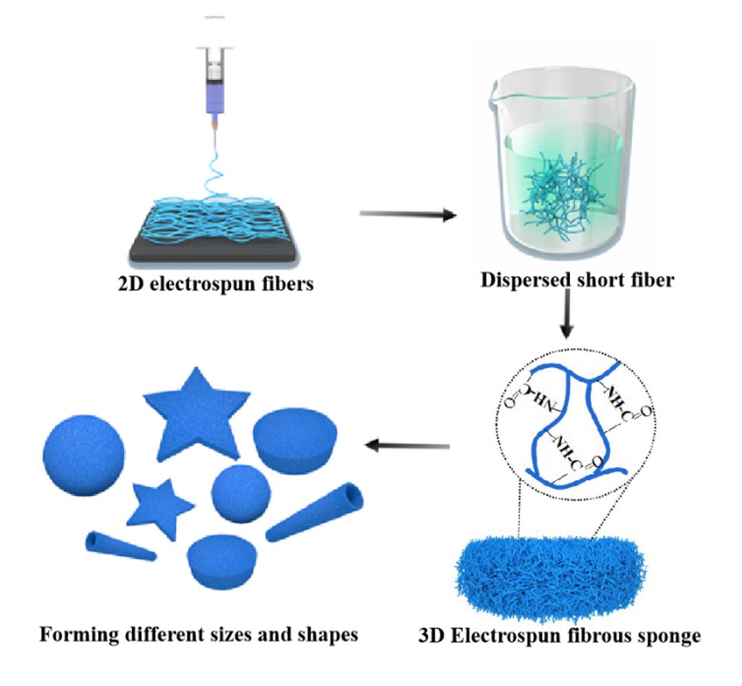
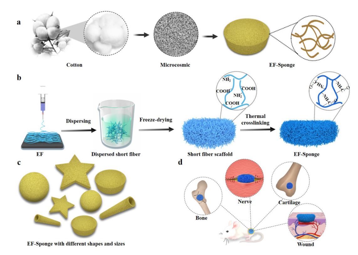
The fabrication of Scaffolds
The schematic of electrospun short fiber sponge scaffolds fabrication was illustrated in Scheme 1b. It was divided into four steps: electrospinning, homogenization, shaping, and crosslinking. Initially, a gelatin/polylactic acid (PLA) solution (12 wt.%) was prepared with a weight ratio of 4:1. Subsequently, gelatin/PLA fibers were prepared using the ordinary electrospinning equipment under suitable conditions, producing fiber membranes with relatively random coverage patterns. The fibers were cut into small fragments and dispersed in a tert-butanol solution by high-speed centrifugation, to ensure the uniform dispersion of the fibers (Fig. 1a). The dispersions were molded and freeze-dried to obtain the uncrosslinked scaffolds. Due to the hydrophilic nature of the gelatin base material, the scaffolds would quickly disintegrate when exposed to water. Therefore, crosslinking of scaffold is necessary and important to form a stable morphology and interconnected networks. To retain the original morphology of nanofibers, chemical reagents were often used for cross-linking[25]. However, chemical crosslinking agents are generally toxic for cell growth and tissue regeneration. Herein, to maintain high biocompatibility of the scaffold, the structure and morphology of the scaffolds were stabilized by thermal crosslinking, and no chemical crosslinking agent was used in the whole preparation process. After heating for 2 h, the structure of the EF-sponge maintained its stability even under intense mechanical agitation (Video 1). Furthermore, no visible damage or physical alteration was caused because of the externally applied forces, confirming the good shape-memory functionality of the scaffold, and the scaffolds of varying sizes and shapes were attainable by altering the molds (Fig. 1e and Video 2). Therefore, it was expected that 3D short fibers would demonstrate optimal applicability in the field of human tissue repair (Scheme 1d).
Morphology and FTIR spectra of scaffolds
In order to investigate the differences in the fiber morphology and fiber density distribution of EF-sponge scaffolds, scanning electron microscopy (SEM) were performed. According to the results, the EFs had a randomly oriented non-woven-fabric-like morphology, with an average diameter of 910 nm. After breaking the EF membrane by using a high-speed homogenization technology, it was observed that the fiber diameters of the EF and EF-sponge samples were same, with an average short fiber length of ~ 27 µm, while exhibiting random fiber distribution. Moreover, the short-fiber density in the EF-sponge sample was discovered to vary with the solid content, i.e., as the solid content increased, the fiber density increased and the sponge structure became dense. More importantly, all scaffolds exhibited fibrous structures, with high resemblance to the natural ECM structures (Fig. 1b). Figure 1c illustrates the thermal crosslinking schematic diagram of the EF-sponge scaffolds. The heat treatment reduced the number of free acids and basic residues in the gelatin, which formed interchain crosslinks and amide bonds after thermal crosslinking. Fourier transform infrared (FTIR) analysis revealed that the absorption intensity peak (1614 cm− 1) in the spectra of EF-sponge, which corresponded to the C − N stretching vibration band, was significantly stronger than that of EF (Fig. 1d).
Water Absorption Capacity of Scaffolds
Wettability of biomaterials is one of the most important factors for cells growth and tissue regeneration. In addition, higher water absorption of scaffold would benefit for skin regeneration. After immersion in water, the EF-sponge was observed to rapidly absorb water (Video 3). The water absorption behaviors of the EF and EF-Sponge are illustrated in Fig. 2a. The maximum water absorption rate of all samples was reached within 10 min. In contrast to the EF sample, the water absorption capacity of the EF-sponge notably varied with fiber density. The increase in water absorption was primarily attributed to the high porosity of the scaffold, in particular, the lower the fiber density, the higher the porosity, and the better the water absorption. The scaffolds with 1% fiber density exhibited the optimal expansion performance (281%), followed by the scaffolds with 2% (106%) and 4% (65.5%) fiber density (Fig. 2b).
The 3D scaffolds exhibited excellent water absorption performance, which is mainly attributed to the porous structure of the scaffolds and the hydrophilicity of gelatin, which is the basic material that comprised the scaffolds. The interconnected porous structure was conducive to the absorption and retention of water. Furthermore, even after several consecutive compressions, the scaffolds still "remembered" their original shapes and quickly recovered it after absorbing water, which may be related to the elasticity and water absorption of the scaffold (Fig. 2c). The enhanced water absorption features of the scaffolds are highly beneficial for biomedical applications. For example, because wound injuries can assume various shapes, the scaffolds can be shaped accordingly to accelerate the absorption of wound effusion from the tissue like a sponge, thus promoting wound healing.
Mechanical analysis of scaffolds
To evaluate the influence of fiber density on the mechanical properties of the EF-sponge, scaffolds with different fiber densities (1%, 2%, and 4%) were fabricated. As shown in Fig. 2d, the 1% EF-sponge exhibited good compression recovery after 20 consecutive compressions. The maximum stress in the first cycle was 6.46 kPa, and was decreased to 6.38 kPa after 20 cycles. The Young's modulus also demonstrated a similar trend, dropping from 0.004 (1st cycle) to 0.003 kPa (20th cycles). The 2% EF-sponge sample exhibited marginally lower compression recovery; the maximum stress in the first cycle was 8.52 kPa, which decreased to 7.55 kPa after 20 cycles. Accordingly, the Young's modulus decreased from 0.007 to 0.006 kPa. The 4% EF-sponge sample demonstrated the highest compression recovery, with the maximum stress decreasing from 11.06 to 9.41 KPa. Accordingly, its Young's modulus decreased from 0.015 to 0.012 KPa. Although the 1% and 2% scaffolds exhibited a comparable compression recovery performance after 20 cycles, their maximum stresses were lower than those of the 4% scaffold. Particularly, from the first until the 20th cycle of the compression tests, the Young's modulus of both samples decreased by approximately two times than that of the 4% scaffold. A plausible theoretical explanation for this behavior was that the internal fibers of the scaffold stuck together after absorbing water; in particular, the higher the fiber density, the more the number of fibers that stuck together, and thus, the stronger the mechanical properties of the scaffold.
Scaffold materials play a key role in determining the evolution patterns of cell proliferation and tissue regeneration. Based on the results for the scaffolds with different fiber densities discussed above, the 2% EF-sponge scaffold was selected for further evaluation and denoted as EF-Sponge.
Biocompatibility and Angiogenesis of HUVECs
To determine the effect of 2D EF membrane and 3D EF-Sponge on cell proliferation, HUVECs were co-cultured with the scaffolds and then tested with the cell counting kit-8 (CCK-8). The experimental results indicated that both the EF and EF-Sponge scaffolds supported the continuous proliferation of HUVECs and there was no significant difference between scaffolds at 1 day. On day 3 and 5, HUVECs showed better proliferation on EF-Sponge than EF (P < 0.01) (Fig. 3b). The results reveal that EF-Sponge could promote HUVECs growth and proliferation in comparison with EF.
Figure 3a showed the results for the hematoxylin and eosin (HE) staining, fluorescence staining, and SEM of the HUVECs after culturing on EF and EF-Sponge for 7 days. The cross-sectional fluorescence staining patterns revealed that the HUVECs adhered and grew along the pores of EF-Sponge and were not simply restricted to monolayer adhesion and diffusion as EF. SEM examination also confirmed their ability to attach on the scaffold surface and proliferate along the nanofibers. However, in contrast to EF, the cell cytoskeleton in the EF-Sponge sample extended further in all directions, inducing the nearly direct contact of the cells. The above behavior confirmed that the scaffold structures not only accelerated cell growth and proliferation but also facilitated cell infiltration and three-dimensional distribution.
In addition to proliferation, angiogenesis of HUVECs is crucial for tissue reconstruction. The protein levels of several key markers, such as the hypoxia-inducible factor 1-alpha (HIF-1α) and vascular endothelial growth factor (VEGF), were quantified via western blotting. The results showed that HIF-1α expression in the EF-Sponge was higher than that in EF, whereas the VEGF gene expression demonstrated the same trend (Fig. 3c-d). HIF-1α targets the downstream genes that regulate cell function and angiogenesis under hypoxic conditions[26], therefore, the high expression of this factor indicated that the EF-Sponge sample provided an oxygen-rich environment for the cells, thus stimulating angiogenesis. As such, it was further affirmed that the porous structure of the scaffolds not only promoted cell proliferation but also facilitated the repair process following endothelial injury.
Evaluation of Diabetic Wound Healing
To determine the healing performance of EF-Sponge for skin wound repair, both EF-Sponge and EF were applied to diabetic full-thickness wounds, and the results were compared with those obtained for an untreated control group. The diabetic model was established one week prior to the animal experiments. The blood glucose levels after one week were all above 16.7 mM, with an average value of 30 mM, indicating the successful establishment of the diabetic model. Figure 4a illustrated the wound healing progress in the three groups of diabetic rats at 0, 7, 14, and 21 days after surgery. Gross observation after 7 days revealed that the wound closure area in the control group had expanded at a slow rate, with incomplete scabs and some yellow pus exudation. However, the wound surfaces of the experimental groups were completely crusted and appeared drier and smaller, which was theoretically attributed to the increased hydrophilicity of the experimental materials that absorbed secretions, thus keeping the wound dry[27]. In addition, compared with the EF group, the EF-Sponge group exhibited enhanced water absorption capacity, which further accelerated wound healing. Consistent with the general observation results (Fig. 4b, c), the wound healing rates of both groups were significantly higher than that of the control group (36% ± 3%), reaching 55% ± 4% and 53% ± 4%, respectively. As the treatment time progressed to the 21st day, the wound healing area of the EF-Sponge group had expanded, and a healing rate of 87% ± 1% was achieved, whereas the wound healing rates of the EF and control groups peaked at 83% ± 2% and 81% ± 2%, respectively. Observation of the final effects of the treatments administered to the rats from each group on day 21 showed that the regenerated skin tissues treated with the EF-Sponge had a smaller scar area (Fig. 4d). Thus, the results confirmed that the EF-Sponge effectively promoted the growth of new tissues and reduced the scar tissue area. In summary, our macroscopic observations affirmed that EF-Sponge was beneficial for promoting wound healing.
Histological Analysis
The pathology of the wound healing process was assessed by HE and Masson trichrome staining tests (Fig. 5a). Pathological sections from all groups were collected on the 7th, 14th, and 21st day of healing for the tests, the results indicated that the wound length of the EF-Sponge group was significantly shorter than that of the control and the wound length of the EF group was in-between (Fig. 5b). In contrast to the control group, new granulation tissue and epidermis were detected on the wound of the EF-Sponge-treated group, along with tissues resembling skin appendages. These observations suggested that during the early stages of diabetic wound healing, the stimulation of skin appendage formation mitigated the regeneration of scar tissues. Furthermore, the number of new hair follicles in the repaired epithelial tissues was counted (Fig. 5c). The number of new hair follicles in all groups increased with the progress of time, particularly in the EF-Sponge group. The number of new hair follicles in the EF-Sponge was significantly higher than that in the control and EF groups (p < 0.01). In summary, histological evaluation of the HE and Masson staining verified the suitability of EF-Sponge treatment for diabetic wound repair based on the acceleration in the formation of granulation tissue and collagen and even skin appendages.
The two main types of skin collagen, type I and type III, are closely related to the mechanism and efficacy of skin injury repair. Figure 6a and c presented the results of collagen I immunostaining. The deposition of collagen I in each group increased with time, and the EF-Sponge group was observed to notably increase. In addition, the expression of collagen Ⅲ in EF-sponge was significantly increased on the 7th, 14th, and 21st days (Fig. 6b and d). It is well known that the amount of collagen type III of scar tissue is relatively lower than that of normal skin, studies have shown that an adequate amount of collagen type III during the early stages of healing significantly benefits the remodeling process, which leads to less scar tissue formation[28]. By associating the pathological results with the formation of skin appendages, the formation of abundant collagen in the healing process was conducive to the remodeling of collagen matrix, promoting the healing of the wound. Therefore, the treatment of EF-Sponge not only promoted collagen deposition and remodeling, but also played a positive role in inhibiting the formation of scar in the healing tissue.
The expression of keratin and collagen at the repair site was evaluated using immunofluorescence staining. Keratin is an important branch of fibrin that is primarily expressed in the hair and epithelial cells[29–30]. Figure 7a and c presented the results of cytokeratin immunostaining. Except for the control group, the expression of cytokeratin was strongly positive in the two treated samples and especially in the EF-Sponge group. These results confirmed that the process of wound tissue re-epithelialization in both treated groups was enhanced and faster than that in the control group, with the EF-Sponge group exhibiting better healing features than the other groups. On the basis of the optimum behavior of the EF-Sponge group in terms of 3D tissue regeneration, it was speculated that this sample could further promote the regeneration of blood vessels. For this purpose, the vascularization ability of the myofibroblast marker alpha smooth muscle actin (α-SMA) was evaluated in vivo at three different time points, by conducting fluorescence expression tests (Fig. 7b and d). Although the number of new vessels was observed to have gradually increased in the control group at all three time points, it was lower than that observed in the experimental groups. The EF-Sponge group demonstrated a significantly higher number of new vessels.
The process of wound repair can be accelerated by enhancing the rate of neovascularization, which in turn provides sufficient nutrients to maintain the newly formed granulation tissue[31]. By promoting the formation of new vessels, the EF-Sponge scaffolds actively contributed to the provision of sufficient nutrients to cells, thereby promoting cell proliferation, which increased the expression of cytokeratin and promoted tissue re-epithelialization. Applying the EF-Sponge in the wound repair treatment process not only stimulated the formation of granulation tissue and new vessels along with collagen remodeling but also contributed to the prevention of scar formation, thus benefiting the proper repair of the diabetic wound.