Optimization of the CV scan potential range
As the material assembled on the electrode and the immunoreaction on the electrode occurred, the distance between the redox peaks gradually became increased according to the characteristics of electrocatalysis. Continuous cyclic voltammetric scans were performed under SPGE conditions. As shown in the Fig. 1, the CV plots of the electrode at the potential ranges of -0.3 to 0.6 V, -0.3 to 0.7 V, -0.3 to 0.8 V, and -0.3 to 0.9 V are shown. As the potential increases, the oxidation peak gradually appears until a clear oxidation peak appears at 0.9V. In order to observe more intuitively the changes brought about by each step on the electrodes, the detection range was chosen to be -0.3 ~ 0.9V.
Scanning circle optimisation
Fig. 2 shows an image of the CV scan turns. The non-closure of the CV curve can be caused by the presence of leakage current and the charging of the double layer; the closure of the response curve indicates that the detection is stabilising. The graph shows that the response curves overlap at the second and third turns, so the number of scanning turns chosen is 2.
Selection of electrolyte solution
Electrolytes, as redox probes, have an important effect on immunosensors. In order to achieve optimal analytical performance of the electrochemical immunosensor, it is necessary to select a suitable electrolyte solution for the experimental procedure. The CV allows comparison of the magnitude of peak current change of the electrochemical immunosensor in different electrolytes, and selection of the optimal electrolyte solution based on the oxidation peak current( ∆Ipc) and the reduction peak current (∆Ipa). In this work, four sets of electrolytes were compared: 0.1 mol/L PBS, 5 mmol/L K3[Fe(CN)6], 5 mmol/L K4[Fe(CN)6] and a 0.1 mol/L KCl solution mixed with 5 mmol/L [Fe(CN)6]3-/4+ (1:1). The OTA solution was dropwise incubated on the electrochemical immunosensor and the response currents were then measured by cyclic voltammetry with a potential range of -0.3 to 0.9 V and a scan rate of 0.5 V/sec. As shown in the Table 1, the oxidation current variation of the 0.1 mol/L KCl solution mixed with 5 mmol/L [Fe(CN)6]3-/4+ (1:1) was 11.86 μA. It showed the greatest change in current before and after the reaction and was therefore used in the next steps of the experiment.
Selection of p-aminobenzoic acid concentration
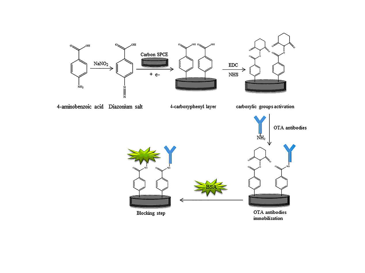
Electrografting is a common method of producing sensors and, due to the controllable nature of the process, the polymer films obtained are good in terms of thickness and stability. In the early stages of electroplating, cations are formed from the p-aminobenzoic acid monomer which forms an intermediate with the other unoxidised 4-aminobenzoic acid monomer. This intermediate can react with both the unoxidised 4-aminobenzoic acid and the p-aminobenzoic acid containing the cation, allowing the reaction to continue. If the concentration of para-aminobenzoic acid is too high, the film will be too thick and the sensitivity of the sensor will be reduced. Conversely, if the concentration is too low, the film is too thin or does not cover the electrode surface at all and can easily peel off, affecting the subsequent modification process.
In the experiments, the concentration of p-aminobenzoic acid was gradually increased and the effect of the modification was determined by the amount of change in response current before and after the modification. After electrodeposition, the electrodes were characterized by cyclic voltammetry and impedance. As shown in Fig. 3, the response current gradually increased with increasing concentration of p-aminobenzoic acid, with the maximum change in response current at 2 mmol/L, followed by a gradual decrease. This was attributed to the insulating property of p-aminobenzoic acid. Therefore, the optimal concentration of p-aminobenzoic acid was chosen as 2 mmol/L.
As shown in Fig. 4 A), the electrical grafting results in the formation of a film with insulating properties, with a reduced oxidation current and an increased reduction current. This result is consistent with the increased impedance value of the EIS plot in Fig. 4 B).
Deposition times of antibodies
To ensure that optimal test conditions were achieved, the deposition time of the self-assembled antibody under direct competition conditions was selected by varying the deposition time and observing the magnitude of the change in the cyclic voltammetry peak current. In this experiment, antibodies were deposited for 30 min, 45 min, 60 min, 75 min and 90 min followed by determination of the electrode CV response current values.
As can be seen in Fig. 5, the amount of change in the current response value increased with increasing antibody deposition time. When the deposition time reached 60 minutes, the amount of change in current reached a maximum value and then decreased. This is due to the fact that more and more antibody is modified on the electrode as the deposition time increases. This leads to an increase in the current response due to antigen-antibody specific binding. However, too long a deposition time will result in reduced antibody activity, limited binding to the antigen and ultimately lower current values. Therefore, 60 minutes was chosen as the antibody deposition time.
Deposition time of BSA
BSA solution is a commonly used blocking solution in immunohistochemistry experiments, which can block the non-specific binding sites and effectively avoid false positives. In the experiment, the peak CV current response was measured after BSA deposition for 10, 15, 30, 45, 60 and 75 minutes. As shown in Fig. 6, the magnitude of the response current change gradually increased as the deposition time increased and the protein film gradually formed. The maximum value was reached when the deposition time reached 60 minutes. Beyond 60 minutes, the non-conducting protein film will reduce the electron transfer rate and cause the response current change value to decrease, so 60 minutes is chosen as the best deposition time.
Electron diffusion in the electrode
CV were obtained at scan rates of 0.05, 0.1, 0.2, 0.25, 0.3, 0.4, 0.5, 0.6, 0.7, and 0.8 V/s using the optimal experimental conditions obtained in the above steps. The relationship between the peak redox current and the square root of the scan rate is shown in Fig. 7A). The peak redox current increases as the scan rate increases, the position of the peak becomes progressively more stable and separated, and the electron transport process is controlled by diffusion. The linear regression equation, as shown in the Fig. 7B).
Linear regression equation for the oxidation peak, y=4.179E﹣4x+2.619E﹣5,R2 =0.9999. Linear regression equation for the reduction peak, y=﹣4.349E﹣4x-4.568E﹣5, R2 =0.9981.
Electrochemical characterization
Characterisation of electron transfer from modified electrodes by cyclic voltammetry. First, each step of the modified electrode was characterised using 5 mmol/L [Fe(CN)6]3-/4+ (1:1) with 1 mol/L potassium chloride. Electrochemical impedance, as a label-free technique, is suitable for studying changes in the electrode surface in rapid bioassays. Therefore, the process of change at each electrode was further characteriszed by ELS. Potassium ferricyanide and potassium ferricyanide are used as electrochemical probes and potassium chloride as electrolyte.
As shown in Fig. 8 A), 4-aminobenzoic acid itself is non-conductive and, when modified on the electrode, creates a barrier to electron transfer at the electrode surface. When the carboxyl group is activated, the conductivity is significantly increased. After modification with Anti-OTA and BSA, the peak current is significantly reduced. After the addition of the ochratoxin solution, the specific binding of the antigen and antibody leads to a further decrease in the electron transfer rate as the contact between the working electrode surface and the redox pairs in the electrolyte is hindered. The reduction peak current gradually increases and the oxidation peak current gradually decreases, while the reduction peak potential shifts negatively, which is a characteristic feature of electrocatalysis.
The semicircular area in the electrochemical impedance diagram is the high frequency region, indicating restricted charge transfer. Its diameter represents the resistance to charge transfer and the value depends on the insulating capacitance between the modified electrode and the electrolyte interface. This is shown in Fig. 8 B). The semicircle in the impedance diagram of the bare electrode is very small, indicating good conductivity of the electrode at this point. When 4-aminobenzoic acid was modified, [Fe(CN)6]3-/4+ had difficulty reaching the electrode surface and the impedance value increased. After subsequent activation of the carboxyl groups on the electrode surface with MES buffer, the electron transfer rate increased and the impedance value decreased significantly. After modification of the antibody on the electrode surface, the impedance increased again. BSA blocking solution was then used to seal the unbound sites and the non-conductivity increased. After dropwise addition of OTA solution, antigen-antibody specific binding inhibited electron transfer and the impedance increased, confirming successful antigen-antibody binding.
Morphological characterization
To study the morphology of the different materials deposited on the electrode surface at each stage, scanning electron microscopy was used to characterise them. Fig.9 A) shows the morphology of the bare electrode surface with the presence of typical flaky graphite particles and large voids. B) and C) show the electrode surface after diazotization, which starts to become rough. This is due to the formation of 4-carboxyphenyl on the electrode surface and the uneven charge distribution leading to the formation of flaky graphite particles of uneven size. D) It shows that the antibody is deposited on the electrode surface. Because the amino group on the antibody forms an amide bond with the carboxyl group on the electrode surface, the antibody can be firmly fixed on the electrode surface. E) It can be seen that the electrode surface sealed with BSA solution has an obvious network structure and the BSA is evenly distributed on the surface. F) The electrode surface after antigen-antibody specific binding is shown, which is obviously different from the surface after sealing.
DPV detection of OTA by immunosensors
A series of ochratoxin standard solutions were prepared using 0.1 mol/L PBS solution at pH 7.4, added dropwise to the electrode and incubated for 20 minutes at room temperature. 5 mmol/L [Fe(CN)6]3-/4+ (1:1) solution containing 0.1 mol/L potassium chloride was used to determine the peak currents by differential pulse voltammetry. A standard curve was constructed from the relationship between the amount of change in current values before and after spiking and the corresponding OTA concentration. As shown in Fig. 10 A).
As shown in Fig. 10 B), the peak current response showed a good linear relationship for OTA concentrations in the range of 2-200 ng/mL. The linear regression equation was ∆i = 1.1196E-7COTA + 2.543E-6, with a linear correlation coefficient of R2 = 0.9970 and a lower limit of detection of 0.5 ng/mL.
Specificity of immune sensors
To test the specificity of the sensors, incubations were carried out using standard solutions of 10 ng/mL OTA, aflatoxin B1 (AFB1), dexamethasone enol (DEX), zearalenone (ZEN) and fumonisin B1 (FB1), respectively. The specificity was evaluated by measuring the DVP current response values. As shown in Fig. 11 A), OTA had the highest current response value and no more than 4.27% change in current among the fungal toxins for which controls were performed. Equal amounts of AFB1, DEX, ZEN and FB1 were then added to the OTA solution and the results are shown in Fig. 11 B). The amount of change in current response values did not exceed 4.35% under the conditions of addition of the many interfering fungal toxins.
The above mycotoxins were configured into different concentrations of standard solutions and the DPV assay was performed with an immunosensor and the results are shown in Fig.11 C). Except for OTA, which showed a linear relationship between the amount of current change and concentration, the changes in several other fungal toxins were not significant. This suggests that the immunosensor has good specificity for OTA.
Stability of immune sensors
In addition to good sensitivity and specificity, the sensor must have a certain degree of stability. The prepared immunosensor was subjected to 15 consecutive CV scans, as shown in Fig.12 A), with a corresponding change in current of no more than 4.62%. In addition, as shown in Fig.12 B), the prepared immunosensor was kept sealed at 4°C and CV scans were performed every 2 days under the same experimental conditions. As shown in Fig.12 C), after 2 weeks the current in the test was essentially unchanged and was 95.77% of the value tested before the electrodes were refrigerated. The reason for the high stability of the immunosensor is that the antibody and activated carboxyl groups are attached to the electrode surface by strong amide bonds, effectively preventing detachment and ensuring detection performance.
Testing of samples
In this experiment, maize, wheat, rice, coffee beans and red wine were selected as the samples for testing. After a simple pre-treatment, the samples were spiked with 10 ng/mL, 20 ng/mL and 50 ng/mL. Three parallel experiments were performed under the same conditions for each group of samples and the results are shown in the table. The recoveries of the five sets of samples at the three concentrations ranged from 90.54 to 100.92%. The electrochemical immunosensor is highly stable and its detection performance does not change significantly within half a month. It was confirmed that the sensor can accurately quantify OTA in cereal and wine samples.