Characterization of silver nanoparticles
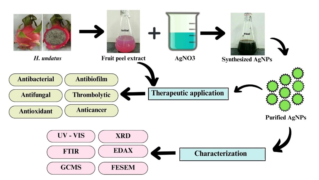
The current study revealed that the H. undatus fruit peel extract to the silver in the form of nitrate. This interaction resulted in the formation of a pale pink to dark brown indicating the presence of silver nanoparticles [24] as shown in Fig. 1. Similar color changes have also been observed in the previous study [16]. The UV-vis spectra were recorded after time intervals of 20 min, 25 min, 30 min, 35 min, 49 min, 45 min, 50 min, 55 min, 60 min, 65 min and 70 min. Figure 2 shows the absorption spectra of AgNPs using H. undatus fruit peel extract. The UV spectrum has exhibited a strong peak at 417 nm which confirms the formation of AgNPs.
The purity of the synthesized NPs was checked by FTIR analysis within the range of 400–4000 cm− 1 [25]. Figure 3 shows the FTIR spectra of biosynthesized AgNPs and the absorption peak bands at 3734.19, 3332.99, 2310.72, 1593.20, 1365.60, 1323.17, 1238.30, 1041.56, 921.97, 821.68, 678.94, 601.79, 559. 36. These absorbance bands are known to be associated with stretching vibrations of O-H (alcohols), O-H (alcohols), C = N (nitriles), C-C (aromatics), C-H (alkanes), C-N (aromatic amines), C-N (aliphatic amines), C-O (alcohols), O-H (carboxylic acids), C-Cl (alkyl halides), C-Br (alkyl halides), C-Br (alkyl halides), C- Br (alkyl halides). H. undatus fruit is primarily composed of betanin, pectin, hylocerenin, betacyanin and phyllocactin [26]. This study has confirmed the presence of a significant amount of phytocompounds and these substances may potentially be responsible for the reduction, capping and synthesis of AgNPs.
The GC-MS analysis of the peel extract of H. undatus was conducted to determine the bioactive chemical compounds present in the extract. Figure 4 displays the GC-MS spectrum with peaks and retention time. The peel extract of H. undatus has shown the presence of the major seven bioactive compounds such as 6-methyl-2, (4-bromophenyl)-7-phenylmethylindolizine, N-[(4,6-Dimethoxynaphthalen-1-yl) methylene], 2,5-dichloro-4-hydroxyphenylamine, Oxacycloheptadec-8-en-2-one, Androstan-17one, 3-ethyl-3-hydroxy, Pyridine-3-carboxamide, 6-chloro-4-trifluoromethyl-N-[2,4-dichloeo-6-methyl]-N-methyl, 1,2-Dipalmitoyl 3-acetyl glycerol Gallic acid, and Triamcinolone acetonide. The peak intensity of N-[(4,6Dimethoxynaphthalen-1-ylmethylene], 2,5-dichloro-4-hydroxyphenylamine was found to be higher with a retention time of 30.12 min followed by Oxacycloheptadec-8-en-2-one with a retention time of 30.74 (Table 1).
Table 1
Chemical composition of H. undatus peel extract
| S.No | RT | Compound name | Molecular formula | Molecular weight | Peak area % |
| 1 | 27.99 | 6-methyl-2,(4-bromophenyl)-7- phenylmethylindolizine | C22H18BrN | 376.289 | 7.991989 |
| 2 | 30.12 | N-[(4,6Dimethoxynaphthalen-1- ylmethylene], 2,5-dichloro-4- hydroxyphenylamine | 𝐶19𝐻15𝐶𝑙2𝑁𝑂3 | 376.233 | 8.799596 |
| 3 | 30.74 | Oxacycloheptadec-8-en-2-one | C16H28O2 | 252.392 | 15.430418 |
| 4 | 30.88 | Androstan-17one 3-ethyl-3- hydroxy | C21H34O2 | 318.49346 | 13.414466 |
| 5 | 31.49 | Pyridine-3-carboxamide, 6- chloro-4-trifluoromethyl-N-[2,4- dichloeo-6-methyl]-N-methyl | C15H10C13F3N2O | 397.606 | 11.854260 |
| 6 | 31.80 | 1,2-Dipalmitoyl 3-acetyl glycerol Gallic acid | C37H70O6 | 610.948 | 11.854137 |
| 7 | 32.00 | Triamcinolone acetonide. | C24H31FO6 | 434.497 | 10.116891 |
The synthesized AgNPs are characterized using XRD to confirm the presence of silver ions and to know the structural information [27]. Figure 5 shows the XRD pattern of AgNPs which confirmed the crystalline nature of synthesized AgNPs. The peaks at 2𝜃 of 27.57, 32.09, 37.88, 44.65, 46.02, 54.59, 57.19, 64.16, 67.32, 72.62, and 76.42 correspond to the crystalline planes of the face-centered cubic structure of metallic silver. The intense peak at 32.09 possibly suggests the presence of silver ions as the major constituent in the biosynthesized AgNPs. A similar result was observed by Phongtongpasuk et al. [16] who identified the most intense peak at 32.5 indicated the presence of AgNPs.
The elemental analysis of the material is depicted in Fig. 6, illustrating the synthesized AgNPs through the EDAX spectrum [28]. This analysis demonstrates a prominent indication of a metallic silver area at 3 KeV and confirms the formation of silver nanoparticles synthesized through the utilization of H. undatus peel (Fig. 6). The Ag peak showed a weight percentage of 35.12 and an atomic percentage of 8.02. A low calcium signal was observed and five moderate signals for carbon, oxygen, sodium, chloride and potassium were detected as a result of the chemicals used in the sample preparation.
FE-SEM images of AgNPs synthesized using the fruit peel extract of H. undatus are shown in Fig. 7. The surface morphology of AgNPs showed a spherical shape with agglomeration. In the present study, the histogram of the particle size ranges from 52–79 nm. The increased concentration of bioactive compounds in the colloidal solution could potentially lead to the formation of nanoclusters. The signals produced from the interaction between the electrons and the sample provide valuable information regarding the sample's external morphology, chemical composition, crystalline structure and material orientation.
Thrombolytic activity
The thrombolytic activity of AgNPs and H. undatus peel extracts was evaluated at three different concentrations. The percentage of thrombolysis was found to be 10, 32.36 and 56.25% for the AgNPs at the concentration of 20, 40 and 80 µg/mL respectively (Fig. 11). Distilled water was taken as a negative control resulting in clot lysis of 6.07% while streptokinase was taken as a positive control exhibiting a clot lysis of 50%. AgNPs may be involved in activating the enzymes that generate plasmin, an enzyme capable of breaking down the cross-links between fibrin molecules and dissolving blood clots [34]. Limited research has been conducted on the use of AgNPs as a thrombolytic agent. The study revealed that as the concentration of AgNPs increased and the percentage of the clot lysis was also increased and it also highlights the potential of AgNPs suggesting that they could have valuable applications in the clinical field for preventing thrombosis and other related disorders.
Figure 11 Thrombolytic activity of AgNPs and H. undatus peel extracts
Antioxidant activity
The antioxidant activity of silver nanoparticles and H. undatus peel extracts was determined by the DPPH method. The AgNPs and H. undatus peel extracts were compared based on their resulting IC50 values. Silver nanoparticles exhibited maximum radical scavenging activity (IC50 3.8 µg/ml), while H. undatus peel extract exhibited the IC50 value of 2.03 µg/ml (Fig. 12). The inhibition % of nitric oxide radical scavenging activity by the AgNPs of H. undatus at different concentrations (20, 40, 60, 80 and 100 µg/mL) was compared based on their resulting IC50 values. AgNPs exhibited maximum radical scavenging activity (IC50 2.8 µg/mL), while H. undatus peel extract exhibited the IC50 value of 2.3 µg/ml (Fig. 13). There is a growing demand for the development of cost-effective, eco-friendly techniques to synthesize metallic nanoparticles with high yields and low toxicity. Several studies have reported the reduction of silver ions into AgNPs using plant extracts. It contains biologically active phytochemicals like terpenoids, flavonoids, vitamins and phenolics which are known for their antioxidant properties [35]. These compounds have a diverse range of biological activities and help to protect cells from damage caused by reactive oxygen species. Our study provides clear evidence that the synthesized nanoparticles and fruit peel extract exhibited radical scavenging activity. AgNPs showed promising antioxidant properties when compared to the fruit peel extract. The presence of numerous bioactive compounds in fruit extract may be the reason for its antioxidant activity [36]. The interaction of plant metabolites with metal ions can lead to the production of enhanced compounds that scavenge free radicals. Negatively charged phytochemicals and positively charged AgNPs work together to enhance the bioactivity of plants through electrostatic attractions [35]. Previous research has also indicated that antioxidant activity tends to increase as the treatment doses increase [36].