Sample collection
This cross-sectional, case-control study was conducted in Modarres hospital, Tehran, Iran, on cases archived between 2008 and 2019 years. A total of 70 blocks of Formalin-Fixed Paraffin-Embedded (FFPE) including 59 samples diagnosed as breast carcinomas, and 11 benign breast lesions as control were collected from the pathology department archives of Modarres hospital. Also, several parameters; such as type of BC, grading of breast cancer, age, and level of education were taken in written format as exclusion criteria and summarized in Table 1. All study procedures are performed in laboratories of Shahid Beheshti Medical School of sciences.
DNA Extraction
The genomic DNA was extracted from FFPE breast tissues. Then we performed a standard polymerase chain reaction (PCR) to detect HPV DNA. To extract DNA, FFPE tissues were cut in 10 μm thickness by a microtome. Deparaffinization was performed by adding 1 ml xylene and spinning. Afterwards, we centrifuged the samples for 5 min, and supernatants were removed. This step was repeated once, then 1 ml of 96% ethanol was added. Microtubes were put in a 50ºC heating block until ethanol was entirely dried up. Then digestion was performed by adding digestion buffer and proteinase K solution. Later, the microtubes were incubated overnight, and the next day they were placed in a 95 ºC heater. Respectively phenol, phenol-chloroform, and chloroform solutions were added to samples, each step followed by centrifugation, and subsequent removal of the supernatant. Ultimately, ethanol was added, then they were kept overnight in an incubator.
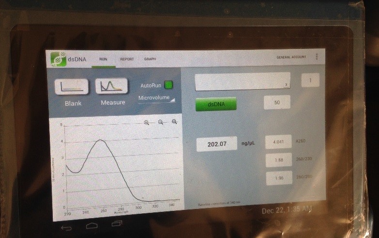
Samples were centrifuged at 12000 g for 30 min on the last day of the extraction. After removing the supernatant, the uncapped microtubes were left in a 37 ºC heater to let the residual ethanol to evaporate. Finally, distilled water was added and the DNA extracts were quantified with a NanoDrop spectrophotometer.
The quality of DNA extracted from paraffin-embedded tissue samples was assessed by a forward (ATGTTCGTCATGGGTGTGAA) and reverse (GGTGCTAAGCAGTTGGTGGT) primer pair targeting a sequence within the GAPDH gene. PCR amplification protocol consisted of 30 cycles of denaturation at 95 °C for 30 s; hybridization at 55°C for 30 s, and elongation at 72°C for 30s. A final elongation step was performed at 72 °C for 10 min.
PCR for HPV and herpes viruses
All samples were screened for HPV L1 conserved region. The nested PCR assay was performed using two sets of primers (MY09/11 and GP5+/6+) for two consecutive amplification reactions. The first reaction was performed in 25 μl using 12.5 μl of master mix (which includes:1X PCR buffer, 2 mM MgCl2, 50 μM of each deoxynucleotide triphosphate (dNTP) and 2 U of Taq DNA polymerase (Takapouzist, Iran), 100-200 ng of template DNA, 10 pmol of each consensus outer degenerate primer MY09(5’-CGTCC(A/C)A(A/G)(A/G)GGA(A/T)ACTGATC -3’) /MY11(5’-GC(A/C)CAGGG(A/T)CTATAA(C/T)AATGG -3’) and distilled water. Thermal cycling (Bio Intellectica) performed with the following program: 5 min at 94 °C, 40 cycles of 1 min at 94 °C, 1 min at 55 °C, and 1 min at 72 °C, with a final extension step at 72 °C for 7 min.
The second reaction was also performed in 50 μl including 25 μl of master mix (which contains: 1X PCR buffer, 3 mM MgCl2, 50 μM each dNTP, 2U Taq DNA polymerase), 100-200 ng of amplified DNA, and 10 pmol each inner consensus primer GP5+ (5’- TTTGTTACTGTGGTAGATACTAC-3’) and GP6+ (5’-AAAAATAAACTGTAAATCATATTC-3’), and distilled water. Thermal cycling used the following program: 4 min at 94 °C, 40 cycles of 1 min at 94 °C, 2 min at 40 °C, and 2 min at 72 °C, with a final extension step at 72 °C for 4 min.
Negative controls containing water instead of DNA were used. We used the HPV18 HeLa cell line as a positive control.
Real-time PCR amplification was performed in a 20 μl volume containing 4 μl 5x HOT FIREPol® Probe qPCR Mix Plus (no ROX) (Solis BioDyne, Estonia), 0, 5 μl of each forward and reverse primers (Pishgam company, Tehran, Iran), 0.5 μl of probes (Sinaclon Co., Tehran, Iran) which listed in Table 2, and 2 μl of each sample or control, and rest of the total volume was obtained by adding distilled water. Amplification and detection were performed by a real-time PCR machine (Rotor-Gene-Q 6000 thermocycler (Corbett, Australia)). Thermal cycles used for quantification of HPV18 E6 gene and HPV type 16 E7 gene were 95°C for 12 minutes, 95°C for 15 seconds, and 60°C for 60 seconds for 40 cycles.
Duplicate reactions for each gene were performed. Probes were labeled with 6-FAM at the 5' end and TAMRA at the 3' end.
Each PCR was performed with negative (DNA free water) and positive controls (genomic DNA of SiHa cells for HPV-16, and genomic DNA of HeLa cells for HPV-18).
The samples were also subjected to a multiplex PCR to detect HSV-1, HSV-2, VZV, and CMV. The PCR reactions were performed in a total volume of 25 μL containing 12.5 μl PCR Master Mix (10X PCR Taq polymerase buffer, 10mM of dNTPs and 1.5 U/rxn of Taq DNA polymerase [Takapouzist, Iran]), 10 pmol of each primer (HSV-1, HSV-2, CMV, and VZV) (Table2), and 50 ng of genomic DNA.
mPCR (multiplex PCR) conditions were as follows: Initial denaturation (95°C for 5min), 35 cycles of denaturation (95°C for 30sec) annealing (60°C for 30 seconds) and extension (72°C for 30 seconds); Final extension was given at 72°C for 10 minutes. Samples were preserved at 4°C and then the PCR product was detected by gel electrophoresis.
Sequencing and Phylogenetic Analysis
The positive PCR samples were sequenced for HPV genotypes. The DNA sequence was determined with the Big-Dye terminator cycle sequencing kit and an ABI 377A sequencer (Applied Biosystems Inc.).
The HPV sequences were edited with the BioEdit program version 7.2.3, and then phylogenetic and molecular analyses were organized using MEGA software program version 6.0.6 [27]. The neighbor joining and the Kimura 2-parameters methods were used for phylogenetic reconstructions that were implemented in the MEGA 6.0.6 program. Statistical significance for the phylogenetic tree was assessed by the bootstrap method (1000 replicates).
Data were analyzed by SPSS statistical software program version 16.0. The correlations were subjected to χ2 (Pearson chi-square) and Fisher’s exact test. Odds ratios and logistic regression were also calculated. Statistical significance was set as a P-value less than 0.05.
Deposit the nucleotide sequence
The nucleotide sequences of HPV isolates that were found out in this study have been settled in the GenBank database [accession numbers QED55703– QED55709]. The GenBank accession numbers for HPV6 types are QED55703 and QED55704, and for HPV18 types are QED55705, QED55706, QED55707, QED55708, and QED55709.