Osteosarcoma (OS) is a primary malignant bone tumor that emerges most often at the wide end of long bones. It usually occurs in children and teenagers but it can also affect adults [1]. The etiology of osteosarcoma includes epidemiologic variables, hereditary disabilities, and environmental factors [2]. The current therapy for osteosarcoma is a combination of therapies including surgery, chemotherapy, and radiation therapy. Although, surgery could be helpful in tumor removal, surgical incision will cause a huge region of bone defects, making it troublesome for the body to repairs itself, leading to serious problems and long-lasting inabilities for patients. A further problem is that the surgery couldn’t ensure expulsion of all tumor tissue. In this manner, osteosarcoma conventional therapy is a big remaining challenge. Therefore, novel methodologies are required to reduce the recurrence and metastasis induced by leftover tumors, and postoperative chemotherapy could be useful [3]. Methotrexate is one of the main effective drugs in the treatment of OS, which is also used as adjuvant chemotherapy [4]. However, the side effects of this drug such as digestive problems, headache, shortness of breath, mouth ulcers and, etc., as well as the low solubility of it can be a problem [5]. The use of microsphere-based drug delivery systems could minimize the side effects of the drug and deliver it to the targeted site in the long term. Poly lactic-co-glycolic acid (PLGA) microspheres are among the effective drug delivery systems in the field of cancer treatment. The high degradability and biocompatibility of this polymer have made it widely used in the field of drug delivery [6].
Bone tissue engineering is a multidisciplinary field of science focusing on developing strategies to overcome the bone defects problem [7]. One of the basic components in tissue engineering is a scaffold. The scaffold have a great role by being placed in the damaged area caused by the tumor and creating a substrate for the placement of healthy cells .[8, 9] It can also be a platform for the drug delivery, including anti-cancer drugs and antibacterial agents [10, 11].
Scaffolds are composed of various materials such as ceramic, metal, polymer and, etc. Sometimes combined material scaffolds are used to take advantage of the appropriate features of each category material [12]. GelMA (Gelatin MethAcryloyl) is used in the category of polymeric scaffolds, which has many advantages including light curing facilities, high degradability and biocompatibility [13, 14]. However, it is hard to use it alone in bone scaffolds due to its low mechanical strength[15]. Utilizing GelMA together with ceramic materials such as hydroxyapatite nanoparticles and nanosilica can lead to the construction of a more favorable scaffold [16, 17]. Alginate also has excellent properties such as high degradability, non-toxicity, and gel-forming ability [18, 19]. Alginate can be modified to have different mechanical properties, making it suitable for a range of applications and an attractive option for tissue engineering applications [18]. Using alginate in bone scaffolds has the potential to improve bone regeneration and repair [20]. Research has shown that the presence of hydroxyapatite nanoparticles in composites stimulates osteogenesis around the tissue and implant. Also, using hydroxyapatite in bone tissue engineering increases the strength of the scaffold [21]. Silica is one of the effective substances in stimulating osteogenesis. The silanol groups presented on the nanosilica surface in the scaffold lead to an increase in its stability in aqueous environments and also provide a platform for the formation of apatite. The electrostatic bond between (Si-O-) and Ca+ causes the formation of a layer rich in positive charge of calcium and creates a bond with the negative charge of phosphate and causes the formation of amorphous calcium phosphate and finally constructing the structure of apatite [22]. Moreover, adding nanosilica to the scaffold increases the biocompatibility of the scaffold [23].
3D printing is considered as a new technology in the science of tissue engineering, which can be used for various therapeutic purposes. This device can prepare a three-dimensional scaffold corresponding to the damaged lesion, which has the desired characteristics [24, 25]. In the current study, 3D printing was used to prepare a bilayer scaffold in order to print the desired scaffolds in the desired shape and dimensions, rapidly and accurately.
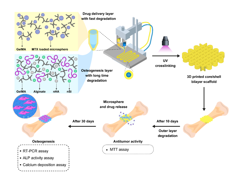
The overall objective of this study was to fabricate an implanted scaffold for osteosarcoma anticancer drug delivery and bone regeneration. To this end, we used 3D printer to prepare a core/shell bilayer scaffold by nano/microspheric hydrogel ink. The scaffolds were assessed for drug release, antitumor activity, cell viability, proliferation, cell adherency, alkaline phosphatase, calcium deposition assay, and osteogenic bone expression.