ADME analysis of the phytochemicals
Natural compounds represent a valuable reservoir for drug development, owing to their vast diversity and intricate structures. This significance is further underscored by the limited number of plant species investigated for pharmacological purposes [42]. ADMET analysis is used to study the pharmacokinetics of these phytochemicals thereby evaluating their risk or negative effect on the human body [43].
In order to select the best, druggable and lead-like phytochemicals for the progression of this study, the selected phytochemicals were subjected to ADME analysis using the pKCSN server.
Subsequent toxicity studies unveiled that all the chosen phytochemicals were predicted to be devoid of hepatotoxic effects and mutagenic potential, as indicated by the negative results in the Ames test which is a key assay designed to identify compounds with mutagenic properties [44]. The determination of the Maximum Tolerated Dose (MRTD) values, representing the highest dose of a phytochemical that can be administered to a patient without inducing undesirable side effects or toxicity (with a cutoff value of 0.477 for categorizing MRTD), was pivotal. All the examined phytochemicals exhibited MRTD values below 0.477, signaling their safety for administration at high doses without causing side effects or toxicity. Specifically, Withaferin A, Withanolide D, and Withanolide F showcased MRTD values of -0.695, -0.867, and − 0.701 log mg/kg/day, respectively, indicating a higher tolerance for elevated doses. In contrast, Catechin, Epicatechin, and Isolariciresinol displayed MRTD values of 0.438, 0.438, and 0.134, respectively, suggesting a comparatively lower tolerance for higher doses (Table 3).
Table 3
Presenting pharmacokinetic properties of Selected Withania somnifera and Saraca asoca phytochemicals
|
Selected Plants phytochemicals
|
Intestinal absorption (human)
|
volume of distribution at steady state (human) (log L/kg)
|
Metabolism
|
Excretion
|
Max. tolerated dose (human) (log mg/kg/day)
|
Hepatotoxicity
|
AMES toxicity
|
|
Withaferin-A
|
85.345
|
-0.131
|
CYP3A4 (cytochrome P450 3A4) substrate
|
Renal OCT2 substrates.
|
-0.695
|
NO
|
NO
|
|
Withanolide-D
|
99.2
|
-0.048
|
CYP3A4 (cytochrome P450 3A4) substrate
|
Total clearance
0.347 (log ml/min/kg)
|
-0.867
|
NO
|
NO
|
|
Withanolide-F
|
95.535
|
0.067
|
CYP3A4 (cytochrome P450 3A4) substrate
|
Total clearance
0.486 (log ml/min/kg)
|
-0.701
|
NO
|
NO
|
|
Catechin
|
68.829
|
1.027
|
NOT METABOLIZED BY CYP2C19 and CYP3A4
|
Total clearance
0.183 (log ml/min/kg)
|
0.438
|
NO
|
NO
|
|
Epicatechin
|
68.829
|
1.027
|
NOT METABOLIZED BY CYP2C19 and CYP3A4
|
Total clearance
0.183 (log ml/min/kg)
|
0.438
|
NO
|
NO
|
|
Isolariciresinol
|
73.973
|
-0.145
|
CYP3A4 (cytochrome P450 3A4) substrate
|
Total clearance
0.177 (log ml/min/kg)
|
0.134
|
NO
|
NO
|
The excretion pharmacokinetic parameter of the phytochemicals was assessed, focusing on the Total Clearance—a metric that signifies the overall rate at which a substance is eliminated from the body, encompassing both renal and non-renal processes. Notably, the total clearance values for Withanolide-D, Withanolide-F, Catechin, Epicatechin, and Isolariciresinol were determined as 0.347, 0.486, 0.183, 0.183, and 0.177 log ml/min/kg, respectively, indicating a comparatively higher rate of elimination from the body. In contrast, Withaferin-A was predicted to undergo renal excretion, supported by its classification as a Renal Organic Cation Transporter 2 (OCT2) substrate. OCT2 is a crucial protein involved in the renal excretion process of compounds categorized as its substrates [45].
The metabolic profile of the phytochemicals was assessed, revealing distinctive patterns. Withaferin-A, Withanolide-D, Withanolide-F, and Isolariciresinol were anticipated to undergo metabolism mediated by cytochrome P450 3A4 (CYP3A4), an enzyme within the cytochrome P450 family known for its involvement in the metabolism of diverse endogenous compounds and drugs [46]. Compounds metabolized by CYP3A4 are classified as CYP3A4 substrates. Conversely, Catechin and Epicatechin were identified as non-substrates for both CYP2C19 and CYP3A4 enzymes. However, it is noteworthy that these phytochemicals could potentially be metabolized by other enzymes, necessitating further investigations to elucidate their complete metabolic pathways.
The distribution profiles of these phytochemicals within the human body were examined, with a focus on the prediction of the volume of distribution at steady state (VDss). The findings, where VDss values below − 0.15 log L/kg are considered high and values above 0.45 log L/kg are deemed low, indicated distinctive distribution patterns. Withaferin-A, Withanolide-D, Isolariciresinol, and Withanolide-F demonstrated VDss values of -0.131, -0.048, -0.145, and 0.067 log L/kg, respectively. These values suggest a propensity for low distribution in tissues and organs, with a higher concentration expected in the bloodstream. Catechin and Epicatechin exhibited predicted VDss values of 1.027 log L/kg each, indicative of high distribution in tissues and organs. These varying distribution profiles underscore the diverse pharmacokinetic behaviors of the studied phytochemicals within the biological system.
The intestine stands out as the principal site for drug absorption from orally administered solutions [47]. In our study, the predicted intestinal absorption of these phytochemicals demonstrated uniformly high rates, ranging between 68% and 99.2%. Notably, Withanolide-D exhibited the highest predicted absorption rate, reaching 95%, followed closely by Withanolide-F and Withaferin-A with absorption rates of 95.5% and 85.3% respectively.
Molecular docking analysis
Molecular docking is an in-silico approach designed to predict and calculate the chemical interactions between macromolecules (proteins) and small compounds (ligands). It has proven to be an exceptionally successful method for screening a diverse range of compounds and discovering novel medicines targeting specific proteins. Of particular interest is protein-ligand docking, widely employed in the pharmaceutical industry [48].
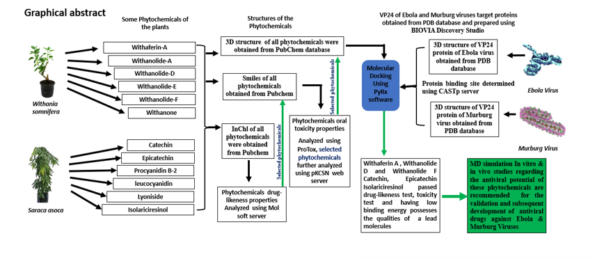
In our investigation, the selected proteins (VP24) play a crucial role in the survival of Ebola and Marburg viruses [30]. Withania somnifera and Saraca asoca are recognized for possessing medicinal properties, including anti-viral effects [21, 49]. To explore their potential as antiviral agents, we focused on six key phytochemicals from each plant, conducting analyses for drug-likeness, oral toxicity, and their binding affinity with the designated target proteins.
The selection criteria ensured that the six chosen phytochemicals adhered to Lipinski's rules and exhibited predicted non-toxicity along with optimal absorption, distribution, metabolism, and excretion profiles. These compounds were then designated as ligands for molecular docking studies with the target proteins. The objective of the docking analysis was to identify ligands capable of binding to the target protein's binding site and to discern their preferred and most advantageous binding poses. This systematic approach aimed to assess the potential therapeutic efficacy of the selected phytochemicals against the targeted proteins crucial for the survival of Ebola and Marburg viruses. The vina docking result generated eight conformations of each ligand that were ranked based on their energies and the conformation with the highest binding affinity was selected for further study.
Based on molecular docking analysis, any active compound with a minimum binding energy of -6.0 kcal/mol is considered to have the potential to induce pharmacological effects [50, 51]. The molecular docking results for Withaferin-A, Withanolide D, Withanolide F, Catechin, Epicatechin, and Isolariciresinol (ligands) revealed low binding energies of -10.0, -10.1, -9.1, -7.4, -7.2, and − 7.2 kcal/mol respectively with the Ebola virus VP24 protein (Fig. 1 and Fig. 2) indicating high binding affinity with the target protein and predicting a potential pharmacological effect (Table 4). Notably, Withanolide D exhibited the lowest binding energy, indicating the highest binding affinity toward the Ebola virus VP24 protein, followed by Withaferin-A and Withanolide F.
The strength of the protein-ligand interaction is intricately shaped by the array of bonds participating in complex formation. Specifically, the formation of hydrogen bonds between the ligand and target protein serves as a pivotal determinant of interaction strength. It follows that a higher count of hydrogen bonds corresponds to a stronger interaction [52], Emphasizing the crucial role of these molecular bridges in strengthening the bond between the ligand and its target protein, our investigation revealed robust hydrogen bonding interactions for all ligands with specific amino acids of the Ebola virus VP24 protein. These interactions involved key residues, such as ARG B:154, ARG A:154, GLU B:46, GLY A:44, GLY B:44, and PRO B:229, all situated at the active site of the protein.
Notably, Withaferin A formed hydrogen bonds with ARG B:154 and GLU B:46, Epicatechin formed bonds with ARG B:154 and GLY B:44, and Isolariciresinol established bonds with PRO B:229 and GLY A:44. Ligands Withanolide-D and Withanolide-F formed hydrogen bonds with ARG B:154 and ARG A:154, respectively, while Catechin formed a bond with GLU B:46. Importantly, ligands Withaferin A, Epicatechin, and Isolariciresinol established two hydrogen bonds, signifying a more potent interaction with the Ebola VP24 protein, compared to the ligands forming a single hydrogen bond. This detailed analysis highlights the specific amino acid residues involved and the varying strengths of interactions across different ligands. Furthermore, various additional bonds, such as Van der Waals, Pi-Anion, Pi-Cation, Alkyl, Pi-Alkyl, and unfavorable Donor-Donor bonds (refer to Table 4) were observed in some complexes, these bonds contribute significantly to the overall stability of the ligand-Ebola VP24 protein complexes. These diverse bonding interactions collectively enhance the binding affinity and structural integrity of the protein-ligand complexes, thereby substantiating the robustness of the predicted interactions.
As observed in our molecular docking analysis, Withaferin-A, Withanolide D, and Withanolide F exhibited notably lower binding energies with the Ebola VP24 protein compared to certain Indonesian natural compound ligands, as reported by Tambunan and Nasution [53]. Specifically, our results indicated − 10.0 kcal/mol, -10.1 kcal/mol, and − 9.1 kcal/mol binding energies for Withaferin-A, Withanolide D, and Withanolide F, respectively, in contrast to the binding energies of some Indonesian natural compounds such as Cycloartocarpin (-7.4847 kcal/mol), Letestuianin B (-6.4799 kcal/mol), Lissoclin A (-6.4729 kcal/mol), Varamine A (-6.4660 kcal/mol), and Lissoclibadin 4 (-6.3958 kcal/mol) in the study conducted by Tambunan and Nasution [53]. This suggests that Withaferin-A, Withanolide D, and Withanolide F could be anticipated to have a higher affinity for the Ebola VP24 protein, potentially leading to a more pronounced inhibitory effect.
The results of drug-likeness and molecular properties of the selected phytochemicals (Withaferin-A, Withanolide D, Withanolide F, Catechin, Epicatechin, and Isolariciresinol) coupled with high binding affinity and the strong interactions formed with the Ebola virus VP2 protein underscore the potential of these ligands in binding with the protein. This suggests a possible hindrance to the normal biological function of the virus protein, making them promising lead molecules for further investigation in antiviral activity through MD simulation, in vitro, and in vivo studies. These studies are crucial for the development of therapeutic drugs targeting diseases caused by the Ebola virus.
There are very few active clinical studies for the development of Marburg virus therapies/vaccines. Some of the prospective studies include modified vaccinia Ankara, chimpanzee adenovirus type 3, Marburg DNA plasmid vaccine, galidesivir and antisense phosphorodiamidate morpholino oligomers. Despite this, there are no licensed vaccines or treatments for the Marburg virus due to viral genetic diversity [54].
The interaction and binding energy of ligands with the Marburg VP24 protein, as presented in Table 5, revealed that Withaferin-A, Withanolide D, Withanolide F, and Catechin exhibited binding energies of -6.2, -6.7, -6.3, and − 6.5 kcal/mol respectively (Table 5), indicating a high binding affinity with the Marburg VP24 protein. Notably, Catechin formed three hydrogen bonds with GLY B:36, THR A:52, and GLU A:226, in addition to Alkyl bonds with PRO B:50, ALA A:225, B:227, and MET A:195, as well as Pi-anion bonds with specific amino acids of the Marburg VP24 protein.
Considering the collective impact of binding energy and the formation of these bonds, resulting in a robust interaction strength, the prediction is for the formation of a stable Catechin-Marburg VP24 protein complex. Thus, Catechin stands out as a potential lead molecule. Although Withanolide-D exhibited the lowest binding energy (highest binding affinity), it formed only an Alkyl bond with PRO B:67. Withanolide-F formed H-bonds with SER B:34 and THR A:55, while Withaferin A formed an H-bond with SER B:34.
However, Epicatechin and Isolariciresinol exhibited binding energies of -5.4 kcal/mol and − 5.2 kcal/mol with the Marburg VP24 protein, respectively, which are relatively higher than those of other ligands, suggesting lower binding affinity with the target protein. Nevertheless, Epicatechin formed three hydrogen bonds with THR A:39, GLU B:68, and PHE B:64, along with two Alkyl bonds with VAL B:66 and VAL A:35, and a Van der Waals bond with PRO A:33. On the other hand, Isolariciresinol formed H-bonds with GLN B:71 and GLU B:68, Pi-Anion bonds with LYS A:237 and GLU B:68, and Alkyl bonds with PRO A:240 and HIS A:48, along with van der Waals bonds with PRO A:240 and GLU A:231. These suggest that despite the lower binding affinity, both Epicatechin and Isolariciresinol have the potential to form strong and stable complexes with the Marburg VP24 protein. The 3D interaction of the ligands with the Murburg virus is illustrated in Figs. 3 &4.
The molecular properties of the ligands exhibit drug-likeness characteristics, and the docking results suggest that these phytochemicals could be considered as lead molecules with a binding affinity for the Marburg VP24 protein. This implies the potential for inhibitory effects, disrupting the normal functioning of the VP24 protein, and subsequently hindering virus replication. To validate and enhance our understanding of the antiviral potential, further investigations such as MD simulation, in vitro, and in vivo studies are imperative. These additional studies will provide comprehensive insights into the efficacy and safety of these phytochemicals, facilitating the development of potential drugs for the treatment of diseases caused by the Marburg virus.