Numerous studies have extensively investigated effects of maternal smoking during pregnancy on long-term health issues, particularly respiratory diseases. Several clinical research studies have found a correlation between maternal smoking and detrimental impacts on lung development, with these impacts subsequently increasing susceptibility to various respiratory disorders such as infections and asthma [35–37]. Consistently, it has been found that maternal smoking during pregnancy in mice can amplify oxidative stress and production of inflammatory cytokines in lungs of the offspring [38]. Additionally, perinatal exposure to CS can exacerbate allergic reactions induced by house dust mites and asthma via immune modulation [39]. Moreover, fetal exposure to second-hand CS can suppress mucus production and augments Th2 polarization, resulting in increased susceptibility to allergic asthma and childhood respiratory infections in mice [40]. Maternal smoking during pregnancy has detrimental effects on offspring mice, impacting not only their lungs, but also other organs. It has been documented that maternal smoking can induce alterations in proteome profile and cause mitochondrial defects in kidneys and livers of the offspring [41–44]. Moreover, maternal nicotine exposure can cause congenital heart defects and coronary artery malformation in fetal mice [45]. Recent studies have emphasized that harmful effects of CS on the fetus during pregnancy are not limited to traditional cigarettes because e-cigarettes also pose risks. It has been shown that maternal exposure to electronic cigarette vapor has particularly evident adverse effects on kidneys and lungs of fetuses [46, 47]. Epigenetic alterations, including DNA methylation, are among reasons why maternal exposure to CS or e-cigarettes has harmful effects on offspring mice [48–50]. In line with these findings, our study further revealed that smoking during pregnancy could impact livers of offspring mice, especially in terms of miRNA expression and associated CYP enzyme levels. Consistently, prenatal CS exposure increased Cyp2a5 gene expression and led to persistent and higher promoter methylation in neonates. This could result in heightened nicotine metabolism, making mice more susceptible to nicotine dependence later in life [51].
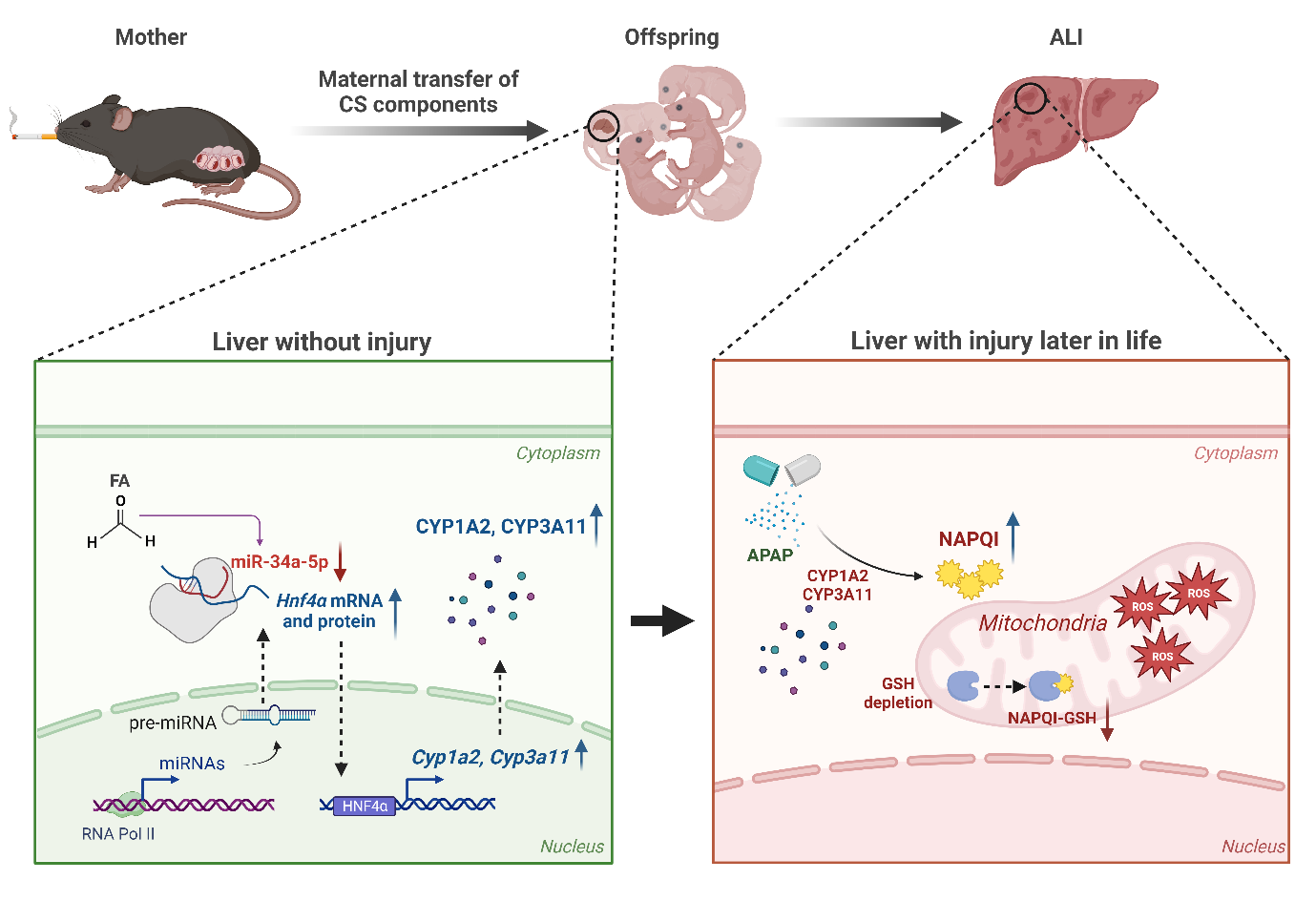
Interestingly, our results also revealed that maternal MSCS exposure made offspring mice more vulnerable to APAP-induced hepatotoxicity by regulating miRNA expression, especially miR-34a-5p. The role of miR-34a-5p has been investigated in various models of acute and chronic liver injury. Considering various results, the function of miR-34a-5p might vary depending on disease models and their progression stages. In non-alcoholic fatty liver disease with iron overload, silencing of miR-34a can increase Sirtuin 1 expression and consequently alleviate TG accumulation [52]. Moreover, miR-34a has been reported to function as a pro-fibrosis factor that can promote alcohol-induced liver fibrosis by reducing senescence of hepatic stellate cells (HSCs) and conversely increasing hepatocyte senescence [53]. However, some studies have shown evidence supporting hepatoprotective effects of miR-34a. Overexpression of miR-34a-5p has been found to be able to mitigate liver fibrosis development and progression by targeting Smad 4 in HSCs [54]. Recent studies have reported that miR-34a-5p can protect the liver from Ischemia/Reperfusion injury through downregulating HNF4α [33]. Furthermore, markedly enhanced liver injuries in mice with hepatocytes-specific miR-34a-5p deficiency in the APAP-induced ALI model strongly suggest that miR-34a-5p might play a critical role in the improvement of ALI [32]. This hepatoprotective function of miR-34a is consistent with our results showing that an increase of APAP-induced ALI by in utero CS exposure was alleviated by miR-34a-5p overexpression.
Mechanistically, we found that overexpression of miR-34a-5p significantly reduced protein levels of HNF4α and CYP3A11, leading to a decrease in the severity of APAP-induced ALI. Our results were further supported by recent studies indicating that HNF4α is a target of miR-34a-5p [55, 56]. Moreover, CYP enzymes including CYP1A2 and CYP3A11 known to play a pivotal role in drug metabolism are regulated by HNF4α [57]. Additionally, overexpression of HNF4α has been found to exacerbate APAP-induced hepatotoxicity [58]. Hence, the miR-34a-5p/HNF4α/CYPs axis might be crucial for drug metabolism and associated liver injuries. Consistent with this notion, our data showed that maternal smoking decreased expression levels of miR-34a-5p in offspring mice. This led to increased expression levels of HNF4α and CYP enzymes in the liver, making the offspring more susceptible to APAP overdose. Conversely, overexpressing miR-34a-5p completely mitigated adverse effects of prenatal exposure to CS on offspring mice owing to reduced levels of HNF4α, CYP1A2, and CYP3A11.
Based on our findings that maternal MSCS exposure could alter miRNA expression and affects subsequent ALI in offspring, it is plausible that many CS components exposed to pregnant mice are transmitted to the offspring, leading to a variety of unwanted side effects. Considering this, we endeavored to identify components that might downregulate the expression of miR-34a-5p in the liver of the offspring. Among many components of CS, we selected DEN and FA, both known to cross the placenta in mice [59–61]. DEN is known for its hepatotoxic effects and its potential to induce liver cancer. It undergoes metabolism by the enzyme CYP2E1, which is implicated in ROS production [62]. FA is recognized as a more potent carcinogen than DEN. It is present in the diet and ubiquitously present in the environment as well as in the CS. It is also found throughout the human body, participating in cellular metabolic processes such as histone and DNA demethylation reactions as well as the one-carbon cycle [63]. Prior research studies have indicated that FA exposure can modify miRNA expression across a range of cells and tissues [64, 65]. In line with this, our findings intriguingly revealed that FA treatment, but not DEN treatment, significantly diminished miR-34a-5p expression in primary hepatocytes. Moreover, while FA treatment enhanced APAP-induced hepatotoxicity, this detrimental effect was counteracted by overexpression of miR-34a-5p. Although FA treatment could not fully mimic effects of maternal smoking and MSCS, our findings indicate that FA can exacerbate APAP-induced hepatotoxicity in vitro, primarily through modulation of miR-34a-5p expression, consistent with in vivo observations. These findings are further supported by changes in protein expression of HNF4α, a downstream target of miR-34a-5p.
It has been already known that males tend to be less affected by hormones than females [66]. There is also a gender difference in basal expression of CYPs involved in APAP metabolism. For example, CYP1A2 and 2E1 are more active in males, whereas CYP3A4 displays an elevated activity in females [67]. Consistently, gender differences exist in susceptibility to ALI following APAP overdose, with male mice exhibiting more severe APAP-induced ALI than female mice [68]. Furthermore, one study has reported that maternal smoking has a greater impact on male fetuses than on female fetuses [22]. This finding is further supported by a recent study showing that maternal smoking has negative effects on lung health of fetuses, with female fetuses seeming to be less affected by adverse effects of maternal smoking than male fetuses [69]. Although we focused on male offspring mice exposed to maternal MSCS in this study, future research should explore potential sex differences in effects of maternal smoking on the onset of ALI in offspring mice.
In conclusion, this is the first study to offer direct evidence that smoking during pregnancy can exacerbate drug-induced liver injury in offspring mice later in life by modulating hepatic miRNAs. Our findings strongly underscore the importance of caution concerning the transmission of toxicants from mother to fetus. We hope that our findings will enhance support for smoking cessation in pregnant smokers.