Changes in the abdominal temperature of the rats
After immersion in 15˚Cseawater for 2 h, compared with the C group, the abdominal temperature of the rats in group H and R dropped significantly at all time points (P<0.05). The abdominal temperature decreased sharply in the first 30 min, and then decreased slowly. After 2 h, the temperature dropped to ~15.6℃, and then fluctuated at this temperature until the 5thh. During this time, some rats died. There were 30 rats in R group (three sub-groups) and 10 rats in H group. During immersing in 15˚C seawater, 2 rats and 8 rats were dead in H group and R group respectively. Fisher exact test shows that there no difference between the two groups (P = 1, odds ratio = 0.75).The number of rats which remained alive at each time period for each group is summarized in Tables SI and SII. After rewarming in a 37˚Cwater bath for 1 h, the abdominal temperature of the rats in group R increased rapidly to ~38˚C, and then dropped to ~35˚C. After rewarming for 2 h, the temperature returned to normal until the 12thh. During the rewarming procedure, no rat died (Table SII). Thus, rewarming in 37˚C water helped the body to warm up quickly and safety. Compared with the 370m (Rewarming in 37˚C water for 0 minute)time point, the abdominal temperature of the rats was significantly increased at all time points (P<0.05;Fig. 1A).Since the temperature was almost constant after immersion in 15˚C seawater for 2 h, the remaining 3 h are not shown in Fig. 1.
Changes in the physiological state of the rats
In the early stages of immersion in the low-temperature seawater, the stress caused a significant increase in heart rate, respiration and amyostasia of rats in the H and R groups. With the core temperature decreasing consistently, the stress exhibited decreased eventually. After immersing in 15˚Ctemperature seawater for 10 min, compared with the C group, the respiratory rate and heart rate of rats in the H and R group increased, although the difference was not statistically significant. After immersion in 15˚C temperature seawater for 10-120 min, compared with the C group, the respiratory rate and heart rate of rats in groups R and H decreased significantly (P<0.05), where it remained stable. The respiratory and heart rates of rats in group R increased significantly after rewarming for 1 h and returned to normal after 2 h. Compared with 370m, the respiratory rate and heart rate of rats was significantly higher at all time points (P<0.05;Fig. 1B and C).
When immersed in 15˚Cseawater for 10 min, compared with the C group, the rats in groups H and R showed notable amyostasia. After 30 min of immersion, the muscle tremor in the R and H groups peaked. With the core temperature decreasing, the amyostasia of R and H groups also decreased rapidly and was absent after 90 min. However, after rewarming for 30 min, the amyostasia of rats in the R group increased significantly, and peaked again after rewarming for 60 min. As the core temperature increased, amyostasia gradually decreased and after 3 h of rewarming, the amyostasia was absent (Fig. 1D).
Based on the above results, rewarming in a 37˚C water bath may recover the vital signs quickly and safely in hypothermic rats.
Expression of intestinal inflammatory factors
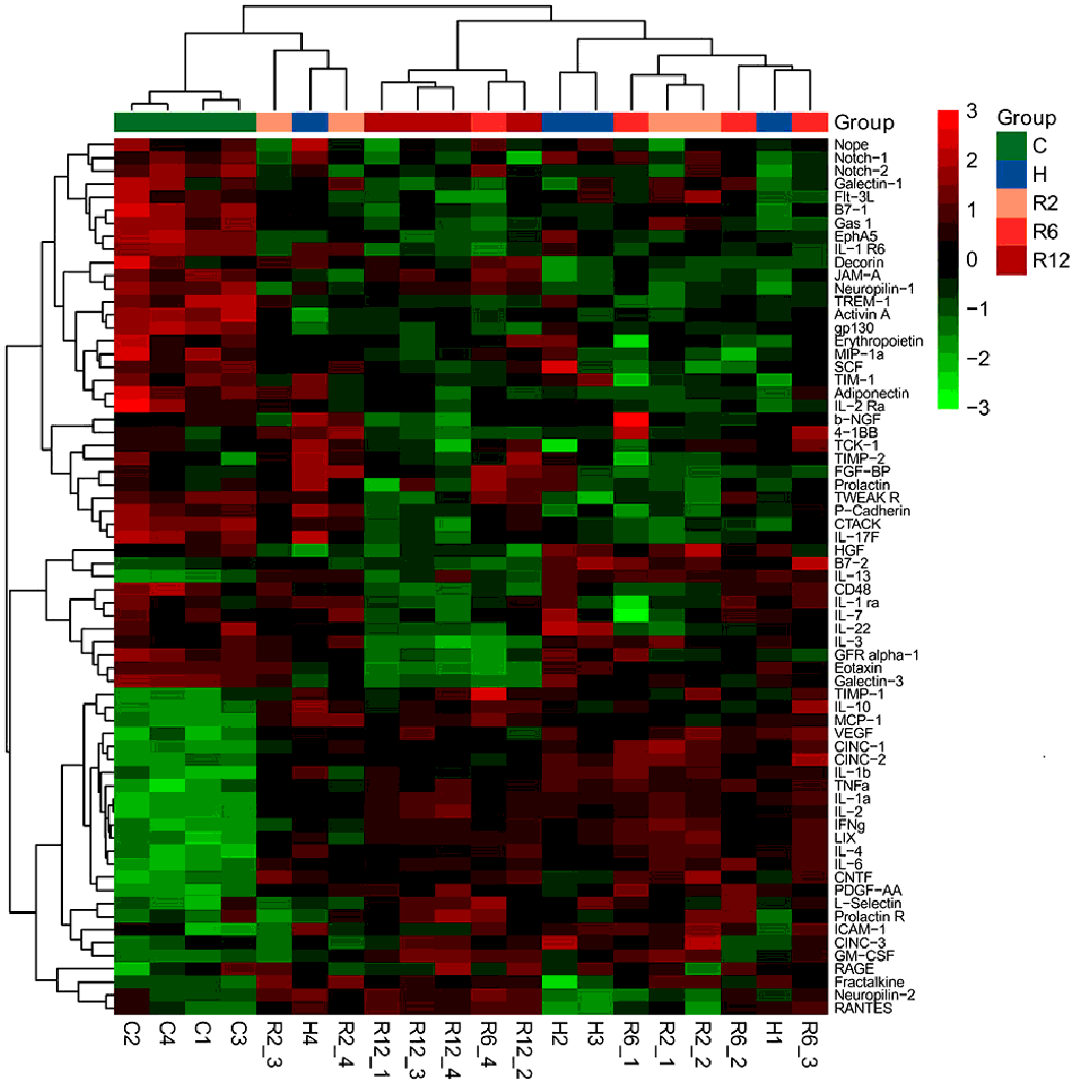
The expression levels of 67 inflammatory factors in the small intestinal were detected using a protein chip. Based on the expression of the inflammatory factors, the C group was clustered into a class via hierarchical clustering, which indicated that hypothermia resulted in notable changes to the body (Fig. S1). The H, R2 and R6 groups could not be clustered into one class; however, the R12 group could almost be clustered into a class, which indicated that rewarming for 12 h could be distinguishable from the H group. Expression of inflammatory cytokines in rats rewarmed for short periods (2 or 6 h) could not be clustered. This suggested that a rewarming period of 12 h was considered sufficient to stabilize the physiology in the rats (Fig. S1).
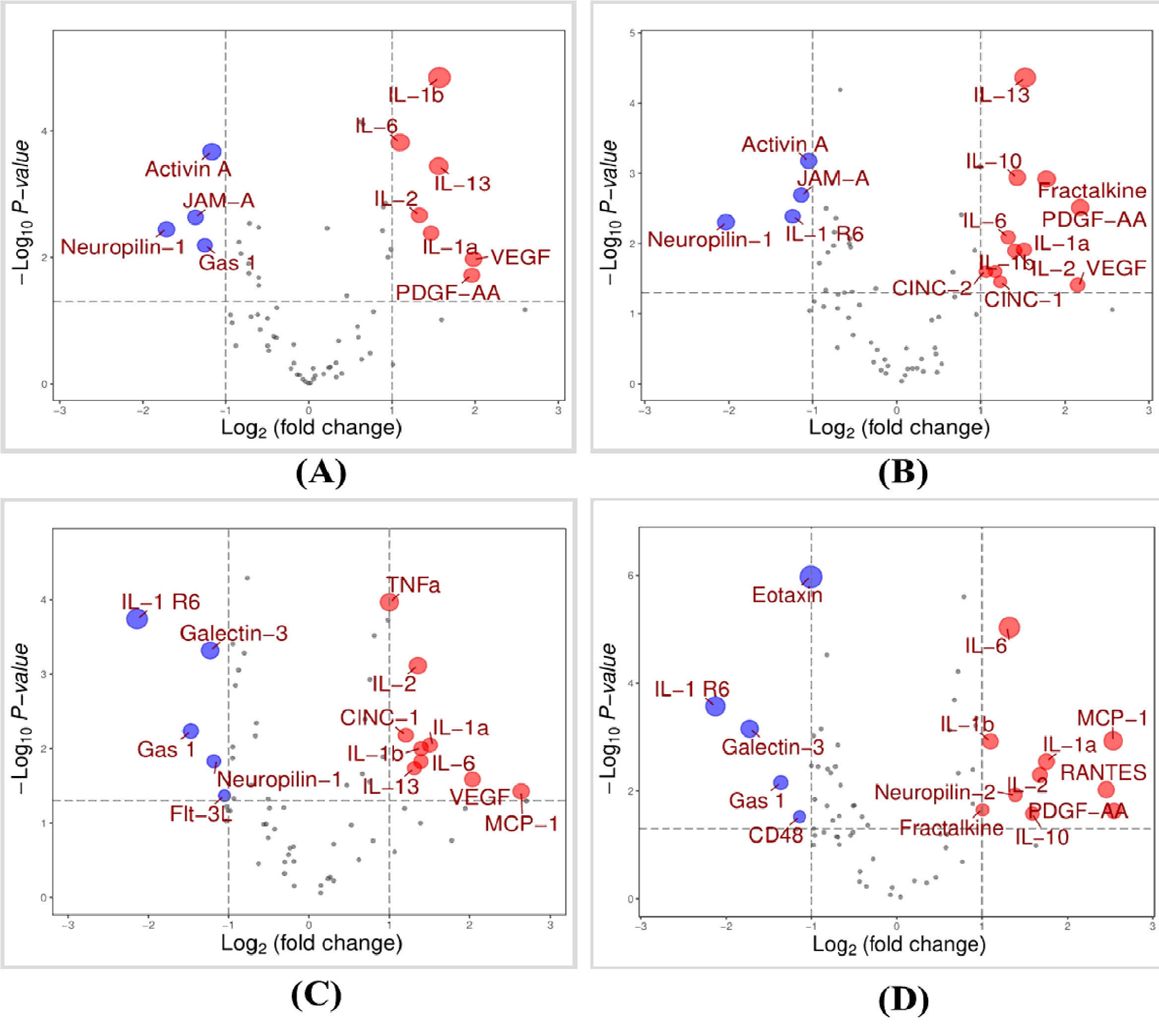
Next, the differentially expressed proteins in the H and R groups (R2, R6 andR12) was compared with the N group (Fig. S2). Compared with the C group, 11 inflammatory factors were differentially expressed, including 7 upregulated and 4 downregulated inflammatory factors, (Fig.S2A). There were 15 and 14 significantly differentially expressed inflammatory factors in the R2 and R6 groups, respectively (Fig. S2B and C).The R12 group exhibited the highest number of differentially expressed inflammatory factors; 15 upregulated and 5 downregulated (Fig. S2D).These changes in inflammatory factors may participate in the development of hypothermia. We selected IL-6, IL-10 and IL-1β for further analysis based on our lab condition (our lab had sufficient conditions to study IL-10 because we had studied IL-10 before) and the differential analysis (IL-1β and IL-6 were the most significant proteins when compared the H group with the C group (Fig. S2A). These two proteins were also significant in the other three comparations (Fig. S2B, C, D).
Plasma levels of IL-6, IL-10 and IL-1β
The expression levels of certain inflammatory factors were altered in the intestine following hypothermia. Thus, whether these changes were also observed in the blood was next assessed. For analysis, three inflammatory factors (IL-6, IL-10 and IL-1β) were selected for further analysis in the blood. Using ELISA, it was shown that following immersion in 15˚C seawater for 5 h, the levels of the three cytokines in the plasma from the H group was significantly higher than that of the C group (P<0.05). Compared with the H group, the levels of the three cytokines in the R group was gradually reduced following rewarming in a warm water bath (P<0.05; Fig.2A-C).The ratio of IL-1β:IL-10 and IL-1β:IL-6 ratios were used to measure the balance between pro- and anti-inflammatory cytokines. The ratio of IL-1β:IL-6 and IL-1β:IL-10in the plasma of the H group rats were increased, and they gradually decreased in the R group with time. However, the increase in IL-1β expression was greater than that of IL-10 and IL-6 in the H and R groups (Fig. 2D).Thus, the levels of inflammatory factors in the blood were also altered. The changes in these three inflammatory factors was similar to that observed in the intestine. Thus, rewarming both recovered the vital status and also restored the levels of inflammatory factors in the blood.
Relative gene expression levels of the cytokines in the gut
Differentially expressed inflammatory factors were found in the hypothermic rats using a protein chip. To verify the change in these factors, IL-1β, IL-10 and IL-6were selected for qPCR analysis. The results showed that the expression levels of IL-1β in the H group was significantly increased ~4.5-fold compared with the C group (P<0.05; Fig. 3A). When rewarming for 2 h, the levels of IL-1β mRNA decreased to normal. However, after rewarming for 6 h, the levels of IL-1β were increased significantly, ~3.5-fold higher than that of the C group. As the rewarming time increased, the expression levels of IL-1β gradually decreased (Fig.3A).Compared with group C, the mRNA levels of IL-10 in group R2, R6 and R12 increased(P<0.05; Fig.3B).The mRNA expression levels of IL-6 in the R2 group increased, whereas it decreased in the H, R6 and R12groups, although the differences were not statistically significant (Fig. 3C).The ratio of IL-1β:IL-6 and IL-1β:IL-10 were analyzed. Compared with the C group, IL-1βexpression was higher than that of IL-10 in the H group and all the R groups (P<0.05; Fig.3D).
Pathological changes in the intestinal tissues
The histological analysis showed that the morphology of intestinal mucosa in the C group was normal (Fig. 4). The villi of the intestinal mucosa and the epithelial cells were neatly arranged. They were free of edema in the interstitial tissue and the villi structure was intact. However, a mass of necrosis was observed in the epithelium of intestinal mucosa villi in the H group, with a large degree of neutrophil and inflammatory cell infiltration, and the shed villi interstitial structure was lost. In the R group, the tight junction structure of the intestinal tissue and the permeability was altered. At the top of the villi, there was increased neutrophil infiltration. These findings suggest that severe inflammation occurred in the intestine following hypothermia.
IL-1βexpression in intestinal tissues based on immunohistochemistry (IHC) analysis
To investigate the expression and distribution of IL-1β in intestinal tissues, IHC was performed. As shown in Fig.5, positive IHC staining, which was considered as brownish yellow or brown particles that could be distinguished from the background cells, was primarily localized in the cytoplasm of the intestinal tissue cells. Compared with the C group, the proportion of positive cells was higher in the H group. In the R2 group, the staining of IL-1βwas decreased compared with the H group. However, the number of positive cells was increased in the intestinal tissue of the R6 group. With a longer rewarming period, the intensity of IL-1β expression gradually decreased. This phenomenon was observed with regard to the amyostasia. When hypothermic or rewarming, amyostasia was first increased and then subsequently decreased (Fig. 1D). However, for IL-1β, there was only one time-point and the end of immersion. A possible explanation for this is that when the temperature is decreasing, the attempts to correct this to maintain the normal body temperatures. However, as the temperature decreases further, the body is unable to maintain a physiological temperature. Thus, perhaps the lower temperature suppresses certain physiological functions. Additionally, this observation may be the result of evolution. When the temperature is too low, the body stops trying to increase the core temperature, instead reserving energy, thus resulting in damage to the body from the colder temperatures.