The long noncoding RNAs have been uncovered to exert an pivotal influence on cell proliferation and apoptosis of AML, the mechanisms of which include altering methylation status of gene promoters[7, 8], recruiting epigenetic complex on gene promoters[29], reshaping chromatin [30, 31], sponging miRNAs to regulate gene expression[32–35], etc. HOTAIR is one of the most studied lncRNAs in AML, which is upregulated in de novo AML patients[36] and predicts an adverse prognosis[37]. HOTAIR locates in HOXC gene cluster on chromosome 12 and exerts biological effect through modulating HOXA family genes. Intriguingly, through analysis of TCGA expression data, we found LINC00649 was also correlated with most of HOXA family genes. Whereas little is known about the role of LINC00649 on pathogenesis and prognosis of AML. HOXA family genes encoded crucial transcription factors in normal hematopoiesis and cell differentiation. The dysregulation of HOXA genes is revealed in several types of solid cancers[38–40]. HOXA genes are also reported to be involved in myeloid cell differentiation, pathogenesis of AML and resistance to chemotherapy[41–43].
In comparison with healthy controls, AML patients have aberrantly lower LINC00649 expression, based on the data derived from TCGA and BeatAML database. Meanwhile, for most of cancers other than AML, expression level of LINC00649 in cancer cells is higher than that of corresponding normal tissues. Furthermore, the survival (OS and PFS), of LINC00649-low group, was significantly worse than that of LINC00649-high group. The unusual expression signature and prognostic value of LINC00649 drove us to explore the possible molecular mechanisms and uncover its biological function.
According to catRAPID algorithm, 9 proteins containing 120 sites were identified to be potentially binding to LINC00649. TIAL1, SRSF9, SRSF2, SRSF3 and RBFOX2 were identified to be associated with HOXA gene expression. TIAL1 is the RNA binding protein, which binds to target sites and splice the pre-mRNA alternatively[44, 45]. While the binding of TIAL1 and HOXA genes has not been previously validated, as well as the association of TIAL1 and AML pathogenesis. SRSF9 involves in constitutive mRNA splicing and can modulate the target of alternative splicing[46]. SRSF9 was reported to be involved in cell proliferation and apoptosis in bladder and cervical cancer[47, 48], and related to prognostic alternative splicing events of renal clear cell carcinoma[49]. SRSF2 and SRSF3 are also splicing factors, which belongs to serine/arginine-rich protein family. Functional mutations of SRSF2 drive the cancer genesis of hematopoietic cells[50] and play a role in myelodysplasia and myeloproliferative neoplasms[51, 52]. SRSF3 is also a multiple cancer related splicing factor, namely glioblastoma[53], colon cancer[54], oral squamous carcinoma[55], etc. Moreover, the expression of SRSF3 is significantly decreased in de novo AML patients in comparison with that of healthy controls, as well as other SR family members, like SRSF1/2/4/5/6/7. RBFOX2 can bind to 5’- UGCAUGU-3’ element of target RNA, exerting alternative splicing. RBFOX2 can modulate erythropoiesis, by promoting alternative selection of exon 16 in protein 4.1R, the product of which is essential for erythrocyte membrane stability[56, 57]. Notably, the expression of RBFOX2 is significantly correlated with all members of HOXA family genes (Supplementary Fig. 1), suggesting potential interaction between them. Furthermore, the pancancer-TCGA expression data was download from UCSC database (https://xenabrowser.net/hub/), the correlation of RBFOX2 and HOXA genes was analyzed by Pearson’s method (Supplementary Fig. 2 − 1/2/3). Notably, the significant association of RBFOX2 and HOXA is a common feature among cancers generated from different tissue, suggesting this relationship may not be coincidental. The expression dataset of normal tissue was downloaded from GTEx database (https://www.gtexportal.org/home/), similar analysis showed that the correlation is insignificant in normal bone marrow (Supplementary Fig. 3 − 1/2/3), which indicated the relationship was a disease-specific feature for AML. All 4 splicing factors and LINC00649 are potential co-regulators for HOXA genes in AML, which has not been explored before.
Based on the results of GESA, the upregulation of PI3K-Akt-mTOR signaling, IL6-JAK-STAT3 signaling, oxidative phosphorylation was identified in LINC00649-low group. PI3K-Akt signaling pathway is frequently activated in AML, activation of PI3K-Akt-mTOR signaling were found in 50% of AML patients[58, 59]. The PI3K-Akt signaling controls leukemic blast cells proliferation and clonogenicity[60, 61]. Aberrantly functional receptor tyrosine kinases drive the activation of PI3K-Akt-mTOR pathway, including IGF1/IGF1R[62, 63], activated FLT3[64] and DEK-NUP14 fusion protein[65]. The inhibitors of PI3K-Akt-mTOR axis have shown preliminary anti-leukemia effects against AML both in vivo and in vitro [66–72], indicating the pathway is a crucial therapeutic target. The IL6-JAK-STAT3 pathway is also activated in LINC00649-low group, which plays a crucial role in oncogenesis of diverse cancers[73]. Constitutive phosphorylation of STAT3 by autocrine secretion of IL6 is revealed in AML cells[74], which can be reversed by TGF-beta 1[75]. Activation of STAT3 is also uncovered revealed in primary pediatric AML samples, and the small-molecule inhibitor of STAT3 can induce apoptosis and inhibitor formation of blast colonies in vitro[76]. The dysregulation of PI3K-Akt-mTOR and IL6-JAK-STAT3 signaling may be attributed to the unfavorable survival profile in LINC00649-low group, which were supported by the success of inhibitors of these pathways. The maintenance of leukemia stem cells depends on BCL2 mediated oxidative respiration, instead of glycolysis as in normal hematopoietic cells[77]. The metformin, targeting oxidative phosphorylation (OXPHOS), induces apoptosis of human leukemia cells in an AMPK-independent way[78]. Cytarabine resistant leukemia cells are characterized by activated OXPHOS, with the high level of reactive oxygen species. Additionally, the resistance can be reversed by agents inducing low OXPHOS status[79]. The activation of OXPHOS in LINC00649-low patients may promoted leukemia cell maintenance and chemo-resistance, leading to inferior survival. The p53 signaling and Hedgehog signaling were found to be suppressed in GSEA. Non mutational p53 dysfunction was common in AML and implicated in diverse inactivating mechanisms[80]. Dysregulation and activation of PI3K-Akt-mTOR signaling pathway can activate MDM2 and interact with NF-kappaB signaling pathway, leading to dysfunction of p53[81]. The activation of PI3K pathway was revealed in LINC00649-low group, which may cause the suppression of p53 signaling and inferior survival considering the central role of p53 in the complex network of AML-associated signaling pathway.
In ORA of LINC00649-associated genes, the enriched biological processes included negative regulation of hematopoiesis and DNA-templated transcription, while molecular function included double strand DNA binding, sequence-specific DNA binding and transcription regulation region DNA binding. The GO analysis mainly implicated in regulation of DNA-binding and transcription, which is consistent with function of HOXA genes. KEGG analysis showed that the genes were enriched in AGE-RAGE, PI3K-Akt, Ras and VEGFR signaling pathways. The association of RAGE and solid cancers has been explored, including renal cell carcinoma[82, 83] and gastric carcinoma[84]. The AGE and RAGE signaling also has been studied in AML, which indicated AGE activated MAP kinase, PI3K and JAK/STAT pathway, leading to proliferation of primary AML samples and AML cell lines[85]. Activation of Ras signaling can also promote the dysfunction of p53 by similar mechanism of PI3K-Akt signaling[81]. VEGRA is reported to be overexpressed and associated with adverse prognosis in AML[86, 87], while the expression of VEGRA is also overexpressed in LINC00649-low group based on our results. The activated VEGFR signaling promoted the proliferation, survival and resistance to chemotherapy of AML blasts[88]. VEGF targeting therapy has been developing and showing preliminary benefit for AML in vitro[89–91]. While the Reactome database provided us with other pathways enriched by LINC00649-associated genes, including signaling by ERBB2 and VEGFR2 mediated cell proliferation. Although mudritinib, an ERBB2 inhibitor, can eliminate AML cell both in vivo and in vitro, the anti-leukemic mechanism is likely not mediated by inhibition of ERBB2 signaling, but by inhibiting ETC complex I[92]. This conclusion is supported by that another ERBB2 inhibitor, lapatinib, showed no anti-leukemia effect in AML patients and expression level of ERBB2 is extremely low in AML cells[92]. The role of ERBB2 signaling has to be further validated. VEGFR2 is a ‘hot’ target in AML, and relevant to chemotherapy-sensitivity, pro-survival effect and angiogenesis in bone marrow[93, 94]. VEGFR2-targeting therapy is being developed in preclinical stage[94, 95]. The dysregulation of all above pathways contributed to the difference of survival between LINC00649-high and low groups.
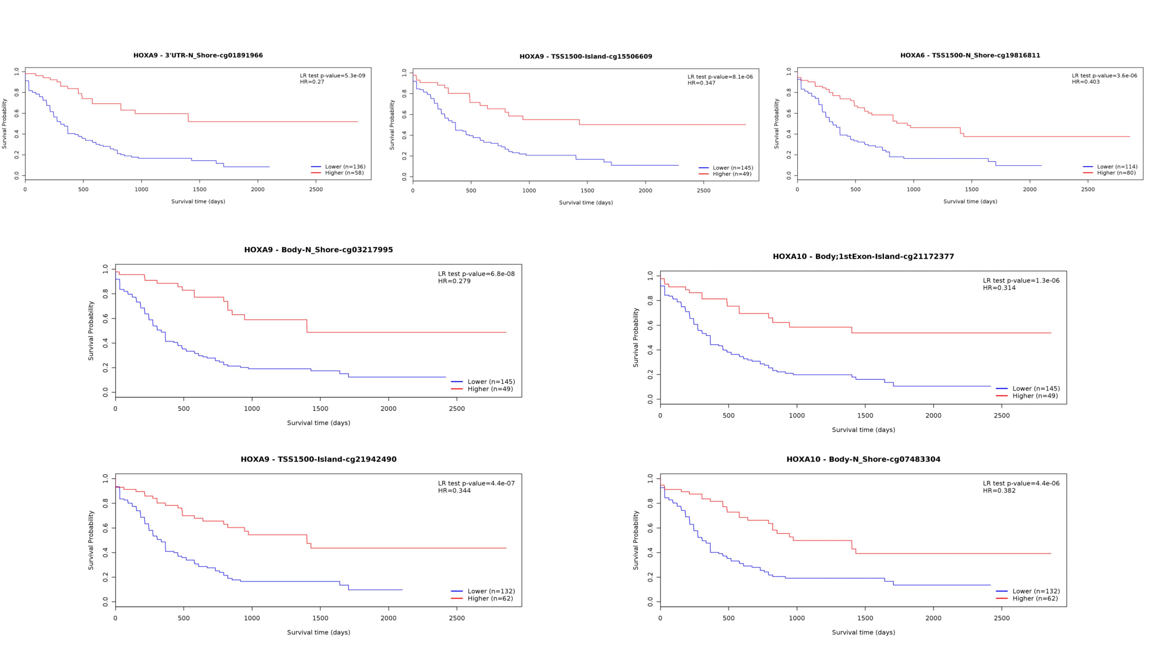
Furthermore, HOXA family genes methylation status was shown to be correlated with LINC00649, which may contribute to the biological impact. The methylation status (beta value) of seven CpG sites involving with HOXA6/HOXA9/HOXA10 (cg21172377, cg07483304, cg05490659, cg02000808, cg19816811, cg16880946, cg18931036) was correlated with expression of LINC00649. Notably, all involved sites were of significance for AML overall survival (Supplementary Fig. 4). Considering that lncRNA HOTAIR can modulate the methylation status of HOXA5 by inhibiting DNMT3B[8], our results suggested similar epigenetic mechanism may implicated in the regulation of HOXA genes.
To improve the diagnostic utility of the prediction model and reveal the key elements in the LINC00649 centric regulation web, we brought in multi-dimension information to establish a prediction model on AML survival. Combining results of correlation analysis and prediction by miRWalk/lncBase database, we established the LINC00649-centered ceRNA network, the sponging miRNA and targeted mRNA of which were inferred as fundamental elements of biological process in AML. Since the lncRNA binding proteins, miRNA-sponging and methylation-alteration are known as the most common ways that lncRNA exert its biological effect, we included expression data of predicted LINC00649 binding proteins, miRNAs/mRNAs in the ceRNA network and methylation data of altered methylated CpG sites into the model. The traditional prognostic markers were also included in the model, namely the patients’ age, gender, race, molecular and cytogenetic risk stratification, count of mutations and WBC. We constructed the LASSO-Cox models fitting OS and PFS. Due to the large scale of included variables and obvious relevance between them, the LASSO regression is a better choice of screening variables into prediction models than COX regression. A few prediction models, including genetic information of AML patients, have been developed previously, including Clinseq-G[96] (AUC for 3-year OS is 0.730), ELN2017 stratification in the validation cohort [96] (AUC for 3-year OS is 0.65), Li Z et al[96] (AUC for 3-year OS is 0.70), Huang R et al[97] (AUC for 1 year OS is 0.666, AUC for 5 year OS is 0.707), Ha M et al[98] (AUC for 5-year OS is 0.613). AUC of our prediction models is far better than all these models, possibly attributing to the integrated multi-dimension information. The higher AUC, the more precisely we identify the risk of individual patients. On the other hand, the Kaplan Meier plots supported the risk stratification using the models to divide patients into high-risk and low-risk group, which well-defined the patients with much better prognosis (median OS of low-risk has not reached). While due to lack of integrated information in one cohort like TCGA database, which included clinical/RNAseq (protein-coding and noncoding RNA)/miRNAseq/methylation datasets, we can hardly validate this model independently. However, the present work brings clues and insights to further studies, by providing potential biomarker and therapeutic targets.
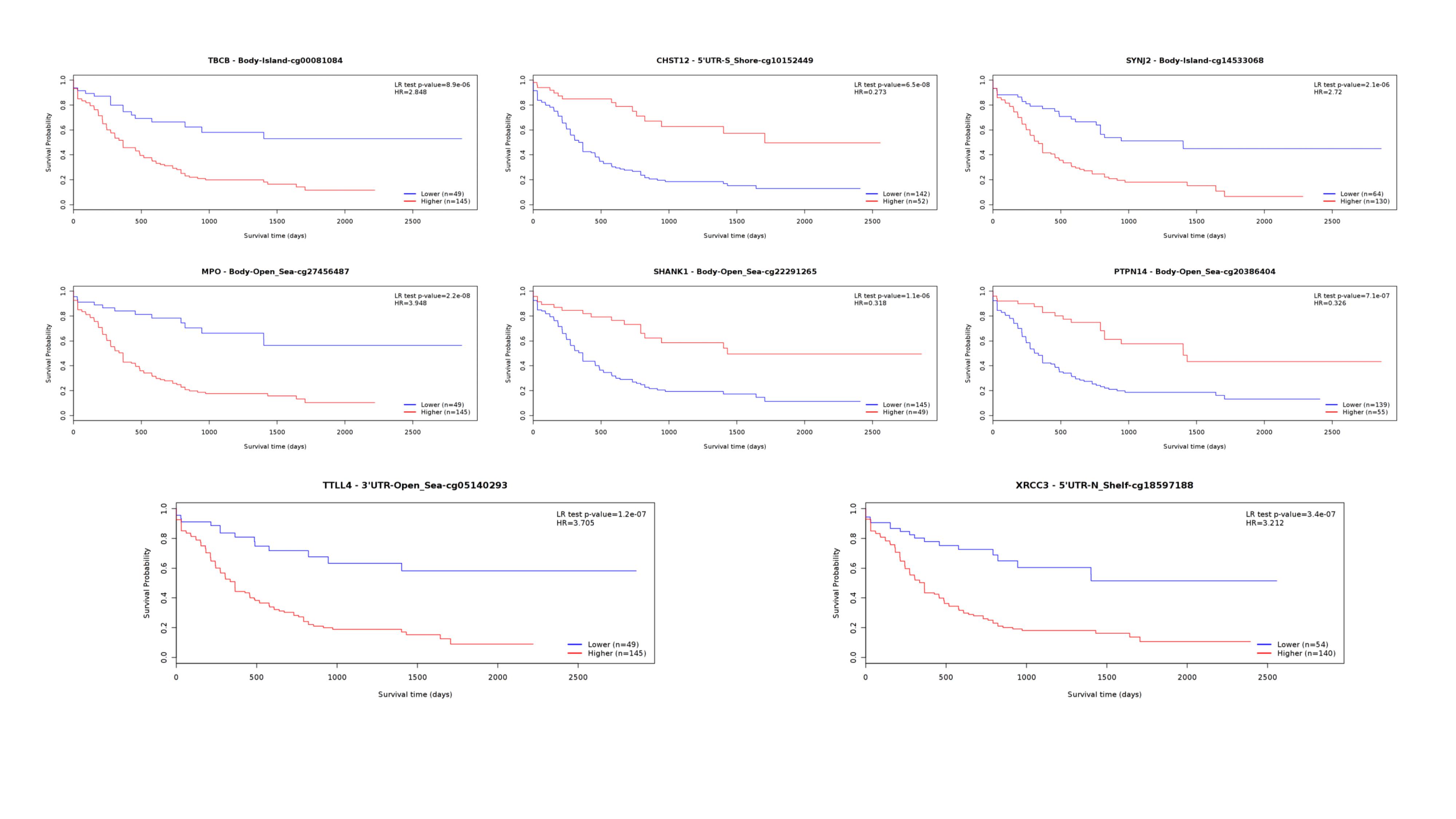
In our OS-prediction model, novel markers were identified (Table 3), which have not been reported to implicated in AML previously. EVPL is a component of the cornified envelope of keratinocytes, the genetic variations of which are associated with several solid cancer types[99–103]. While the association of other protein-coding genes (KIF26A/SERINC5/SMAGP/CD320) or noncoding genes (miR-502-3p) with either hematological or solid malignancy has not been investigated. Among the included methylation positions, individual methylation status of cg27456487 (MPO), cg05140293 (TTLL4), cg10152449 (CHST12), cg22291265 (SHANK1), cg18597188 (XRCC3), cg14533068 (SYNJ2), cg00081084 (TBCB) and cg20386404 (PTPN14) were significantly associated with AML survival, according to MethSurv online tools (https://biit.cs.ut.ee/methsurv/) (Supplementary Fig. 5). A low expression ratio of MPO has been reported as a deleterious marker for AML, indicating a lower complete remission rate[104] and shorter PFS [105]. In untreated AML patients, hypermethylation status of MPO is detected and correlates with MPO expression, which can be induced by demethylating agents[106]. The alteration of MPO is demonstrated as an indicator for DNA methylation pattern implicating downregulation of DNMT3B[107], our results supported its significance in pathogenesis of AML. The included methylated sites other than MPO have not been reported to implicated in AML. In PFS prediction model (Table 3), no genetic variables (expression or methylation status of genes) were described in relation with AML previously. Notably, KIF26A was included in both OS and PFS model, which belongs to kinesin superfamily and is reported as an oncogenic marker for breast cancer[108] and pancreatic ductal carcinoma[109].