The phytoconstituents of aqueous leaf extract of Costus afer (ALECA) is shown on Table 1.
Table 1
Quantitative phytochemical screening (mg/100g) of the aqueous leaf extract of Costus afer (ALECA)
|
Chemical constituents
|
Content
|
|
Alkaloids
|
4.3 ± 0.10
|
|
Phenolic compounds
|
1.23 ± 0.05
|
|
Saponins
|
2.9 ± 0.08
|
|
Tannins
|
2.72 ± 0.11
|
|
Flavonoids
|
25 ± 0.13
|
-
Rats treated with with the trace metal mixture TMM only, had a considerable increase in liver weight (p < 0.05) compared to the normal control rats (received only deionized water). Rats that received both aqueous leaf extract of Costus afer (ALECA) and trace metal mixture (TMM)
had decreased liver weight (Table 2). The absolute and relative weightsof liver of rats that received only trace metal mixture (TMM) was 9.4 ± 1.35 g and 3.62 ± 0.47 % respectively while the rats administered with only deionized water had 5.08 ± 0.95 g and 2.45 ± 0.41 % respectively.
Table 2
Effect of aqueous leaf extract of Costus afer on the absolute and relative weight of liver treated with trace metal mixture (TMM)
|
Treatment
|
*Absolute (g)
|
Relative (%)
|
|
Deionized H2O
|
5.08 ± 0.95a
|
2.45 ± 0.41a
|
|
TMM
|
9.4 ± 1.35c
|
3.62 ± 0.47c
|
|
TMM + 750 mg/kg ALECA
|
8.48 ± 1.13bc
|
3.52 ± 0.42bc
|
|
TMM + 1500 mg/kg ALECA
|
8.36 ± 0.52bc
|
3.15 ± 0.17bc
|
|
TMM + 2250 mg/kg ALECA
|
7.86 ± 0.22b
|
3.06 ± 0.08b
|
| *Values = Mean ± SD, N = 5., data with different superscripts (a, b, c) are significantly different from each other (p < 0.05), data with the same superscripts are not significantly different; TMM = Trace metal mixture |
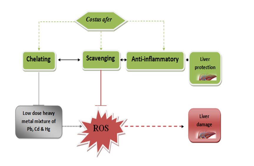
The tests for liver function were done to evaluate the likely protective role of Costus afer treatment from metal mixture exposure. Treatment with trace metal mixture (TMM)caused significant elevation in aspartate aminotransferase, alanine aminotransferase and alkaline phosphatase levels, bilirubin (total and direct), and a decrease in total protein and albumin, whereas the rats that received both aqueous leaf extract of Costus afer (ALECA) and trace metal mixture (TMM) showed reduction in the liver enzyme markers and a rise in total protein and albumin in comparison to metal mixture-treated albino rats (Fig. 1). The aspartate aminotransferase, alanine aminotransferase and alkaline phosphatase, total bilirubin, direct bilirubin, total protein and albumin levels in rats treated with only trace metal mixture (TMM) were significantly different (172 µ/l, 77.8µ/l, 222.2µ/l, 32.6 mg/dl, 15.2 mg/dl, 43.8 g/l, 25.4 g/l, p < 0.05) respectively, from the groups that received both aqueous leaf extract of Costus afer (ALECA) and trace metal mixture (TMM).
Figure 2 is the parallel coordinates plot showing clustering of liver function parameters interaction in different treatment groups. The Fig consists of three classes; class 1 (gp 1 and 5), class 2 (gp 2 and 3), and class 3 (gp 4). The factor loading of liver function variables on significant principal components which was done sequel to the Varimax rotation is presented in Table 3 whereas the correlation plot is shown in Fig. 3. An illustration of the differentiation of parameters and the interactions among liver function parameters in different groups is depicted in Fig. 4. A 3-component system showing 95.35% of total variance was obtained after statistical principal component analysis.
The co-ordinate plot of liver function parameters showing the association between the variables consists of 3 classes. The result showed that class 1 rats had high total protein and albumin levels with low aspartate aminotransferase, alanine aminotransferase and alkaline phosphatase, total bilirubin and direct bilirubin,, while class 2 rats had high aspartate aminotransferase, alanine aminotransferase and alkaline phosphatase, total bilirubin and direct bilirubin levels with low total protein and albumin levels. Class 3 rats were of the intermediate class showing that they had midrange values for all the liver function parameters.
The Principal component analysis (PCA) provided a better interpretation of the data, having grouped the connected variables as one. The male albino rats administered with trace metal mixture (TMM) of Pb, Cd and Hg, seven principal factors (F1-F7) explained 100% cumulative variations in the liver function parameters. The first two components (PC1 and PC2) accounted for more than 92% of total variance, while PC3 gave rise to only 2.80% of the total variance. Factor 1 (F1) had Eigen value > unity explaining 87.34% of total variation of the data set and was regarded as being significant, while factors 2 and 3 (F2 and F3) had Eigen values < unity explaining 7.95% of the total variation. (Anyanwu et al. 2020a).
Strong positive correlations (≥ 0.88) were perceived between these parameters (aspartate aminotransferase, alanine aminotransferase and alkaline phosphatase, total bilirubin and direct bilirubin) whereas a weak association (r = 0.6617) was observed between the Total protein and albumin. Also, both factors had negative correlations with aspartate aminotransferase, alanine aminotransferase and alkaline phosphatase, total bilirubin and direct bilirubin which is an indication that a reduction in total protein and albumin would likely lead to an increase in aspartate aminotransferase, alanine aminotransferase and alkaline phosphatase, total bilirubin and direct bilirubin and vice versa. All the variables were printed in bold in PC1 (Table 2) whereas aspartate aminotransferase had the highest loading on PCI after Varimax rotation. The principal components were extracted with the aid of a 3D graphing software to plot the correlated variables.
Table 3
Loadings of liver function variables on significant principal components after Varimax rotation
|
Liver function parameter
s
|
*PC1
|
PC2
|
PC3
|
|
aspartate aminotransferase (U/L)
|
0.9105
|
0.0017
|
0.0280
|
|
alanine aminotransferase (U/L)
|
0.9443
|
0.0023
|
0.0129
|
|
alanine phosphatase (U/L)
|
0.9071
|
0.0020
|
0.0036
|
|
total bilirubin
|
0.9192
|
0.0075
|
0.0004
|
|
direct bilirubin (mg/dl)
|
0.9258
|
0.0008
|
0.0093
|
|
total protein (g/l)
|
0.8008
|
0.0787
|
0.1166
|
|
ALBUMIN(g/l)
|
0.7059
|
0.2677
|
0.0250
|
|
Eigen value
|
6.1136
|
0.3607
|
0.1958
|
|
Variability (%)
|
87.3368
|
5.1532
|
2.7973
|
|
Cummulative (%)
|
87.3368
|
92.4900
|
95.2873
|
| *Bold figures are highly correlated coefficients; and thus, the principal factors. AST = aspartate aminotransferase, ALT = alanine aminotransferase, ALP = alanine phosphatase, T.BIL = total bilirubin, D.BIL = direct bilirubin, T.Protein = total protein |
Variables (axes F1 and F2: 92.49%)
An assessment of the inflammatory status after treatment with trace metal mixture TMM was done by evaluating the pro- and anti-inflammatory cytokines level in the liver. The co-treatment using Costus afer significantly decreased the levels of pro- and also increased (p < 0.05) the anti- inflammatory cytokines (IL-6 and IL-10) in the liver tissue in comparison to in the trace metal mixture TMM-treated group (Fig. 5), suggestive of anti-inflammatory activity of aqueous leaf extract of Costus afer (ALECA). The pro- and anti-inflammatory cytokine levels (IL-6 and IL-10 respectively) of rats administered with the trace metal mixture TMM only was significantly different (61.8 Pg/g tissue and 14.7 Pg/g tissue, p < 0.05) from the inflammatory cytokines seen in rats co-treated with Costus afer.
The oxidative status in the hepatocyte subsequent to the metal mixture treatment using the lipid peroxidation marker, MDA level was evaluated. The 90-day treatment which was done with the following metal mixture in the following dosage PbCl2- 20mg/kg, CdCl2- 1.61mg/kg, HgCl2 – 0.40mg/kg body weight induced oxidative reaction in the organ (liver tissue). The MDA level increased significantly (p < 0.05) in the hepatocyte of trace metal mixture TMM -treated rats in comparison to those of the normal control group (Fig. 6). A significant decrease in oxidant level was observed in the rats treated with Costus afer and metal mixture compared to those treated with only the metal mixture.
With respect to how the treatment affects the non-enzymatic glutathione (GSH) and enzymatic superoxide dismutase and catalase (SOD and CAT) activities in the liver tissue. Treatment with trace metal mixture TMM resulted a significant decrease (p < 0.05) in glutathione and superoxide dismutase and catalase levels in comparison with control. Rats that received aqueous leaf extract of Costus afer (ALECA) plus trace metal mixture TMM had elevated levels of glutathione and superoxide dismutase and catalase in comparison to rats that received only trace metal mixture TMM. s
Heavy metal concentration on the liver of the rat samples
The concentration of trace metals (lead, cadmium and mercury) in the liver were notably elevated (p < 0.05) in the liver of the rats treated with the trace metal mixture TMM- in comparison to the control (Table 4). Treatment of rats with aqueous leaf extract of Costus afer (ALECA) plus trace metal mixture TMM resulted in significant reduction in trace metals (lead, cadmium and mercury) levels in comparison to rats that received only trace metal mixture TMM In addition, the group exposed to the trace metal mixture TMM only, had the highest level of trace metals (lead = 90.992 ± 13.284, cadmium = 0.78 ± 0.133 and mercury = 0.305 ± 0.0439) in comparison to the control group. Pearson’s rank correlation analyses indicate the inter-trace metal relationship among trace metals in liver of rats showed strong positive correlation (r > 0.90) between metals such as: Pb and Cd, Pb and Hg, as well as Cd and Hg. All correlations were significant at p < 0.01 (Fig. 7).
Table 4
Levels of trace metals (mg/kg) in the liver
|
Treatment
|
Cadmium (Cd)
|
Mercury (Hg)
|
Lead (Pb)
|
|
Deionized H2O (only)
|
< 0.001 ± 0.000a
|
< 0.001 ± 0.000a
|
0.392 ± 0.552a
|
|
TMM (only)
|
0.782 ± 0.133c
|
0.305 ± 0.039c
|
90.992 ± 13.284c
|
|
TMM + 750 mg/kg ALECA
|
0.143 ± 0.046b
|
0.051 ± 0.020b
|
31.007 ± 6.017b
|
|
TMM + 1500 mg/kg ALECA
|
0.033 ± 0.024a
|
0.009 ± 0.001a
|
8.857 ± 4.849a
|
|
TMM + 2250 mg/kg ALECA
|
0.003 ± 0.002a
|
0.001 ± 0.001a
|
3.500 ± 1.141a
|
| *Values are expressed as (Mean ± SD). Values in the same column with different superscripts are significantly different from each other (p < 0.05) and those with the same superscripts in the same column are not significantly different; TMM = Trace metal mixture |
Histopathology of the liver
The liver sections of six different groups were sectioned and presented in (Fig. 8 (G1), (G2), (G3), (G4), and (G5) representing groups treated with deionized H20, metal mixture (MM), (MM + 750 mg/kg ALECA, MM + 1500 mg/kg ALECA, and MM + 2250 mg/kg ALECA.
Regression Model Development
XLSTAT 2016 software was employed for the model development in this study. Lead, Cadmium, Mercury, and ALECA were considered as the input data and were used for the calibration of the model using multiple linear regressions. Zero coefficients for x2 and x3 were observed as outputs and thus subsequent calibrations were done without the 3 constant parameters (input data).. In other words, y is made a function of ALECA variable for a given set of the heavy metal variables namely: Lead, Cadmium, and Mercury. A plot of catalase concentration vs ALECA concentration was modelled using linear, quadratic and exponential options (Table 5). The R2, MSE and RMSE values were determined and shown in Table 6 for the process.
For verification of he calibrated models, the observed model was compared with predicted model on the liver catalase level (Fig. 9) with corresponding R2 = 0.935 .
The quadratic and exponential models for the liver catalase level was R2, MSE and RMSE of (0.935, 1.513 and 1.230) and (0.880, 1.022 and 1.011) respectively. The linear model best predicted the process with R2, MSE and RMSE values of. 0.935, 0.760 and 0.872 respectively. A repetition of the regression models was done for other liver parameters and based on R2, MSE and RMSE, the best models were selected. To verify the calibrated models, it was important to compare observed against predicted liver catalase level with corresponding R2 = 0.999. The modeled liver catalase values for ALECA plus TMM treated groups at 0 mg/kg, 750 mg/kg, 1500 mg/kg and 2250 mg/kg were 2.680, 4.770, 6.860 and 8.950, while the observed values were 2.360, 5.640, 6.080 and 9.180 respectively. Similarly, the verification was equally done on other parameters in the liver.. These models could help predict the residual values of liver function parameters, antioxidant profile and inflammatory cytokines in the liver of male albino rats at any treatment dose using ALECA to a high precision provided the levels of lead, cadmium, and mercury remains constant as considered in this work.
Table 5
Regression models of catalse in
|
Type of Model
|
Equation
|
R2
|
MSE
|
RMSE
|
|
Exponential
|
y = 2.801e0.000x
|
0.880
|
1.022
|
1.011
|
|
*Linear
|
y = 0.002x + 2.68
|
0.935
|
0.760
|
0.872
|
|
Quadratic
|
y= -8E-08x2 + 0.003x + 2.635
|
0.935
|
1.513
|
1.230
|
| NB: * The model with the best fit. |
| A replication of regression models was done on other biomarkers and the best with respect to R2, MSE and RMSE values was selected and summarized as shown in the Table 6 below; |
Table 6
Model equations for antioxidant biomarkers, inflammatory cytokines and other test parameters in the liver
|
Biomarkers
|
Type of Model
|
Model Equations
|
R2
|
MSE
|
RMSE
|
|
SOD
|
Quadratic
|
y = 8E-08x2 + 7E-05x + 0.237
|
0.999
|
0.000
|
0.011
|
|
GSH
|
Quadratic
|
y = 1E-07x2 + 0.000x + 0.814
|
0.999
|
0.000
|
0.018
|
|
MDA
|
Quadratic
|
y = 3E-08x2- 0.000x + 0.645
|
0.996
|
0.001
|
0.025
|
|
IL-6
|
Quadratic
|
y = 4E-06x2-0.032x + 62.33
|
0.996
|
5.618
|
2.370
|
|
IL-10
|
Exponential
|
y = 13.98e0.000x
|
0.991
|
5.387
|
2.321
|
|
AST
|
Quadratic
|
y= -9E-06x2-0.013x + 171.7
|
0.999
|
1.152
|
1.073
|
|
ALT
|
Linear
|
y= -0.021x + 80.32
|
0.978
|
14.904
|
3.861
|
|
ALP
|
Exponential
|
y = 222.6e − 2E−0x
|
0.996
|
7.730
|
2.780
|
|
T. Bilirubin
|
Quadratic
|
y= -8E-07x2- 0.007x + 32.75
|
0.998
|
0.450
|
0.671
|
|
D. Bilirubin
|
Exponential
|
y = 15.53e− 5E−0x
|
0.995
|
0.174
|
0.417
|
|
T. Protein
|
Quadratic
|
y = 2E-06x2 + 0.006x + 43.72
|
0.999
|
0.128
|
0.358
|
|
Albumin
|
Quadratic
|
y= -7E-07x2 + 0.007x + 25.56
|
0.995
|
0.512
|
0.716
|
| Where y = concentration of the parameter analyzed, x = Costus afer dose, MSE = Mean Squared Error, RMSE = Root Mean Squared Error, AST = aspartate aminotransferase, ALT = alanine aminotransferase, ALP = alanine phosphatase, T.BIL = bilirubin(total), D.BIL = bilirubin (direct), T.Protein = total protein(total), SOD = superoxide dismutase, GSH = reduced glutathione, MDA = malondialdehyde, IL-6 = interleukin-6, and IL-10 = interleukin-10 |