Rheumatoid arthritis (RA) is a chronic autoimmune illness mainly associated with progressive joint disability and cartilage damage due to the release of multiple inflammatory mediators. It has been well reported that females between the age of 40 and 50 have a significant risk for the development of RA than males 5. Current treatment therapies such as NSAIDs, DMARDs, etc., have their limitations of well-known side effects, variations in efficacy, and high cost 2. Thus, the various researcher has explored the safety and effectiveness of various therapeutic moieties from plant origin for RA management. Madhuca indica is a traditional medicine rich in various phytoconstituents dominant with the presence of flavonoids. It has been widely used to manage various inflammatory disorders due to its inhibitory potential against histamine, serotonin, prostaglandin, and COX-2 25. Thus, in the present investigation, we have evaluated the anti-arthritic potential of isolated phytoconstituent (3,5,7,3′,4′- Pentahydroxy flavone, i.e., QTN) from methanolic extract of Madhuca indica Leaves in female Wistar rats after subplantar administration of FCA. QTN exerts its antiarthritic potential via inhibition of oxido-nitrosative stress, pro-inflammatory cytokines (TNF-α, IL-1β, and IL-6), and NF-kβ, Ikβα, COX-2, and P2X7 expressions.
It has been reported that cachexia, i.e., marked decrease in body weight, is a characteristic feature of many chronic diseases, including cancer, heart or renal failure, diabetes, and Crohn’s disease 26–31. Clinically it has been shown that rheumatoid arthritis (RA) exhibited hypermetabolism and accelerated protein breakdown, which is a major reason for increased morbidity and premature mortality in those patients 32. In the present study, there was a significant reduction in the body weight recorded in AIA control rats even before the manifestation of external signs of the illness, such as destruction of joint integrity and function disability. The result of the present study is in line with the findings of the previous researcher 33. It has been reported that decreased body weight affected by immune inflammation and elevated levels of pro-inflammatory cytokines (such as TNF-α and IL’s) are thought to play a vital role in the regulation of leptin activity 34. In the present study, rats administered with FCA showed a significant reduction of body weight, which might be via a decrease in leptin levels, whereas administration of QTN showed the significant attenuation in FCA induced decreased body weight which might be due to inhibition of the release of inflammatory mediators.
During the inflammatory insult, the release of pro-inflammatory mediators (such as prostaglandin E2) and pro-inflammatory cytokines are responsible for the initiation of pain promotes nociceptor sensitization resulted in a decrease threshold 35, 36. Most anti-inflammatory agents possess analgesic, i.e., reduction of allodynia and hyperalgesia as an essential ancillary property, which is widely utilized to increase pain threshold in various animal models 37–39. It has been well established that AIA-induced arthritis is associated with altered allodynia and hyperalgesia 2, 10, 40, and in the present investigation also intradermal administration of FCA resulted in a significant reduction of allodynia and hyperalgesia evaluated by an array of behavioral assessment using Randall-Selitto, von Frey hair, and Hargreaves test. However, the administration of QTN showed significant attenuation in FCA induced alteration of allodynia and hyperalgesia by virtue of its anti-inflammatory potential.
The researcher showed that elevated paw thickness is evidence of arthritis induction 2, 10, 40, 41. Determination of paw thickness using a plethysmometer is the well-established and standardized method during AIA-induced arthritis 2, 10, 40. In the present study, the inflamed rat’s edematous hind paw was estimated using a plethysmometer. It was further subjected to a constant force to assess the pain threshold that was examined by the Randall-Selitto assay method. Treatment with QTN significantly decreased paw thickness, which might be due to inhibition of the release of inflammatory mediators, indicating its anti-inflammatory potential in FCA-induced arthritis. This potential of cytokine blockage in pain nervous fibers by QTN might be responsible for increased pain threshold to exert its basis of analgesic effect. The presence of flavonoid moiety in the methanolic extract of Madhuca indica could be responsible for its anti-inflammatory, analgesic, and anti-nociceptive activities. The present investigation results corroborate with the findings of the previous investigator where phytoconstituents isolated from methanolic extract of Madhuca indica showed anti-inflammatory potential via inhibition of TNF-α and IL-1β 16.
It has been reported that AIA is associated with the diminished Hb, and RBC levels represent the anemic condition of arthritic rats, which is a common diagnostic feature in patients with chronic arthritis 20, 42. This decreased Hb, and RBC levels in AIA rats may be due to either sequestering or insufficient iron in the reticuloendothelial system and synovial or failure of bone marrow response erythropoietin along with the destruction of premature red blood cells 42. It has been well documented that the release of inflammatory cytokines such as IL-1β played a vital role in this vicious cycle to bring about decreased Hb and RBC levels during acute phase response 26, 43–46. Furthermore, a moderate rise in WBC count occurred during arthritic conditions due to the release of IL-1β and mediated increase in the colony-stimulating factors. Spleen played a major causative role in the shortened half-life of RBCs and subsequently anemia in AIA rats, which might cause splenic atrophy 2, 40. Erythrocyte sedimentation rate (ESR) serves as an index of suspension stability of RBCs in plasma, and it’s an indirect measurement of acute phase response for determining the disease activity in RA 40, 42. In the present investigation, AIA rats exhibited decreased Hb and RBC levels along with increased ESR that is in line with the findings of the previous researcher 40, 42. Thus, the reduction in the ESR and improvement in RBC and Hb count brought about by QTN treatment indicate the significant recovery from the anemic condition and further support its anti-arthritic effect.
In the present investigation, a battery of serum chemistry tests was assessed to determine the functionality of the vital organs like the liver after administration of FCA. There were significant alterations in liver functions after chronic oral administration of FCA, reflected by an increase in albumin, ALT, AST, and ALP levels. It has been reported that albumin corresponds to 50% of the total protein 47. An elevated level of serum albumin after the FCA administration corroborated with an increased level of complete protein in synovial tissue. AST and ALT are the two cytoplasmic enzymes present in abundance in the liver 48, representing the liver function, and alteration in their levels reflects hepatic toxicity 49. Administration of FCA caused a significant elevation in the AST and ALT level, thus produces hepatotoxicity. A recent study has documented that arthritis patients are associated with primary liver disease clinically 50. Findings of the present investigation also suggest that FCA induced arthritis is associated with hepatotoxicity, which was reflected by elevated AST and ALT levels. However, the administration of QTN significantly attenuated these elevated levels of hepatotoxicity markers suggesting its hepatoprotective role, which might contribute to its antiarthritic potential.
Oxidative stress plays a central role in the induction and maintenance of painful arthritis 50. It has been documented that increased production of reactive oxygen species such as hydrogen peroxide, hydroxyl, and superoxide radicals contributes to elevated oxidative stress 4, 10, 20. This elevated oxidative stress further depletion of protective antioxidant moieties (SOD and GSH) that resulted in the elevation of lipid peroxidation (MDA), causing damage to the macromolecules in vital biomembrane 22, 37, 51. In the present study, the synovial SOD and GSH were significantly decreased, whereas the MDA level increased significantly after the FCA administration. However, treatment with QTN significantly attenuated FCA-induced decreased SOD and GSH in the synovial tissue suggesting its antioxidant potential that might support its antiarthritic mechanism. The result of the present investigation is in line with the findings of the previous researcher, where QTN isolated from Madhuca indica exerts its potential via inhibition of elevated oxidative stress 17.
The researcher has well-identified CRP as a vital marker during various inflammatory diseases. Moreover, rheumatoid arthritis patients also exhibit increased serum CRP levels associated with inflammation and tissue destruction 40. Furthermore, a couple of inducible inflammatory enzymes, including nitric oxide (NO) and COX-2, play a key role in the activation of an inflammatory network of mediators 43. Numerous finings documented the linkage between elevated nitric oxide and the release of pro-inflammatory cytokines (TNF-α and IL-1β) in local synovial fluid 6, 43, 52. Furthermore, COX-2, an isoenzyme, is abundantly present in activated macrophages responsible for the synthesis of prostaglandins that mediate various inflammatory reactions 53. Thus, dual inhibition of these inducible inflammatory enzymes (NO and COX-2) would be important in terms of symptomatic relief from pain and inflammation. In the present investigation, the activity of NO and COX-2 significantly decreased after administration of QTN, which might, in turn, inflammation and exerts its anti-nociceptive potential in FCA induced rats to modulate the paw withdrawal latency.
Cytokines such as TNF-α and IL’s play a vital role in RA’s pathogenesis 2. The release of these pro-inflammatory cytokines in response to antigen-stimulated immune response cause recruitment, activation, and deposition of polymorphonuclear neutrophils (PMNs) into the joint space 9, 54. Further, these PMN’s caused the elevated response of ROS, which damages cartilage and joint 55. The researcher reported that differentiation and proliferation of T and B cells as well as their resorption into bone inducted by TNF-α and IL-6 whereas IL-1β responsible for modulation of immune response via production of nitric oxide (NO) and prostaglandin 7, 56. Recent evidence demonstrated an elevated response of pro-inflammatory cytokines in RA patients 57. Thus, measures have been oriented towards the administration of anti-TNF-α antibodies to manage RA 2. In the present investigation, the FCA administration resulted in an elevated response of these pro-inflammatory cytokines in the synovial fluid. In contrast, treatment with QTN ameliorated this influx of cytokines. The results of the present investigation are in accordance with the findings of the previous researcher. In contrast, the administration of Madhuca indica significantly inhibited the elevated response of TNF-α and IL-1β 17, thus exerts its anti-inflammatory potential to modulate the pathogenesis of disease.
Numerous researchers suggested that the Nuclear Factor kappa-light-chain-enhancer of activated B cells (NF-kB) plays a central dogma role in the induction and maintenance of immune-inflammatory disease modulation of various biomolecules such as COX-2 and pro-inflammatory cytokines 10, 53. During the resting state, the NF-kB remains unstimulated and retain in an inactive state in the cytoplasm. Whereas, IκB kinase, which is an enzyme complex, plays an essential role in the upstream NF-κB signal transduction pathway, and its phosphorylated activation leads to subsequent ubiquitination and degradation of 26S proteasome 58. This cascade leads to NF-kB translocation from cytoplasm to nucleus, where it modulates the expression of various genes, including pro-inflammatory cytokines 58. In the present investigation, activated expression of IκBα and NF-kB significantly up-regulated in synovial of AIA control rats after administration of FCA whereas QTN treatment significantly down-regulated these expressions of pro-inflammatory cytokines via inhibition of phosphorylation of IκBα and thus inactivation of NF-kB.
Purinergic Receptor-X 7 (P2X7), a protein-coding gene from the purinoceptors family, has been implicated in the induction and maintenance of various diseases associated with bone and cartilage, including rheumatoid arthritis 59. The researcher documented its vital role in bone remodeling via the release of various pro-inflammatory factors such as prostaglandins and IL-1β into the synovial fluid 59. It has been suggested that activation of ectonucleotidases degrade extracellular ATP, which results in the formation of active molecules such as adenosine or pyrophosphates 60. These active molecules further promote the activation of alternative macrophages and thus initiate the release of pro-inflammatory signaling 60. Therefore, extracellular metabolism of ATP by P2X7 modulate the sequence of inflammatory influx and thus initiate the pathogenesis of RA 59. In this view, inhibition of P2X7 receptor activation would be beneficial for the management of RA. In the present study, treatment with QTN significantly inhibits the activation of P2X7, which might reduce the bone and cartilage damage in RA.
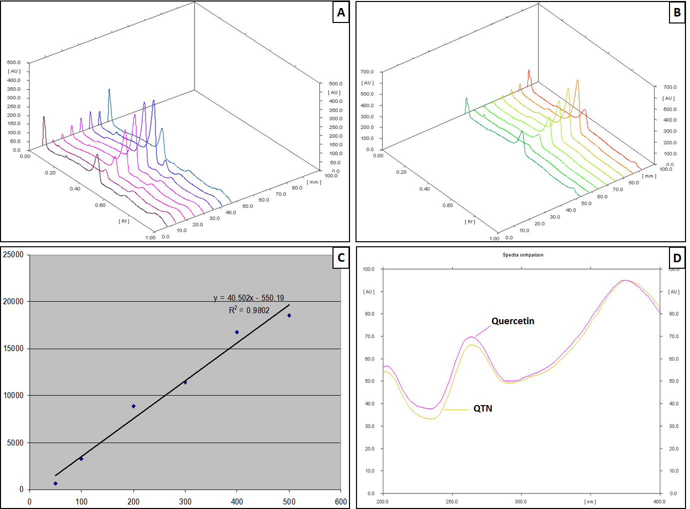
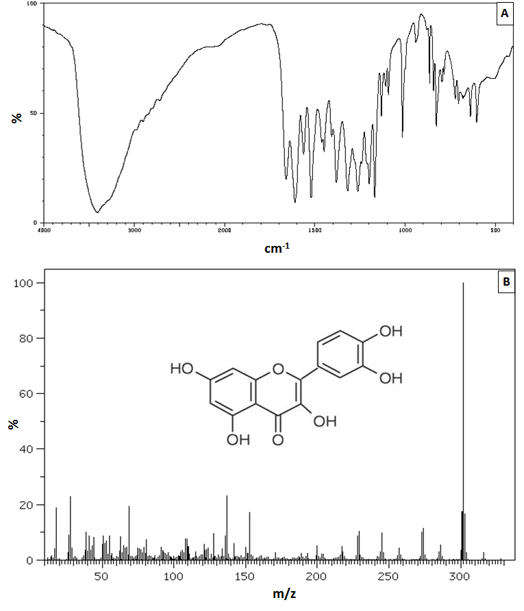
Recently an array of isolated phytoconstituents from herbal origin has been implicated in the management of arthritis clinically. Studies investigated the potential of various moieties such as Pycnogenol® from Pinus pinaster Aiton), Curcuminoids from Curcuma longa, Bromelain from Ananas comosus, etc. for the symptomatic relief of RA 61. Furthermore, a researcher suggested that the moiety bearing carbonyl group at C-4 and a hydroxyl group at C-3 or C-5 in their structure have a chelation ability with metal ions to exert its antioxidant potential 24. In the present investigation, isolated moiety from methanolic extract of leaves of Madhuca indica, i.e., QTN (3,5,7,3′,4′- Pentahydroxy flavone) also possesses such hydroxyl and carbonyl groups in its structure, holding promising antioxidant potential. Thus, QTN can be considered as a potential therapeutic moiety for the management of RA clinically.