DAHP plays a crucial role in the formation of CPNSs
CPNSs obtained by heating a solid mixture of Alg and DAHP in 1:5 mass ratio at 120, 150, 180, 210, and 240°C for 3 h were denoted as CPNSs-120, CPNSs-150, CPNSs-180, CPNSs-210, and CPNSs-240, respectively. The Alg/DAHP mixture was colorless and showed a mild color change to off-white at 120°C. The mixture experienced mild dehydration at 150°C, resulting in light brown hue, and showed higher degree of carbonization at 180°C and above displaying brown or black color (Fig. 1A). The TEM images in Fig. 1B show that the Alg/DAHP mixture without heating had a gel-like structure, and the morphology changed to polymeric form at 120°C. From 150°C and above large sheet-like formation can be observed. At 180°C, the mixture forms 2D layered CPNSs. The resulting CPNSs-180 have sizes ranging between 200 and 500 nm, with a thickness of approximately 1.43 ± 0.25 nm and surface roughness was calculated to be 0.23 nm, as determined by atomic force microscopy (AFM) (Fig. 1C). This thickness is considerably greater than that of single-layered graphene, which ranges from 0.4 to 1.0 nm, and GO, with a range of 0.7−1.2 nm38. The surface roughness (0.23 nm) is higher than that reported (0.2 nm) for single-layer free-standing chemically modified GO39, which suggests the presence of polymeric alginate fragments on the CPNSs. At higher carbonization temperatures of 210 and 240°C, sheet-like structures were formed; however, along with other carbonized products with different morphology and their aggregated forms were adsorbed onto the sheets (TEM images in Fig. 1B).
Alg suspended in sodium phosphate buffer (5 mM, pH 7.4) exhibits a negative charge, characterized by a zeta potential of ca. −50 mV (Supplementary Table 1). The zeta potential slightly changed as the synthesis temperature increased during the preparation of CPNSs to ca. −46 mV for 180°C. However, with subsequent temperature increments, the zeta potential decreased significantly to ca. −11 mV, probably due to a higher degree of carbonization resulting in elimination of carboxylate functional groups. As the synthesis temperature increased, the hydrodynamic size of the CPNSs increased significantly for 210 and 240°C. The thermal-driven dehydration and cross-linking of Alg lead to the formation of sheet-like structures. The significant carbonization at higher temperatures results in the stacking of CPNSs and other carbonized particles to form larger aggregates.
The UV-vis absorption spectra of the CPNSs prepared at different temperatures are presented in Supplementary Fig. 1. CPNSs obtained at 180°C and above showed a band at ca. 285 nm and extending to the visible region of the spectra attributed to the π→π* electronic transition of the aromatic sp2 domains of the C = C and n→π* transition of C = O and N-containing functional groups, respectively40. The baseline of the spectra in the entire wavelength region increased with the increase in the synthesis temperature due to the formation of larger nanosheet structures. Alg exhibited an X-ray diffraction (XRD) pattern with peaks at 2θ of 13.7°, 21.6°, and a broad band around 39.0° corresponding to the (110) plane of polyguluronate unit (G), (200) plane of polymannuronate (M), and amorphous halo, respectively (Supplementary Fig. 2)41. The crystallinity of Alg is due to inter and intramolecular hydrogen bonding. When heated at 150°C and higher, the XRD peak corresponding to guluronate and mannuronate structure disappeared. Instead, a broad peak emerged, indicating the presence of carbonized nanostructures with disordered carbon phases42. The Raman spectra of Alg and CPNSs synthesized at different temperatures were compared with that of GO (Fig. 1D). Alg did not show D and G bands, whereas, CPNSs prepared at temperatures 180°C and above showed the D band around 1350 cm–1 and G band around 1600 cm–1, and their intensity and shape gradually increased and sharpened, respectively, with synthesis temperature. Nevertheless, they are still not well defined as that of GO and therefore, do not reflect structures identical to GO, due to a low degree of graphitization and ultrasmall size of the graphene-like domains43. Heating the precursors such as sodium alginate and sucrose at high temperatures (> 500 oC) and inert atmosphere (e.g., N2 and Ar) produces carbon with well-defined D and G bands44,45. However, in this work, no high temperature or inert atmosphere was used. The G band is due to the formation of in-plane stretching of carbon-carbon bonds in the aromatic rings of graphene-like structures46, revealing that DAHP assists in the alignment of polymer chains and carbonization of Alg to form CPNSs; whereas, the D band indicates the amorphous and disordered nature of graphene-based materials47. The oxygen (O), nitrogen (N), and phosphorous (P) contained functional groups and dopped in the CPNSs disrupt the periodicity and long-range order of the graphene lattice, leading to a loss of crystallinity. Therefore, the CPNSs must be carbonized alginate having carbon-based structures with distinctive polymeric characteristics.
To verify whether Alg could form nanosheets in the presence of other ammonium-, phosphate- or sulfate-containing compounds upon heating at 180°C, we carbonized Alg in the presence of ammonium dihydrogen phosphate ((NH4)H2PO4), phosphoric acid (H3PO4), ammonium hydroxide (NH4OH), NH4OH/H3PO4 mixture, and disodium hydrogen phosphate (Na2HPO4) (Supplementary Fig. 3). It is noteworthy to mention that CPNSs were obtained only with (NH4)H2PO4. Alg did not form nanosheet structures in the presence of H3PO4, NH4OH, H3PO4 and NH4OH mixture, or Na2HPO4. Also, CPNSs were not formed with Alg in the presence of other sulfates, sulfite, and ammonium-related salts such as ammonium sulfite ((NH4)2SO3), ammonium sulfate ((NH4)2SO4), ammonium chloride (NH4Cl), and sodium sulfite (Na2SO3) in the same mass ratio (Supplementary Fig. 4). Thus, we conclude that solid-state heating at 180°C and DAHP have a crucial role in the formation of perfect CPNS structures.
Phosphate diester linkages mediate the formation of CPNSs
In order to investigate the process of CPNSs formation, we performed a time-course analysis of Alg/DAHP mixture that was heated at 180°C for a duration of 3 h (Fig. 2). As the heating progressed, notable changes occurred. Within 5 min, the color of the mixture shifted to a pale brown hue, followed by a transition to dark brown at 15 min (Fig. 2A). Eventually, within 30 min, the mixture turned black due to carbonization. Concurrently, the time-course TEM analysis demonstrated the formation of supramolecular structures by Alg within the initial 5-minute heating period (Fig. 2B). At 15 min, it tended to form thick and large sheet-like structures, which subsequently became thin at 30 min. After 3 h of heating thin-layered clean sheets of varying sizes were formed, likely due to fragmentation during the later stage of the thermal process. The time-course XRD pattern of the products and Alg are presented in Fig. 2C. Over time, a noticeable alteration in the crystallinity of Alg becomes apparent. The crystallinity in Alg arise from the arrangement of G (2θ = 13.7°) and M (2θ = 21.6°) units and the amorphous halo (broad peak centered at 2θ = 39°), which are disrupted and realigned during the heating. The amorphous halo completely disappeared after 15 min. The appearance of a broad peak centered at 2θ of 26.2° indicates very small graphene domains in the carbonized products with highly disordered carbon42,43. This transformation, denoting carbonization, can be observed from 30 min onwards. The hydrodynamic diameter of the CPNSs by heating for different time intervals shows an increase in size to as high as ca. 2200 nm at 30 min, which decreases to ca. 440 nm after 3 h due to fragmentation of the polymer sheets during carbonization (Supplementary Table 2). The TEM-energy-dispersive X-ray spectroscopy (EDS) mapping of CPNSs-180 displayed in Supplementary Fig. 5A confirms the presence of nitrogen and phosphorus, and the HRTEM image and the selective area electron diffraction (SAED) pattern suggest the low crystalline nature of the CPNSs (Supplementary Fig. 5B), in agreement with the XRD pattern. If the 2D structures are not carbon nanosheets, they might instead be black phosphorous nanosheets. It is a thermodynamically stable allotrope of phosphorus with 2D structure of atomic arrangement very similar to that of graphite48. Black phosphorus is highly crystalline and has orthorhombic structure49. However, the XRD patterns in Fig. 2C and Supplementary Fig. 2 and SAED pattern in Supplementary Fig. 5B did not show any crystalline properties corresponding to black phosphorus. Furthermore, black phosphorus is formed only at very high temperature and pressure48. Thus, the possibility of 2D phosphorus allotropes can be ruled out, and we believe the 2D nanostructures obtained by heating a mixture of sodium alginate and diammonium hydrogen phosphate as shown in Fig. 1B must be CPNSs.
The molecular structural changes occurring during the heating were further studied by Fourier-transform infrared spectroscopy (FTIR) (Supplementary Fig. 6). The FTIR spectrum of Alg exhibited specific vibrational modes such as –OH stretching at 3200−3400 cm–1, asymmetric stretching of –COO– at 1610 cm–1, the symmetric stretching of –COO– at 1412 cm–1, and the C–O–C (ring) vibrational modes at 1081 cm–1 of the pyranose rings50. The C–O(H) symmetric vibration peak appeared at 1306 cm–1, and stretching vibration of the C–O–C glycosidic linkage in alginate polymer appeared at 1036 cm–1. FTIR spectra of Alg show significant changes in peaks at lower wavenumber region, 500–1700 cm–1 for different durations of heating with DAHP. Notably, the C–O(H) peak at 1306 cm– 1 decreased significantly with heating time, probably due to the dehydration process. The –C–O(H) symmetric vibration peak at 1306 cm–1 began to disappear within 5 min, meanwhile a new P = O asymmetric stretching peak emerged at 1246 cm–1 and then started disappearing after 30 min. A peak at 1739 cm–1 appeared after 5 min and disappeared after 30 min, indicating some new carbonyl groups of esters are formed and then degraded with time51. After 15 min of heating, new peaks emerged at 1054 cm–1, corresponding to P–O–C stretching in the phosphate ester bond. The Alg after reaction with DAHP and without heating (i.e., 0 min) showed an O–P–O bending vibration peak of the phosphate ester at 546 cm–1, which slightly shifted to 515 cm–1 after 5 min onwards and decreased significantly after 1 h. A new peak emerged at 1054 cm–1, corresponding to P–O–C stretching in the phosphate ester bond. It can be inferred that Alg polymer chains are cross-linked via phosphate diester bonds. During the heating, (NH4)2HPO4 undergoes thermal decomposition to form various chemical species, such as NH3 (g), (NH4)H2PO4 (s), and H3PO4 (l)52. The phosphoric acid reacts with Alg to form esters, which is in agreement with a similar work reported by Marcilla et al., in which the various acid species react with different compounds in tobacco to form esters. Meanwhile, a portion of the NH3 formed by the thermal degradation of (NH4)2HPO4 could form quaternary ammonium salts of the carboxylic acid group52.
We conducted the 31P nuclear magnetic resonance (NMR) spectroscopy analysis of the time course formation of CPNSs-180 (Fig. 2D). The 31P NMR peak of phosphate group appeared at a chemical shift of 0.87 ppm for the purified, non-heated Alg/DAHP mixture, indicating the adsorption of phosphate on the Alg. Upon heating for 1 min at 180°C, the peak shifts a little downfield (to 1.21 ppm), probably due to the formation of monoesters53,54. With a further increase in heating time to 7.5 min, peak resonances toward an up field of the central peak were observed (–5.39, − 8.50, and − 9.26 ppm), depicting the formation of phosphate diesters and pyrophosphate structures. After 15 minutes of heating, the peaks for pyrophosphate (–8.50 and − 9.26 ppm) disappeared, and with further heating the peaks 1.21 and − 5.39 ppm depicting phosphate esters, including monoesters and diesters remained.
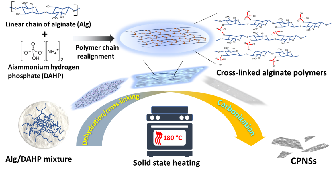
The plausible mechanism of the formation of CPNSs is illustrated in Scheme 1. The formation of diester in solid-state will form bridges between the alginate polymer chains to form 2D polymer sheets. The previous report reveals that dry phosphorylation of starch using orthophosphate occurs through the reactive hydroxyl groups of the starch molecules to form brown-colored products at temperatures of 170°C and higher55. Investigation on the phosphorylation of Alg using urea/phosphate system using various NMR spectroscopy techniques revealed that the most probable site for phosphorylation is the equatorial hydroxyl group of mannuronic acid units in the polymeric chain50. Therefore, it is evident that heating of Alg with DAHP in a solid-state initially leads to the formation of sheet-like structures formed by the cross-linking of Alg polymer chains via phosphate diester linkages. Since this reaction system contains a mixture of monoesters, diesters, and unreacted alginate chains, the carbonized product contains CPNSs along with polymeric Alg featuring nonspecific shapes. The molecular arrangement in pure Alg is mainly due to the solid-state intra- and inter-molecular hydrogen bonding, which are disrupted upon heating above 170°C and produce carbonized products without specific shape56,57. However, the formation of cross-linking among the alginate polymer chains by phosphate ester bonds dominates the formation of a stable 2D structure, resulting in carbonized polymeric nanosheets.
The elemental composition of Alg and the products obtained at various time intervals is presented in Supplementary Table S3. The carbon content (weight percentage) of pure Alg has been found to be 29.16%, which is close to the values reported by previous studies, and some report reveals that it varies with the harvesting season of the algae57–59. The carbon and oxygen contents of Alg after reacting with DAHP (i.e., CPNSs-180 0 min) is determined to be 19.94% and 43.34%, respectively. The carbon content increased up to 38.73% and the oxygen content decreased to 33.93% after being heated at 180°C for 3 h, indicating carbonization to form slightly carbonaceous nanomaterials (i.e., CPNSs). A previous report also suggests that P/N-doped carbon dots synthesized at low temperature (90°C) possess a low degree of carbon content with a weight percentage of 8.6260. Carbonization of precursors in semi-closed atmosphere and temperatures as high as 900°C has been reported to yield carbonized products with higher carbon content (as high as ~ 69%) and low oxygen content (10%)61. The carbonized polymer products have nitrogen doping, and the final CPNSs obtained after 3 h possess 6.8% nitrogen by weight. Alginate polymer has high affinity toward phosphate ions62 and it forms phosphate ester after reacting with the DAHP during the heating process, resulting in high phosphorous content (12.45%) in the CPNSs as determined by inductively coupled plasma optical emission spectroscopy (ICP-OES). The initial heating of Alg/DAHP mixture at 180°C leads to the crosslinking of the hydroxyl groups of the DAHP with the side chain hydroxyl group of the Alg to form phosphate ester bond63. With increase in the reaction time, carbonization progressed, resulting in the decrease in P content. At 3 h of heating intense phosphorylation and phosphorus doping occurred in the CPNSs due to the presence of phosphoric acid and P2O5 in the system thereby increasing the P content to 12.45%. Thus, the CPNSs obtained from Alg by carbonization in the presence of DAHP are N and P co-doped. The C1s, O1s, N1s, and P2p XPS spectra of the CPNSs synthesized at 180°C for 3 h are presented in Supplementary Fig. S7. The CPNSs have oxygen-containing functional groups and N-doping in the form of pyridinic (399.1 eV), graphitic (399.85 eV), and pyrrolic (400.66 eV) nitrogen. The deconvoluted P2p spectra of the CPNSs show peaks at 132.49, 133.01, 133.63, and 134.42 eV corresponding to the presence of P–C, P–O, P–O–C, and P = O bonding, respectively, which confirm phosphorous is incorporated in the CPNSs64.
CPNS-modified filter paper for efficient removal of bacteria
The CPNSs were tested for their antibacterial activity toward Gram-negative (E. coli) and Gram-positive (S. aureus) bacteria and toward V. parahaemolyticus, a Gram-negative bacteria that poses a significant risk in aquaculture. V. parahaemolyticus can rapidly multiply and infect cultured seafood species, which not only damages the health of these species but also increases the risk of foodborne illnesses when these products are consumed65. Different from the GO nanosheets which exhibit antibacterial activity through different mechanisms such as direct interaction with the bacteria through their sharp edges or by wrapping on the bacterial cell66,67, the CPNSs having 2D structure do not show antibacterial activity (Supplementary Fig. 8). The difference in the antibacterial behavior of the CPNSs may be ascribed to only adsorbing bacteria but could not further disrupt the bacterial membranes due to polymeric structures on the CPNSs' surfaces. It has been reported that the functional groups on GO play a crucial role in its antimicrobial activities68. However, these CPNSs, when modified on filter paper could effectively remove bacteria from contaminated water. Incubating the filter paper with CPNSs resulted in the effective coating of the nanosheets on the fibers, as evident from the SEM images in Fig. 3A, however, it did not form a separate layer above the filter paper. The CPNS-modified filter paper was effective in removing V. parahaemolyticus (105 CFU mL–1) from contaminated seawater samples, with CPNSs-180 and CPNSs-210 showing superior effects (> 90%) (Fig. 3B). Notably, neither the bacteria's morphology nor the bacterial membrane was disrupted after passing through the CPNS-modified membrane (Fig. 3C). Adsorbing bacteria without membrane disruption is advantageous, preventing toxin release into the filtrate. For instance, disruption of V. parahaemolyticus’ cell membrane releases toxins like PirA and PirB proteins, which induce necrosis and functional loss in the hepatopancreas of shrimp69.
We further evaluated CPNSs-180-modified filter paper for the removal of E. coli and S. aureus. The removal efficiency for E. coli was less than 30%, and that for S. aureus was around 80% (Fig. 4). The difference in the bacterial removal efficiency of CPNSs-modified filter paper may be attributed to the different bacterial shapes and membrane structures21,24,70. The SEM image of the CPNS-modified filter paper after passing the V. parahaemolyticus bacteria solution clearly shows bacteria trapped on the membrane (Fig. 3D). In contrast to our previous work, graphene oxide@carbon nanogels (GO@CNGs)-modified membrane reported for the removal of bacteria from contaminated water, where the efficiency of the membrane decreases with an increase in water flux21, the efficiency of the CPNS-modified membrane was not affected by the increase in water flux (Supplementary Fig. 9).
Combating V. parahaemolyticus
The efficiency for the removal of V. parahaemolyticus at a higher concentration (107 CFU mL−1) was further evaluated in aquarium condition (2 L water in the aquarium tank) using the CPNS-modified filter paper with a larger surface area (17.34 cm2). The CPNSs-modified filter paper was effective in eliminating > 98% V. parahaemolyticus within 2 h of circulation (Fig. 5A), and no leakage of bacteria was observed from the membrane even after 24 h. The uncoated filter paper (Ctrl) showed removal of ca. 70% within 1 h; however, it decreased with time and down to ~ 17% after 24 h, which shows that though the filter paper can adsorb bacteria, upon continuous passage of water, the bacteria are washed from the membrane back to the solution, due to the large pore size of the membrane and weak affinity toward V. parahaemolyticus. It is noteworthy that the bacterial removal efficiency for the GO-coated filter paper was ~ 74% after 4 h and remained stagnant beyond that time, probably due to the clogging of pores due to fouling of the membrane71; which shows the superior efficacy of our CPNSs-modified filter paper. The decrease in water flux and removal efficiency of the membrane due to the clogging of pores of the membrane is a major drawback in membrane-based filtration systems. Therefore, we further performed the shrimp challenge experiments with the CPNSs-modified filter (Fig. 5B). After challenging white leg shrimp (Litopenaeus vannamei, 10 no. in 2 L sea water) with V. parahaemolyticus (106 CFU mL−1), CPNSs-180-modified filter paper was loaded onto a filter holder, and the aquarium water was circulated through it, and the results were compared with that of the control filter (filter paper without CPNSs-180 coating) and control (without any filter paper or membranes). The shrimps in the CPNSs-modified filter paper group showed 100% survival even after 48 h, while that of the other two groups decreased to < 50%. After 72 h, the survival rate of the shrimp decreased to 10% and 20% for the control and the control filter paper group, respectively; >50% survival rate was observed in CPNSs-modified filter paper group. Therefore, we hope that the CPNS-modified filter may serve its use for filtering out even a very high concentration of bacteria contaminated in aquarium water. Notably, replacing the CPNSs-modified paper every 48 h after filtration could remove the bacteria completely without affecting the survival rate of the shrimp upon Vibrio infection (Supplementary Fig. 10). Though the control filter paper was also replaced every 48 h, only 10% survival was observed after 96 h.
In summary, the methodology presented in this study, which employs low-temperature carbonization of Alg with DAHP, presents a unique approach to prepare 2D carbonized nanomaterials for filtering out pathogens from water. The advantage of this procedure is that it bypasses the need for high temperatures, sophisticated equipment, or hazardous solvents, making it more sustainable and potentially cost-effective. The decomposition of DAHP yields a phosphorylating agent, PO4 3–, which facilitates the phosphorylation of the equatorial hydroxyl group of the mannuronic units within the alginate polymer chain. This results in the creation of phosphate diester linkages between the chains, which are more robust than the solid-state hydrogen bonds in the polysaccharides. Consequently, 2D polymer sheets are formed at the early stage of heating and are subsequently carbonized to produce CPNSs. Moreover, the study effectively demonstrates a practical application of the synthesized CPNSs, using them to enhance the survival rate of shrimp in aquaculture by removing the bacterial strain V. parahaemolyticus. These CPNSs demonstrate notable bacterial adsorption capabilities, particularly towards strains like V. parahaemolyticus. The adsorption property facilitates the development of an efficient bacterial filtration system using ordinary filter paper. Notably, our shrimp challenge experiments indicate an enhanced survival rate among shrimp exposed to V. parahaemolyticus after passing through the CPNSs-modified filter paper. This is an important contribution as it suggests the potential for Alg-CPNSs to improve aquaculture health and productivity. V. parahaemolyticus is responsible for causing acute hepatopancreatic necrosis disease (AHPND) in shrimp, a condition that leads to severe damage and dysfunction in the hepatopancreas. AHPND was accountable for a massive loss of US$44 billion in the global shrimp farming sector between 2010 and 201672. As of the latest estimates, the annual economic losses attributed to AHPND now stand at USD 7 billion 73 . Although this study represents a promising advancement in the synthesis of CPNSs and their application in aquaculture, further exploration and validation of the findings are necessary to understand this approach's potential benefits and drawbacks fully.
We have used Alg as a source of polysaccharide in this study. It would be interesting to explore whether other marine or non-marine polysaccharides can also be used as precursors in preparing CPNSs, which could potentially widen the applications and versatility of the method. On the other hand, our study shows that Alg-derived CPNSs have a high bacterial adsorption capability, but it could benefit from a more detailed examination of the mechanism behind this adsorption. Understanding this mechanism could aid in improving the efficiency and specificity of the CPNSs for bacterial adsorption. While the study focuses on the removal of specific bacterial strains, it would be valuable to examine the effectiveness of these CPNSs in adsorbing other types of contaminants in aquaculture water in the future, which could determine the broader applicability of the approach. The long-term efficacy of these Alg-CPNSs in a realistic aquaculture setting, including their durability and the need for replacement over time, is also essential in the future. While this study observes an immediate increase in the survival rate of shrimp, it might be beneficial to look at the long-term effects of using Alg-CPNSs in the future. This could include the potential impacts on the health and growth of the shrimp over time, as well as any potential environmental impacts. Finally, it would be valuable to assess the environmental impact of using Alg-CPNSs, including a lifecycle analysis, considering the source of the polysaccharides, the process of synthesizing the CPNSs, their use, and eventual disposal.